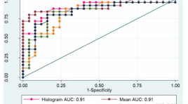
Khảo sát giá trị của các số đo đậm độ trên chụp cắt lớp vi tính trong chẩn đoán adenoma tuyến thượng thận
05/05/2021 17:03:06 | 0 binh luận
SUMMARY: Objective: To describe the characteristics of common adrenal lesions on CT and to compare the diagnostic performance of different methods of measurement in diagnosing adenoma. Materials and Methods: We conducted a retrospective study in 63 patients with adrenal lesions who were performed preoperative CT and had histologic results at the University Medical Center Hospital between January 2016 and January 2020. A radiologist who had more than 10 years of experience of CT measured: lesion size, mean and standard deviation (SD) attenuation value, pixel histogram, calculated p10 based on histogram or following formula P10=mean – (1,282 x SD). The diagnostic performance of mean value, histogram analysis, calculated p10, absoulte and relative washout index were compared. Results: The study was composed of 32 patients with 32 adenomas and 31 patients with 33 non-adenoma lesions. Adenomas were more common in female (n = 26 [81.3%]) than in male patients. However, non-adenoma were almost equally distributed between male and female patients (n = 15 female patients [48. 4%]). The mean age of patients in the adenoma group was 44.3 ± 12.6 years (range, 12-75 years), which is not significantly different from the mean age of 41.2 ± 15.1 years for patients in the non-adenoma group (range, 13-77 years) (p = 0.26). Adenoma group had the mean diameter smaller than non-adenoma lesions (p <0,001) and more homogeneous attenuation pattern (p=0.001). The sensitivity, specificity and accuracy of the mean attenuation analysis, histogram, calculated p10, absoulte and relative washout were (37.5%, 100.0% and 66.7%, cutoff 10 HU), (84.4 %, 82.8% and 83.6%, cutoff 10%), (78.1 %, 82.1% and 80%, cutoff 0), (88 %, 72% and 80%, cutoff 60%), and (84.0 %, 65.4% and 74.5%, cutoff 40%), respectively. Conclusion: The mean attenuation analysis, histogram and calculated p10 play a crucial role in differentiation adrenal adenoma from non-adenoma lesion. The accuracy of histogram analysis and calculated 10th percentile outperformed the mean attenuation as a diag-nostic criterion for adenoma. Keywords: adrenal gland, adenoma, computed tomography
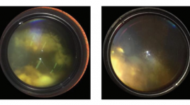
Đánh giá hiệu quả bước đầu điều trị u nguyên bào võng mạc bằng phương pháp truyền hóa chất qua đường động mạch
05/05/2021 12:48:16 | 0 binh luận
SUMMARY Retinoblastoma is the most common malignant tumor eye tumor in children which can cause blindness and even mortality if diagnosis and treatment are delayed. Intra-arterial chemotherapy (IAC) under the guidance of radiology is a new method for the treatment of intraocular retinoblastomas. Purpose: To evaluate the initial outcomes and safety of intra-arterial chemotherapy in treatment retinoblastoma. Materials and Methods: 15 patients diagnosed with intraocular retinoblastoma were clinically and subclinically examined, made imaging diagnosis (ultrasound and MRI), then define retinoblastoma stage by the 2003 international classification, whether or not it has been combine with other methods , and then is indicated for IAC, from October 2017 to June 2019. Each intervention is 3-4 weeks apart. The patients were followed immediately after treatment for the clinical symptoms and after 3-4 weeks after intervention to reassess the tumors and treatment results. Results: 15 patients (including 6 males and 9 females) corresponded to 15 study eyes with an average 35,5 ±20,8 age of months (11 months to 84 months). According to the International classification, there are 3 patients (20%) of group B retinoblastoma, 6 patients (40%) of group C retinoblastoma, 6 patients (40%) of group D retinoblastoma , no patients in group A and group E. The total number of tumors in 15 eyes is 27, which has been treated with intravenous chemotherapy combined with one or two local treatments (laser and cryotherapy) . The total number of interventions was 29, each patiens was treated from 1 to 3 times. As a result, 4 patients (26,7%) had good treatment results, 8 patients (53,3%) had average results, 3 patients (20%) had bad results, then had to enucleate; No patient were distant metastases or death during the follow-up. As a result, we saved 12/15 eyes at the risk of enucleation, 2/15 patients had complications of the treatment process, 1 patient had erythema of the eyelid and forehead skin area, 1 patient had choroidal - retinal atrophy due to occlusive vasculopathy. Conclusions : The method of intra- arterial chemotherapy in the treatment of retinoblastoma has promising, safe, and effective initial results in maximally salvaging the eye. Keyword: Retinoblastoma, Intra-arterial chemotherapy (IAC).
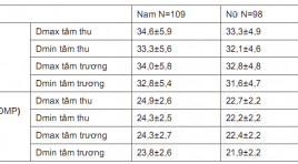
Khảo sát mối liên quan độ giãn động mạch chủ ngực với mức độ tổn thương động mạch vành trên cắt lớp vi tính 256 dãy
05/05/2021 12:24:42 | 0 binh luận
SUMMARY: Objective: The relationship between the thoracic aortic dilatation and the degree of coronary artery injury by 256-slice CT scanning. Subjects and methods: Descriptive cross-sectionalnstudy was performed on 207 patients who was performed coronary angiography by 256-slice CT scanning at Bach Mai hospital from January 2019 to August 2019. Results : 207 patients in study, 107 males (52,7%), 98 females (47,3%). Average age is 62,2±10,3. There was 34 patients with thoracic aortic dilatation (16,4%). There was the significant difference of the Dmax, Dmin diameter at levels (sinuses of Valsalva, sinotubular junction, ascending aorta, descending aorta) and Dmin diameter at level of annulus between the systolic phase and diastolic phase (p<0,001. There was relationship between dilatation of thoracic aorta with valve calcium score (p<0,001). There was not relationship between dilatation of thoracic aorta with coronary artery calcium score and degree of coronary artery stenosis due to atheroma. Conclusion: This study showed significant differences of the thoracic aortic dimensions, between systolic phase and diastolic phase. Dilatation of thoracic aorta was associated with aortic valve calcium score but not with degree of coronary artery injury. Key words: thoracic aortic dilatation, coronary artery, 256-slice CT.
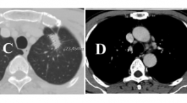
Đặc điểm hình ảnh cắt lớp vi tính 256 dãy trong chẩn đoán và theo dõi điều trị ung thư phổi biểu mô tuyến có đột biến EGFR
05/05/2021 12:40:07 | 0 binh luận
SUMMARY: Purpose : The characteristics of 256-slice computer tomography in patients with EGFR-mutated lung adenocarcinoma and the tumor response to targeted therapy according to RECIST 1.1 criteria were taken into investigation in this study. Methods: 32 patients with EGFR- mutated lung adenocarcinoma received TKI (tyrosine kinase inhibitor) were underwent 256-slice CT scanner before treatment and 3 months, 6 months of treatment, from July 2017 to July 2019 at Friendship Hospital. Results: Before therapy, on 256-slice CT scanner in patients with EGFR-mutated lung adenocarcinoma, we observed tumors on the right in 56.3% of patients, tumors in the upper lobe in 56.3%, tumors size larger than 3 cm in 81.3%, lobulated or spiculated margin in 100%, pleural effusion in 50%, air bronchogram in 34.4% and cavitation in 3.1%. Metastases was present in lymph nodes in 68.8%, followed by metastatic deposits in lung (56.3%), bone (53.1%), brain (9.4%), adrenal gland (9.4%) and liver (6.3%). After 3 months of treatment , the percentage of partial response was 34.4%, stable disease was 59.4% and progressive disease was 6.3%; after 6 months, these ratio were 40.6%, 43.8% and 15.6% respectively. Conclusion: Common CT scanner features in patients with EGFRmutated lung adenocarcinoma were lobulated or spiculated margin, size larger than 3cm and pleural effusion; cavitation was rarely noticed. Metastases usually presented in lymph node, lung and bone. The disease control rate at 3 months and 6 months of therapy were 93.7% and 84.4% respectively. CT scanner is a potential tool for evaluating tumor response and improving effective treatment in patients with lung cancer received TKI. Keywords: lung adenocarcinoma, EGFR mutation, computed tomography.

Vai trò của chuỗi xung 3D TOF MRA trong đánh giá rò động tĩnh mạch màng cứng nội sọ
05/05/2021 17:25:06 | 0 binh luận
SUMMARY Objective: We evaluate the role of 3D TOF MRA in diagnosis of the fistula location and cortical venous drainage of intracranial dural arteriovenous fistula (DAVF) in comparison with Digital Subtraction Angiography Subjects and methods: Prospective study between 1/2015 and 4/2019, 93 patients (35 male, 58 female), aged from 11 to 88 (mean 55), diagnosed of DAVF on conventional MRI with 3D TOF MRA and then underwent DSA for confirming the diagnosis. In three cases, 3D TOF MRA field of view is not enough to evaluate the fistula site but enough to evaluate the present of abnormal hyperintensity in cortical veins. Results: In our study, source images from 3D TOF MRA showed high sensitivity and positive predictive values (up to 100%, 97,6% respectively) in diagnosis of DAVF (n=90) and detected cortical venous drainage (n=93) with high specificity and high positive predictive value (100%). Cohen's Kappa coefficient showed very good agreement between 3D TOF MRA and DSA in detecting the location of DAVF. In 4 falsepositive cases, 1 case showed high-intensity area in the transverse-sigmoid venous sinous on 3D TOF MRA due to thrombosis and 3 other cases showed high-intensity area in the cavernous sinus causing misdiagnosis of DAVF. Conclusion: The use of 3D TOF MRA source images is valuable in diagnosing the location of fistulas and cortical venous drainage in intracranial DAVF. False-positive cases in this study suggested that MRA with contrast could ameliorate the limitation of 3D TOF MRA. Keywords: DAVF, dural arteriovenous fistula, cortical venous reflux, cortical venous drainage, 3D TOF MRA, DSA, false-positive.
Đặc điểm lâm sàng và siêu âm xoắn tinh hoàn chu sinh báo cáo loạt ca và hồi cứu y văn
02/06/2020 09:46:31 | 0 binh luận
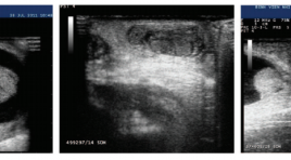
Perinatal testicular torsion clinical and sonographics findings SUMMARY Objective: Perinatal testicular torsion either occurring prenatally in utero or postnatally in the first month of life, is surgical emergency with hope salvaging of testis. A challenge to clinicians and radiologist. With sonographics findings as small size of testis, heterogenous echostructure, thickened tunica albuginea with rim hyperechoic (calcification), to suggest testicular torsion. Methods: Cases report Results : From January 2015 to May 2019, we had 11 patients with perinatal testicular torsion introduced into the study batch. The average age is 8.2 days. One song twists on two sides, on the left 7 shifts. The time of detection after birth, an average of 1.5 days, no cases of prenatal ultrasound were detected. The average time of hospital admission is 17 days. 100% normal birth, full month.Ultrasound signs: testicular big size 7/12 (58.3%), heterogeneous parenchyma structure 11/12 (91.7%), calcareous membrane calcification 2/12 (16.6%), hydrocephalus, heterogeneous fluid, 7/12 fibrin (58.3%), enlarged stalks, edema 3/12 (25%). Mark Whirpool positive 8/12 (67%), central blood loss 11/12 (91.7%). The rate of testicular removal is 10/12 (83.3%). Conclusions: Twisted perinatal testicular, rare surgical emergency, causes purple swelling of the scrotum and requires early diagnosis and surgical intervention. High-value color doppler ultrasound determines testicular twisting and eliminates the causes of swelling and pain in the scrotum. Keywords: testicular torsion, neonate, ultrasound.
Siêu âm đàn hồi trong chẩn đoán ung thư tuyến tiền liệt: đánh giá bước đầu qua 101 trường hợp
02/06/2020 09:42:08 | 0 binh luận
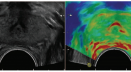
Transrectal sonoelastography in detection of prostatecancer: initial assessment of 101 cases SUMMARY Purpose Evaluate the efficiency of transrectal strain elastography (SE) and transrectal B-mode ultrasonography (TRUS) in determining prostate cancer. Material & Methods From 20/6/2015 to 10/9/2015, There are 101 patients with PSA level of higher 4ng/ml have been selected. Abnormal echo regions in prostate were found via conventional TRUS in those patients, then Abnormal echo regions would be evaluated by real-time strain ultrasound elastography. Patients have undergone six core biopsies by transperineal approach. Experimental studies Results Comparising between 3 methods: B-mode TRUS, strain elastography and DRE about sensitivity, specificity, positive predictive value (PPV) and negative predictive value (NPV). (table) Table.Comparising between 3 methods: B-mode TRUS, strain elastography and DRE. Sensitivity (%) Specificity (%) PPV (%) NPV (%) B-mode 88.5 55.1 67.6 81.8 Strain-elasto 94.2 65.3 74.2 91.4 DRE 69.2 98 98 75 Conclusions: Sonoelastographyprovidesmore information to detect prostate cancer and biopsy guidance. SE reached a higher sensitivity and specificity than B-mode US in the detection prostate cancer.Strain ultrasound elastography can be used as routine as colour Doppler. Key words: sonelastography, strain ultrasound elastography, prostate cancer, ultrasound.
Siêu âm chẩn đoán bệnh lý cường tuyến cận giáp nguyên phát: nhân 26 trường hợp được phát hiện tại hệ thống y khoa medic TPHCM và Cần Thơ
02/06/2020 09:26:46 | 0 binh luận
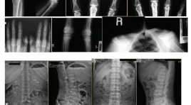
SUMMARY Primary hyperparathyroidism is a rare disease, difficult to diagnose early due to non specific clinical findings. The radiographic signs often misinterpreted. X-ray signs often show a condition of osterosporosis, fractures or urinary tract stones. Biochemical examination are still insufficient at some hospital, so a majority of cases were detected too late with severe skeletal sequelae, and sometimes irreversible. Retrospective study of 26 cases diagnosed at Medic Medical system and follow-up after treatment, we observed that: - 26 Patients: 8 Man (31%) 18 Female (69%), 15YO-54YO, Average:38.5YO - Clinical symtoms are often nonspecific, often showing pathology of the skeletal system 92%, urinary stones : 46%, digestive tract: 77%- 85%. - Radiographic skeletal specific sign of of osterosporosis77% , fractures 42%, Osteolytic 52% or urinary tract stones.46%. suggest diagnosing hyperparathyroidism. - PTH (Parathyroid hormone) value are elevated in 100% of cases. - Neck ultrasound: 100% of patients (26/26) have parathyroid adenoma, most on one side of the lower lobe is the main cause of hyperparathyroidism. - Primary hyperparathyroidism are radically cured by surgically removal of the adenoma, so the early diagnosis of this
Bạn Đọc Quan tâm
Sự kiện sắp diễn ra
Thông tin đào tạo
- Những cạm bẫy trong CĐHA vú và vai trò của trí tuệ nhân tạo
- Hội thảo trực tuyến "Cắt lớp vi tính đếm Photon: từ lý thuyết tới thực tiễn lâm sàng”
- CHƯƠNG TRÌNH ĐÀO TẠO LIÊN TỤC VỀ HÌNH ẢNH HỌC THẦN KINH: BÀI 3: U não trong trục
- Danh sách học viên đạt chứng chỉ CME khóa học "Cập nhật RSNA 2021: Công nghệ mới trong Kỷ nguyên mới"
- Danh sách học viên đạt chứng chỉ CME khóa học "Đánh giá chức năng thất phải trên siêu âm đánh dấu mô cơ tim"


