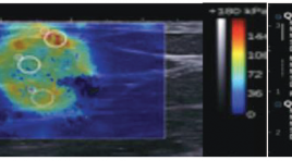
Đánh giá giá trị chẩn đoán ung thư vú của siêu âm đàn hồi nén và sóng biến dạng
05/05/2021 17:50:03 | 0 binh luận
SUMMARY Objective : Evaluating the value of B-mode ultrasound and elastography ultrasound in the diagnosis of breast cancer. Methods: Breast lesion patients were classified BIRADS from 3 to 5 after underwent B-mode ultrasound and elastography ultrasound examination and done biopsy to have histopathological results at Bach Mai Hospital from July 2019 to February 2020. Results: The cut-off value of fat-to-lesion ratio is 28,4 with sensitivity (Se), specificity(Sp) and accuracy (Acc) were 76,9%; 93,3%; 85,7% respectively. The cut-off value of Elasto/B-mode ratio is 1 with Se, Sp, Acc were 100%, 73,3%; 85,7%. Se, Sp and Acc of shear-wave elastography were 100%; 97,8% and 97,5% respectively with the cut-off value is 36 kPa. Sp, Se and Acc of Tsukuba score were respectively 84,6%; 88,9%; 86,9%. B-mode ultrasound combine with shear-wave elastography has highest Se and Sp were 100%; 91,1% respectively. Conclusion: Elastography ultrasound combine with B-mode ultrasound can upgrade or downgrade the BIRADS level, so they can increase accuracy to diagnose breast cancer especially BIRADS 3 or 4a lesions.
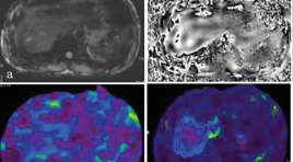
Cộng hưởng từ đàn hồi gan: nguyên lý, kỹ thuật và ứng dụng lâm sàng
05/05/2021 17:35:14 | 0 binh luận
SUMMARY Magnetic resonance elastography of liver is a technique for evaluation of liver stiffness and is currently considered the most accurate non-invasive imaging technology for evaluation of liver fibrosis. MRE used a phase-contrast sequence which examinate the propagation of mechnical waves in the liver parenchyma to generate to images depicting the stiffness of tissure. Hepatic fibrosis increases the stiffness. MRE can detected early fibrosis and evaluated stage of liver fibrosis through changes of the stiffness of liver parenchyma. MRE is more accurate than US elastography in the evaluation liver fibrosis. MRE has been used commonly in the word. Choray hospital is one of the first hospital to implement MRE in Viet Nam. In this lecture, we will introduce the basic principles, techniques and clinical applications of liver MRE. Keywords: Magnetic resonance elastography, liver, stiffness, fibrosis, cirrosis, principle, technique, applications
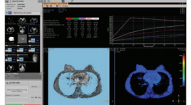
Nghiên cứu đặc điểm hình ảnh và giá trị của cộng hưởng từ 1.5 TESLA trong chẩn đoán và định hướng điều trị ung thư vú
05/05/2021 17:32:49 | 0 binh luận
SUMMARY Objective: Study imaging characteristics and values of 1.5 Tesla Magnetic Resonance Imaging in diagnosis and treatment of breast cancer. Material and Methods: A prospective study on 52 patients with breast cancer underwent( received) breast MRI before treatment Results: The common signs is mass shape (88,5%) with irregular margins or dendrites (100%), heterogeneous contrast enhancement (96,2%) strong penetration in the first 2 minutes after contrast injection (84,6%), kinetic curve is mainly type II or type III (88,4%). Multiple lesion has 13 (25%). Invasive ductal carcinomas is 49 (94,2%) Conclusion: General features of breast cancer MRI are mainly mass with irregular margins or dendrites, and heterogeneous contrast enhancement, mainly strong penetration in the first 2 minutes after contrast injection , no case has poor absorbtion, plateau kinetics and washout kinetics are major. Key Words: Magnetic resonance imaging, breast cancer.
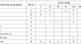
Nghiên cứu đắc điểm hình ảnh chụp cắt lớp vi tính trong chẩn đoán u nhầy mũi xoang
05/05/2021 17:27:41 | 1 binh luận
SUMMARY Objective: Our aim was to describe computed tomography image characteristic of paranasal sinus mucoccele. Method: A retrospective and prospective, axial-descriptive study in paranasal sinus mucoccele patients who were treated at National Otorhinorarynology from December 2016 to July 2019. Results: 32 patients were enrolled. Mean age was 52.9 (22-83) with M/F=1. Involved sinus distribution including 37.5% frontal-ethmoid, 31.3% frontal, 9.4% ethmoid, 6.3% ethmoid-maxillary, 6.3% sphenoid and 9.4% maxillary sinus. 96.9% tumors were hyperdense or isodense (compared to brain tissue) in pre-contrast CT Scanner. In the postcontrast image: 84.4% of tumors did not marked enhance while another 15.6% had rim enhance which could be explained due to patients clinical acute symtoms of infection. In characteristic, 87.5% tumors had erosion of sinus bone (65.6% lamina papiracea, 25% orbital roof and 25% ethmoidal roof). Regarding to the spread of mucocele: 68.75% tumors had intraorbital extension while 15.6% had intracranial extension. No record of nerve or cavernous sinus invasions. Conclusion: A sinus computed tomography scan with contrast material was highly valuable in diagnosis of paranasal sinus mucocele and contribute to the planning of surgery. Key words: paranasal sinus mucocele, computed tomography
Đánh giá hiệu quả bước đầu trong loại bỏ các tổn thương vú lành tính bằng sinh thiết vú có hỗ trợ hút chân không tại Trung tâm Điện quang Bệnh viện BẠCH Mai
05/05/2021 17:23:21 | 0 binh luận
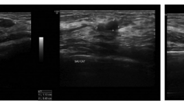
SUMMARY Objective: A experimental research was performed in radiology center of Bach Mai hospital to evaluate the initial efficacy in the removal of breast benign lesions by vacuum-assisted biopsy. Subjects and methods: There is a prospective intervention study in 32 female patients with 45 breast benign lesions with needle aspiration vacuum-assisted biopsy under ultrasound guidance from January 2018 to December 2018. Results: The mean age is 36.5 years old. The 20-40 years old group is most common (60.0%). The average size of the lesions measuring on ultrasound is 12.9mm. The average number of samples is 13.2 with the average time of cutting is 14.5 minutes. The most common abnormality pathology is breast fibroadenoma (62.2%). Fibrocystic breast disease accounts for 17.8% of all lesions, which is second highest rate. The main complications after biopsy are pain and hematoma in tissu. 78.8% of patients after treatment don’t have to take Paracetamol. The average size of hematoma after 1 month with 10G needle is 4.8mm; with 8G needle is 6.3mm. Conclusion: Vacuum-assisted breast biopsy is an effective and safe method for removal benign breast lesions. This method is also highly aesthetic. The anapathology results based on this method are reliable, especially for small lesions. Key words: Vacuum-assisted biopsy, mammotome, breast benign lesions.

Đặc điểm hình ảnh siêu âm của ung thư tuyến vú tại Bệnh viện Bạch Mai
05/05/2021 17:17:10 | 0 binh luận
SUMMARY Objective: Describer the ultrasoundgraphic appearance of breast cancer. Methods: Data was collected from 49 patients who underwent breast ultrasound, guided interventional and operated procedures from august 2017 to june 2019, diagnosis of breast cancer. Study the imaging findings based on guidline of ACR BI-RADS 2013. Results: Ultrasoundgraphic appearance: 100% breast cancer lesions are mass, 85,7% tumors having irregular shape. There are 75,5% tumor having not orientation parallel to skin surface. Spiculated and angular margin 63,3%, 73,5% tumors having hypoechoic, 40,8% tumor having calcification in mass. Conclusion: Most of breast cancer tumors having mass finding, irregular shape. The findings as orientation not parallel to skin surface, speculated or angular margin, hypoechoic and having calcification in mass are suspected to breast cancer. Keywords: Breast cancer, ultrasound.
Ứng dụng X quang cắt lớp vi tính trong đánh giá mối tương quan giữa kích thước mạch máu gan với các yếu tố tuổi, giới tính và dạng giải phẫu
05/05/2021 17:13:29 | 0 binh luận
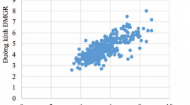
SUMMARY Objective: The study aimed to determine the correlation between dimensions of hepatic vessels (including hepatic arteries, portal and hepatic veins) and aging, gender and anatomical variants factors, using multidetector computed tomography (MDCT). Design: We conducted a retrospective study of 611 adults (344 Male, 277 Female, mean age 55.0 ± 13.1 years), who were clinically examined and underwent abdominal MDCT with iodinated contrast agents for different complaints at the University Medical Center between August 2017 and August 2018. MDCT images were stored on the picture archiving and communication system (PACS) and processed to create multiplanar reformation (MPR), curved planar reformation (CPR), maximum intensity projection (MIP), volume rendering (VR) images which were subsequently used to measure the length and diameter of hepatic arteries, portal, and hepatic veins. Also to determine the correlation between dimensions of hepatic vessels and aging, gender and anatomical variants factors. Results: The average diameter of common hepatic artery in the variant group was smaller than in the normal group. The length of common hepatic artery increased with age (P<0.05). There was a strong correlation (R = 0.77, P <0.05 ) for the diameters between the common hepatic and proper hepatic arteries. The diameters of main portal vein, right portal vein and left portal vein decreased with age (P<0.05). The male group had diameters of hepatic artery and portal vein greater than the female one. There is no correlaton between the dimension of hepatic veins and some factors of anatomy, age and gender. Conclusions: MDCT might be considered a safe and accurate imaging modality with high sensitivity in assessing the correlation between dimensions of hepatic vessels and the factors of age, gender and anatomical variants.
Khảo sát hình thái mạch máu gan và các biến thể giải phẫu bằng hình chụp x quang cắt lớp vi tính
05/05/2021 17:08:09 | 0 binh luận
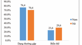
SUMMARY Aims: To identify the prevalence of normal anatomy and vascular variants of hepatic vessels by using multidetector computed tomography (MDCT). Methods: We conducted a retrospective study of 611 adults who, for different reasons, came to university medical center Hochiminh city, all underwent abdominal MDCT with contrast material. From the data stored in PACS, using image processing applications (MPR, CPR, MIP, VR) to investigate anatomy of the hepatic artery, the portal vein and the hepatic vein systems. Results: From 08/2017 to 08/2018 at University Medical Center, HCMC, a total of 564 of the 611 patients had the common hepatic artery originated the celiac axis, anatomic variations were seen in 7.7% of patients. 9 of 10 anatomic types (criteria laid by Michels ‘s classification) were identified in our study, type 1 (the typical type) seen in 73.6% cases, we also found 5 other types of the hepatic artery not mentioned in Michels classification (3.1%). Normal anatomy of portal vein was identified in 84.1% of cases. Trifurcation - the most popular type of portal vein variants was seen in 11.3% of patients. The common trunk between the left hepatic vein and the median hepatic vein was seen in 58.6% cases. The accessory right hepatic veins were identified in 45.5% of the patients. There were not correlation between kinds of hepatic vascular variants (p>0.05). Conclusion : Hepatic vascular anatomy plays an important role in hepatectomy, pancreaticoduodenectommy and living liver transplantation. Because of high prevalence of vascular variants, knowledge of these abnormalities and their frequency is of major importance for the surgeon * Bệnh viện ĐHYD TP. HCM. as well as the radiologist.
Bạn Đọc Quan tâm
Sự kiện sắp diễn ra
Thông tin đào tạo
- Những cạm bẫy trong CĐHA vú và vai trò của trí tuệ nhân tạo
- Hội thảo trực tuyến "Cắt lớp vi tính đếm Photon: từ lý thuyết tới thực tiễn lâm sàng”
- CHƯƠNG TRÌNH ĐÀO TẠO LIÊN TỤC VỀ HÌNH ẢNH HỌC THẦN KINH: BÀI 3: U não trong trục
- Danh sách học viên đạt chứng chỉ CME khóa học "Cập nhật RSNA 2021: Công nghệ mới trong Kỷ nguyên mới"
- Danh sách học viên đạt chứng chỉ CME khóa học "Đánh giá chức năng thất phải trên siêu âm đánh dấu mô cơ tim"


