


ỨNG DỤNG HÌNH ẢNH FDG PET TRONG ĐỘNG KINH
23/01/2024 11:52:21 | 0 binh luận
TÓM TẮT Một số trường hợp bệnh nhân động kinh cục bộ không đáp ứng với điều trị bằng thuốc thì việc phẫu thuật có thể mang lại nhiều lợi ích. Hình ảnh cắt lớp phát xạ positron (positron emission tomography - PET) với thuốc phóng xạ 18F-Fluorodeoxyglucose (FDG PET) đánh giá chuyển hóa glucose của não là một kỹ thuật đã được ứng dụng rộng rãi trong việc đánh giá trước phẫu thuật ở những bệnh nhân động kinh kháng trị mà có hình ảnh cộng hưởng từ và điện não đồ không tương hợp trong việc xác định vùng sinh động kinh. FDG PET giúp phát hiện vùng sinh động kinh, phân vùng động kinh, giúp xác định mức độ lan rộng của vùng sinh động kinh. FDG PET còn cung cấp thêm thông tin quan trọng về tình trạng chức năng của phần còn lại của não. Ngoài ra, các đặc điểm chuyển hóa trên hình PET cũng góp phần tiên lượng hiểu quả phẫu thuật. Ghi hình PET thường được thực hiện ngoài cơn. Sử dụng hình ảnh MRI chụp trước đó hòa trộn với hình PET để xác định vị trí giải phẫu. Bài báo cáo này nhằm giới thiệu quy trình ghi hình FDG PET não, cách phân tích hình ảnh và vai trò của FDG PET trong đánh giá trước phẫu thuật động kinh trong điều kiện nguồn lực còn hạn chế ở Việt Nam. Từ khóa: FDG PET, động kinh kháng trị

KẾT QUẢ BƯỚC ĐẦU ĐIỀU TRỊ UNG THƯ TUYẾN GIÁP THỂ BIỆT HÓA SAU PHẪU THUẬT BẰNG 131I TẠI KHOA XẠ TRỊ VÀ Y HỌC HẠT NHÂN - TRUNG TÂM UNG BƯỚU BỆNH VIỆN E
23/01/2024 10:55:27 | 0 binh luận
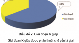
TÓM TẮT Mục tiêu: 1. Đánh giá kết quả điều trị bệnh nhân ung thư tuyến giáp thể biệt hóa sau mổ bằng 131I tại Khoa Xạ trị và Y học hạt nhân, Trung tâm Ung bướu Bệnh viện E. 2. Đánh giá một số yếu tố liên quan đến kết quả điều trị. Đối tượng và phương pháp nghiên cứu: Nghiên cứu mô tả cắt ngang tiến hành trên 67 bệnh nhân ung thư tuyến giáp thể biệt hóa được điều trị Khoa Xạ trị và Y học hạt nhân, Trung tâm Ung bướu Bệnh viện E Trung ương. Kết quả: Tuổi trung bình của bệnh nhân là 41,3 ± 12,7, nữ : nam = 3,01 : 1, 91% ung thư tuyến giáp là thể nhú, trong đó 43,3 % chưa có di căn, 55,2 % di căn hạch cổ, 1,5 % có di căn xa. Ở giai đoạn I, II và III chiếm tỷ lệ 98.5%. Bệnh nhân đáp ứng hoàn toàn sau điều trị 1, 2, 3 liều 131I là 82 % ,94 %, 97,5 % . Tổng liều điều trị trung bình chúng tôi đã sử dụng là 112,5 ± 31,2 mCi, số lần điều trị trung bình là 1,03 ± 0,3 lần. Ở bệnh nhân chưa có di căn xa, liều 131I từ 30 – 50 mCi có giá trị hủy mô giáp còn sót tương tự như liều 100 mCi. Kết quả điều trị ở bệnh nhân < 55 tuổi tốt hơn ≥ 55 tuổi, bệnh nhân chưa có di căn và di căn hạch cổ tốt hơn có di căn xa. Giai đoạn I, II, III tốt hơn giai đoạn IV. Bệnh nhân có Tg < 10 ng/dL tốt hơn Tg ≥ 10 ng/dL, A-Tg < 100 IU/mL tốt hơn A-Tg ≥ 100 IU/mL. Kết luận: Bệnh nhân nhận liều 131I thấp (30 –50 mCi) có hiệu quả điều trị tương tự liều cao (100 mCi) ở bệnh nhân chưa có di căn xa. 82,0% bệnh nhân có kết quả tốt ngay sau 1 liều 131I. Với liều điều trị trung bình cho một bệnh nhân là 102,5 ± 33,6 mCi, sau 1,03 ± 0,3 lần điều trị, 92 % bệnh nhân đáp ứng hoàn toàn sau 2 liều I131, 97% bệnh nhân đáp ứng hoàn toàn sau 3 liều điều trị I131. Bệnh nhân có tuổi nhỏ hơn 55 tuổi, chưa có di căn xa, ung thư ở các giai đoạn sớm, nồng độ Tg, A-Tg thấp có đáp ứng với phương pháp điều trị tốt hơn. Từ khóa: Ung thư tuyến giáp, Ung thư tuyến giáp thể biệt hóa, 131I, Thyroglobulin, Anti Thyroglobulin, Xạ hình toàn thân với I-131.
BƯỚC ĐẦU NGHIÊN CỨU HIỆU QUẢ SỚM CỦA PHƯƠNG PHÁP ĐIỀU TRỊ UNG THƯ BIỂU MÔ TẾ BÀO GAN TIẾN TRIỂN...
22/01/2024 16:01:55 | 0 binh luận
TÓM TẮT Mục tiêu: Đánh giá kết quả sớm của phương pháp đặt cổng truyền hóa chất động mạch gan bằng phác đồ Lipiodol kết hợp với Cisplatin và 5 FU trên bệnh nhân UTBMTBG giai đoạn tiến triển tại chỗ. Đối tượng và phương pháp nghiên cứu: Chúng tôi thực hiện can thiệp lâm sàng tiến cứu, có theo dõi dọc trên đối tượng là 10 BN UTBMTBG giai đoạn tiến triển. Mỗi bệnh nhân được đặt cổng truyền hoá chất dưới da phục vụ truyền hoá chất động mạch gan. Phác đồ điều trị bao gồm Lipiodol, Cisplastin và 5FU. Sau 1 tháng điều trị, các bệnh nhân được thăm khám, chụp CLVT và phân loại đáp ứng thành đáp ứng hoàn toàn, đáp ứng một phần, bệnh ổn định và bệnh tiến triển theo tiêu chuẩn mRECIST. Kết quả : 6 BN (60%) có đáp ứng sớm sau điều trị, trong đó 1 BN (10%) có đáp ứng hoàn toàn và 5BN (50%) đáp ứng một phần. Một bệnh nhân có bệnh tiến triển sau điều trị, và 2 bệnh nhân bệnh ổn định. Kích thước phần ngấm thuốc của khối u giảm từ 102±41mm xuống còn 83±53mm, có ý nghĩa thống kê với p <0.05. Ngoài ra, HKTMC thoái triển trên 2 BN, và chức năng gan được cải thiện trên 2 BN. Có 3 BN xuất hiện biến chứng, bao gồm nhiễm trùng, tụ máu, tắc và vỡ cổng truyền. Kết luận: Truyền hoá chất động mạch gan với phác đồ Lipiodol kết hợp Cisplastin và 5FU đạt tỷ lệ đáp ứng sớm cao, có thể gây thoái triển HKTMC và cải thiện chức năng gan.
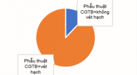
ĐÁNH GIÁ HIỆU QUẢ PHẪU THUẬT VÉT HẠCH TÁI PHÁT Ở BỆNH NHÂN UNG THƯ TUYẾN GIÁP THỂ BIỆT HOÁ SAU ĐIỀU TRỊ 131I TẠI VIỆN Y HỌC PHÓNG XẠ VÀ U BƯỚU QUÂN ĐỘI
18/10/2023 09:46:18 | 0 binh luận
SUMMARY Objectives: to describe the clinical and subclinical symptoms and evaluate the response after recurrent lymph node dissection of differentiated thyroid cancer patients after 131I treatment. Subjects: 50 differentiated thyroid cancer patients after 131I treatment, recurrence for the first time. Study methods: Retrospective and prospective description, data collection through patients’ medical records. Results: the mean age of the study patients was: 43.5 ± 12.6, the most common age is <55 years old with the rate of 80%, the female/ male ratio=3.5/1. The group of patients with Tg < 1 ng/ml accounted for the majority with 33/50 patients (66%). The median Tg before and after surgery was 2.02 ng/ml and 0.35 ng/ml, respectively. After surgery, response achieved in 98% of patients, mainly complete response and incomplete response in biochemical, accounted for 40%, 36%. Lymph node dissection reduced Tg in 78% of patients. Reccurent lymph nodes mainly observed in group IV, group III and group VI. The recurrence of lymph nodes was mainly in the central and cervical regions on the same side of the primary tumor. Conclusion: Surgery is the preferred first-line treatment for the reccurrent lymph node thyroid cancer patients. It changes response assessment, improve prognosis for reccurence patients after 131I treatment. Keywords: Differentiated thyroid cancer, response after recurrent lymph node dissection, Tg
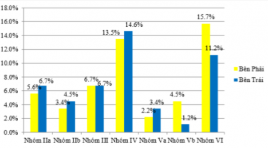
NGHIÊN CỨU GIÁ TRỊ CỦA KỸ THUẬT CHỌC HÚT TẾ BÀO BẰNG KIM NHỎ DƯỚI HƯỚNG DẪN CỦA SIÊU ÂM TRONG CHẨN ĐOÁN HẠCH DI CĂN Ở BỆNH NHÂN UNG THƯ TUYẾN GIÁP THỂ BIỆT HÓA
16/10/2023 17:08:53 | 0 binh luận
SUMMARY Background: Ultrasonic-guided fine needle aspiration cytology is a minimally invasive diagnostic technique with the highest accuracy and sensitivity in the diagnosis of lymph node metastasis. Objectives: The study consists of 2 objectives: 1) Describe clinical and subclinical characteristics in treated patients with differentiated thyroid cancer; Evaluation of the role of ultrasound-guided fine needle aspiration cytology in the diagnosis of lymph node metastasis in patients with differentiated thyroid cancer. Materials and methods: A cross-sectional descriptive study design was conducted on 89 patients diagnosed with differentiated thyroid cancer and found that the cervical lymph nodes were potentially malignant under ultrasonography and indicated fine-needle aspiration cytology under ultrasound guidance. Results: Among 89 research subjects, small cell aspiration of lymph node groups, group IV and VI lymph nodes with suspicion of malignancy accounted for the highest proportion of 25/89 nodes (28.1%) fine needle aspiration cytology, in which positive accounts for 30.1%. Sensitivity, specificity, negative predictive value, false positive rate, false negative rate, and cytology accuracy were 90%; 100%; 100%; 52.9%.0%, 10.1%, 91.01%. Conclusion: Ultrasonic-guided fine needle aspiration cytology for the diagnosis of lymph node metastasis in differentiated thyroid cancer has a low false-positive rate, high accuracy, accurately reflecting the presence or absence of underlying cervical lymph node metastasis. Avoid complications and thoroughly treat metastatic disease for patients. Keywords: Ultrasonic-guided fine needle aspiration cytology, metastatic lymph nodes, differentiated thyroid cancers
NGHIÊN CỨU MỐI LIÊN QUAN GIỮA NỒNG ĐỘ THYROGLOBULIN, THỜI GIAN NHÂN ĐÔI THYROGLOBULIN VỚI TỔN THƯƠNG TÁI PHÁT, DI CĂN TRÊN HÌNH ẢNH 18F-FDG PET/CT Ở BỆNH NHÂN UNG THƯ TUYẾN GIÁP THỂ BIỆT HÓA KHÁNG 131I
16/10/2023 17:03:32 | 0 binh luận
SUMMARY Background: 18F-FDG PET/CT is method with high sensitivity and specificity in the diagnosis of recurrent lesions in patients with radioiodine refractory differentiated thyroid cancer ((RAI-R- DTC). The aim of this study was to evaluate the relationship between Tg, Tg – DT and rucurrent/metastatic lessions detected in 18F-FDG PET/CT imaging in RAI-R DTC patients. Methods: 119 RAI-R DTC patients underwent 18F-FDG PET/CT. At least two consecutive Tg measurements under the thyroid hormone replacement therapy (TSH ≤ 0,1 uIU/ml) to calculate Tg - DT before 18F-FDG PET/CT scan. We analyzed the relationship between PET/CT imaging and clinical characteristics, risk of recurrence, Tg and Tg – DT. Results : 18F-FDG PET/CT imaging detected recurrent lesions in 88 patients (73,9 %) and distant metastasis in 29 patient (24,3 %). Tg-DT was significantly lower in the positive PET/CT group compared to negative PET/CT group (median 12.9 vs 166 months, p< 0.001). Serum Tg concentration in the patients with distant metastasis was higher than those without distant metastasis (334 ng/ml vs 114.6 ng/ml, p < 0.001). In univariate and multivariate logistic regression analysis, Tg-DT was an independent predictor of positive PET/CT and serum Tg concentration was a predictor of distant metastasis. The ROC curve determines the optimal threshold of Tg - DT in predicting positive PET/CT is 34.75 months and Tg level at 218.1 ng/ml predicting distant metastasis Conclusion: Tg - DT is reliable value in predicting positive 18F - FDG PET/CT and serum Tg concentration is valuable for predicting the outcome of distant metastasis in RAI – R DTC patients. Keywords : thyroglobulin, thyroglobulin doubling time, differentiated thyroid carcinoma, 18F - FDG PET/CT.

TÍNH TOÁN CHE CHẮN CHO PHÒNG XẠ TRỊ GAMMA DÙNG PHẦN MỀM MONTE CARLO CODE EGSNRC
16/10/2023 16:54:04 | 0 binh luận
SUMMARY This study applies the Monte Carlo simulation method EGSnrc Code with two dedicated codes: BEAMnrc code is used to simulate the beam emitted from the accelerator head and DOSXYZnrc code is used to calculate the dose emitted from the accelerator. From there, evaluate the beam attenuation of radiation emitted from the accelerator through the layers of shielding material. Initial study results showed that dose limited at staff area (area Control) is 0,11 mSv/ week (5,5 mSv / year) and in the public area (area Uncontrol) is 0,022 mSv/week (1,1 mSv/year). Initial research results show that the effectiveness of the application of the EGSnrc simulation program with two specialized codes, BEAMnrc and DOSXYZnrc, shows that the radiation dose in the study area for medical staff is safe, within the allowable limits. Keywords: Accelerator, Radiation Safety, MCNP, EGSnrc, DOSXYZnrc, and BEAMnrc
ĐẶC ĐIỂM HÌNH ẢNH SIÊU ÂM CỦA HẠCH VÙNG CỔ Ở BỆNH NHÂN UNG THƯ TUYẾN GIÁP THỂ BIỆT HÓA ĐÃ ĐIỀU TRỊ
16/10/2023 16:33:28 | 0 binh luận
SUMMARY Background: Ultrasound is a minimally invasive diagnostic technique with the highest accuracy and sensitivity in the diagnosis of lymph node metastasis. Objectives: The study consists of objectives: Describe the ultrasound imaging characteristics of cervical lymph nodes in treated patients with differentiated thyroid cancer. Materials and methods: A cross-sectional descriptive study design was conducted on 89 patients diagnosed with differentiated thyroid cancer and found that the cervical lymph nodes were potentially malignant under ultrasonography and indicated fine-needle aspiration cytology under ultrasound guidance. Results: Among 89 study subjects, the main reason for admission was lymphadenopathy, accounting for 95.5%. The results of papillary pathology accounted for 96.6%. Time after thyroid surgery when cervical lymph node metastasis is detected is 1-3 years (27%). The time after thyroid surgery when cervical lymph node metastasis was detected was 6-36 months (38.2%). Oval shape on ultrasound has 73%, cortex on ultrasound accounts for 65.2%, decreased lymph node on ultrasound accounts for 64%, loss of hilar lymph nodes on ultrasound accounts for 62.9%. Conclusion: Ultrasound plays an important role in diagnosing the stage of the disease, fully and accurately describing the lesions and stage of the disease, helping to increase the effectiveness of treatment. Keywords: ultrasonic imaging, lymph nodes, differentiated thyroid cancers.
Bạn Đọc Quan tâm
Sự kiện sắp diễn ra

THÔNG BÁO SINH HOẠT KHOA HỌC 19/01/2024: CẬP NHẬT CHẨN ĐOÁN VÀ ĐIỀU TRỊ UNG THƯ TRỰC TRÀNG
19-01-2024 09:47:01 0 bình luận

Thông tin đào tạo
- Những cạm bẫy trong CĐHA vú và vai trò của trí tuệ nhân tạo
- Hội thảo trực tuyến "Cắt lớp vi tính đếm Photon: từ lý thuyết tới thực tiễn lâm sàng”
- CHƯƠNG TRÌNH ĐÀO TẠO LIÊN TỤC VỀ HÌNH ẢNH HỌC THẦN KINH: BÀI 3: U não trong trục
- Danh sách học viên đạt chứng chỉ CME khóa học "Cập nhật RSNA 2021: Công nghệ mới trong Kỷ nguyên mới"
- Danh sách học viên đạt chứng chỉ CME khóa học "Đánh giá chức năng thất phải trên siêu âm đánh dấu mô cơ tim"


Đơn vị hợp tác

