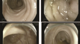
UNG THƯ PHỔI DI CĂN ĐẠI TRÀNG: BÁO CÁO CA BỆNH VÀ ĐỐI CHIẾU Y VĂN
17/10/2023 16:02:52 | 0 binh luận
SUMMARY Lung cancer is one of the leading causes of cancer related mortality worldwide. The brain, liver, adrenal glands, and bone are the most likely site of metastatic disease in patients with lung cancer. Gastrointestinal (GI) metastasis from primary lung cancer is very rare. Only few reports have been published and the majority of described metastatic sites involved the small instestine. In the present study, we report a case of lung cancer with colonic metastasis and also review the published literature of primary lung cancer with colonic metastasis. Keywords : lung cancer, gastrointestinal metastasis, colonic metastasis.
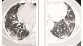
NHẬN XÉT ĐẶC ĐIỂM LÂM SÀNG VÀ HÌNH ẢNH CẮT LỚP VI TÍNH PHỔI Ở BỆNH NHÂN SAU NHIỄM SARS-COV-2 TẠI CÁC CƠ SỞ KHÁM CHỮA BỆNH CỦA MEDLATEC
13/10/2023 12:01:05 | 0 binh luận
SUMMARY A cross-sectional descriptive study was conducted on 1436 patients after SARS-CoV-2 infection who visited MEDLATEC medical facilities from March 1, 2022 to March 31, 2022. The results showed that 39% were male and 61% female, with a mean age of 39 and the majority of patients in the working age range from 18-60 years old. 74% of patients infected with SARS-CoV-2 have clinical symptoms, of which cough is the most common symptom, followed by chest pain and shortness of breath. The group of patients with a healthy history accounted for 76% of cases; the remaining group of underlying diseases with hypertension accounted for the most, followed by the group with chronic lung disease. Up to 49% of patients have suspected COVID-related lung lesion; with the most important lesions being interstitial thickening and most of them being mild according to the CT-score scale. Keywords: clinical characteristics, CT scanner, SARS-CoV-2
GIÁ TRỊ DỰ BÁO TỬ VONG CỦA ĐỘ NẶNG TỔN THƯƠNG PHỔI TRÊN X-QUANG TẠI THỜI ĐIỂM NHẬP VIỆN Ở BỆNH NHÂN COVID-19
13/10/2023 11:44:33 | 0 binh luận
SUMMARY Background: We aimed to investigate the performance of a chest X-ray (CXR) scoring scale of lung injury in prediction of death among patients with COVID-19 admitted at Vinmec Central Park hospital (HCM City, VN) during the peak epidemic in 2021. Method: Retrospective design; X-ray images and clinical data were collected from all hospitalized patients with SARS-CoV-2 PCR positive from July to September 2021. Three radiologists independently assessed the CXR score at admission which consists of measuring both severity and extent of lung injuries on four lung quadrants (scale o to 24). Association between CXR and mortality risk was estimated using a survival regression with log-log distribution. Result: The study included 219 patients (mortality rate = 28). There was a high consensus for CXR scoring among 3 radiologists (κ = 0.90; CI95%: 0.89-0.92). PCA analysis revealed that CXR has a similar role as CRP score for predicting risk of mortality. CXR score was the strongest predictor of mortality (tdAUC 0.85 CI95% 0.69–1) within the first 3 weeks after admission, compared with other conventional clinical features. After adjusting for the patient’s age, there was a significant effect of increased CXR score on mortality risk (HR = 1.15, CI95%:1.04-1.27, p=0.009). At a critical threshold of 16 points, the CXR score allows for predicting inhospital mortality with good sensitivity (0.82; CI95%: 0.78 to 0.87) and specificity (0.89; CI95%: 0.88 to 0.90). Conclusion: The day-one CXR score is an independent and effective predictor of the risk of death in COVID-19 and could be used to identify high-risk patients in countries like Vietnam where CXR is more readily available than CT scans. Keywords: COVID-19, Chest X-ray, Lung injury score, Mortality prediction
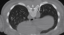
SARCOMA CƠ VÂN DI CĂN PHỔI TỔNG QUAN TÀI LIỆU VÀ BÁO CÁO CA BỆNH
12/10/2023 15:49:15 | 0 binh luận
SUMMARY Sarcoma is a general term for a type of cancer found in the connective tissue cells (the cells of the mesenchyme). There are many types of sarcoma because connective tissue cells are present everywhere in the body (bone, cartilage, blood vessels ...) but this type of cancer is divided into 2 main groups, bone sarcoma, and soft tissue sarcoma. Rhabdomyosarcoma (RMS) belongs to the group of soft tissue sarcoma of skeletal muscle, is a common malignancy, and is one of the leading causes of cancer death in children. Alveolar rhabdomyosarcoma (ARMS) is a subtype of RMS that is extremely rare in adults. We present a pediatric case of ARMS, primary in the butt area with pulmonary metastases, confirmed by histopathology and immunohistochemistry. Reported data and literature review will help physicians have a better diagnostic approach when encountering similar cases. Keyword: Rhabdomyosarcoma; alveolar rhabdomyosarcoma, soft tissue tumor lung metastasis.
CHẨN ĐOÁN VÀ ĐIỀU TRỊ TRÀN DỊCH DƯỠNG CHẤP MÀNG PHỔI TỰ PHÁT
12/10/2023 15:45:14 | 0 binh luận
SUMMARY Nontraumatic chylothorax (NTC) is the presence of chylous fluid in the pleural cavity. Unlike traumatic chylothorax, diagnosis and interventional treatment has very high rate of success while nontraumatic chylothorax has many difficulties in both diagnosis and treatment. The most important in management of NTC is depicting the lesion of lymphatic vessel which is varying from patient to patient. The current understanding of the anatomy of the chylous circulation allows for successful diagnosis and treatment in some patients, but the incidence is still limited. This article reviews the causes of chylothorax and approaches to diagnosis and treatment of patient with NTC. Keywords: chylothorax, thoracic duct embolization *
NỐT ĐƠN ĐỘC BÁN ĐẶC ÁC TÍNH PHỔI TỔNG QUAN TÀI LIỆU VÀ BÁO CÁO CA BỆNH
12/10/2023 14:25:41 | 0 binh luận
SUMMARY A Solitary Pulmonary Nodule (SPN) is a focal opacity on chest radiographs or CT with a clear border; at least partially covered by lung parenchyma; usually spherical; diameter equal to or less than 3 cm; can be solid (Solid Nodule – SN), Non-solid (Ground Glass Opacity – GGO) or Semi-solid (Part Solid – PS). One-third of lung cancers present as solitary masses or nodules and the majority are of the adenocarcinoma type. WHO's updated histopathological classification 2021, Adenocarcinoma is divided into the following types: Minimally Invasive Adenocarcinoma (MIA); Invasive Non-Mucinous Adenocarcinoma (INMA); Invasive Mucinous Adenocarcinoma (IMA); Colloid Adenocarcinoma (CA); Fetal Adenocarcinoma (FA) and Enteric Adenocarcinoma (EA). Minimally invasive adenocarcinoma (MIA) usually has the main characteristic components, which have Lepidic Predominant Adenocarcinoma (LPA). We report a case of LPA, PS type, was discovered incidentally, diagnosed, and operated at the National Lung Hospital, initially with excellent results. Keywords: Adenocarcinoma; Lepidic predominant adenocarcinoma; Minimally invasive adenocarcinoma; Lung cancer; Lung cancer staging
ĐẶC ĐIỂM HÌNH ẢNH X QUANG NGỰC Ở BỆNH NHÂN COVID-19 TỬ VONG TẠI PHÒNG CẤP CỨU BỆNH VIỆN CHỢ RẪY
12/10/2023 12:49:46 | 0 binh luận
SUMMARY Background: COVID-19 is a global pandemic with very high mortality. Patients are hospitalized with rapid disease progression and early death in the Emergency Department. Chest X-rays play an important role in the diagnosis and prognosis of mortality. Purpose: (1) describe the chest X-ray findings of deceased covid-19 patients; (2) evaluate the relationship between Brixia score to mortality of COVID-19 patients in the Emergency Department. Methods: Retrospective study of COVID-19 patients who died at the Emergency Department, Cho Ray Hospital from August 1, 2021 to August 31, 2021, diagnosed with COVID-19 by RT-PCR technique. Clinical features and radiographic features were collected. Results: There are 226 cases in our study. We found that almost lesions were distributed in the bilateral lung (99%), in all 6 lung regions (78.3%), and diffuse distribution (94,2%). Interstitial, alveolar and consolidation lesions accounted for 69.5%, 61.9% and 94.7%, respectively. Pleural effusion occurs in 10.9% of cases. Pneumothorax and pneumomediastinum accounted for only 0.9% and 1.3% of cases. The mean Brixia score was 15. The mild, moderate, and severe subgroups of the Brixia score were 6 (2.7%), 31 (13.8%), and 188 (83.5%) cases, respectively. There is a relationship between Brixia score and gender. Conclusions: Characteristics of chest X-rays in deceased COVID-19 patients mostly had consolidation, multifocal, many lung fields involved, and diffuse distribution in both lungs. Other features such as pleural effusion, pneumothorax, subcutaneous emphysema, and pleural thickening can be observed. Most of the patients who died had high Brixia scores. Keywords: Chest X-ray, COVID-19, deceased, Emergency Department, Brixia score.
GIẢ PHÌNH ĐỘNG MẠCH PHỔI: NHÂN 3 TRƯỜNG HỢP KHÁM VÀ ĐIỀU TRỊ TẠI BỆNH VIỆN PHỔI TRUNG ƯƠNG
11/10/2023 15:21:47 | 0 binh luận
SUMMARY Pulmonary artery pseudoaneurysms (PAPs) is a rare abnormality of the pulmonary artery system. Beside, PAPs have no specific clinical presentation or may be asymptomatic, which may be present in congenital anomalies or in a variety of conditions such as pneumonia, pulmonary neoplasm, pulmonary tuberculosis, or lung fungus... [1]. We present 3 cases of PAPs examined and treated at the National Lung Hospital with PAPs appearing on the background of 3 different diseases. Both of these 3 cases have nonspecific symtoms as cough, chestpain and dyspnea with 2 cases of hemoptysis. They are all detected by computed tomography (CT) angiography. There are 2 cases treated by surgery that have good results and 1 case who continued the TB regimen received medical treatment. Keywords: pulmonary artery pseudoaneurysms, pulmonary artery, hemoptysis.
Bạn Đọc Quan tâm
Sự kiện sắp diễn ra
Thông tin đào tạo
- Những cạm bẫy trong CĐHA vú và vai trò của trí tuệ nhân tạo
- Hội thảo trực tuyến "Cắt lớp vi tính đếm Photon: từ lý thuyết tới thực tiễn lâm sàng”
- CHƯƠNG TRÌNH ĐÀO TẠO LIÊN TỤC VỀ HÌNH ẢNH HỌC THẦN KINH: BÀI 3: U não trong trục
- Danh sách học viên đạt chứng chỉ CME khóa học "Cập nhật RSNA 2021: Công nghệ mới trong Kỷ nguyên mới"
- Danh sách học viên đạt chứng chỉ CME khóa học "Đánh giá chức năng thất phải trên siêu âm đánh dấu mô cơ tim"


