Chần đoán rò động mạch vành trên cắt lớp vi tính đa dãy
06/05/2021 15:51:18 | 0 binh luận
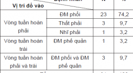
SUMMARY Objective : Evaluate the characteristics of coronary artery fistulas (CAFs) by multidetector computed tomography (MDCT). Material and methods: During 21 months (between January 2019 and September 2020), study on 31 patients were diagnosed with CAFs on MDCT at Radiology Centre of Bach Mai hospital, prospective descriptive study. Result: We enrolled 31 patients (11 male, 20 female, mean age 56 years) with CAFs on MDCT. 18 patients had multiple fistulas (58,1%), 13 patients had single communication (41,9%). 6,5% originated from the right coronary, 35,5% arose from the left coronary artery system and 58,5% from both right and left coronary artery. 87,1% of fistulas drain to the right side of the circulation(74,2% drain to pulmonary artery). 1 patient (3,2%) had fistula drain to the left side of the circulation (bronchial artery). 3 patients (9,7%) had fistulas drain to both right and left side of the circulation (pulmonary artery and bronchial artery). 10 patients had large fistulas (32,3%), 21 patients had small fistulas (67,7%). 19 patients had aneurysm of fistulas (61,3%), most of them drain to pulmonary artery (73,7%). 38,7% of patients were diagnosed with CAFs by echocardiography (38,7%). 6 patients were examined by DSA: 2 patients were not detected origin of fistulas by DSA, 3 patients were not detected drainage of fistulas by DSA. Conclusion: DSCT is a noninvasive and useful modality for diagnosis of CAFs.
Vai trò của cộng hưởng từ trong chẩn đoán và theo dõi điều trị lao não, màng não
06/05/2021 15:55:20 | 0 binh luận
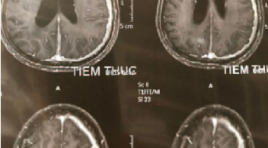
SUMMARY Purpose: Characteristic description image and evaluate the role of CHT in diagnosis and follow up and treatment of meningitis tuberculosis. Subjects and methods: Prospective and retrospective description 45 patients meningitis tuberculosis with evidence of TB bacteria in the cerebrospinal fluid undergoing MRI before and after treatment from July 2019 to September 2020. Comparison of lesions on MRI before and after tuberculoma, tuberculosis meningitis treatment. Result: In a total of 45 study patients, the average age is 28.8, male / female = 1.5, the rate of patients developing lesions on MRI before treatment (88.9%), The most common signs of damage in patients with tuberculosis of the brain, the common meningitis before treatment include: sign of meningitis enhancement (84.4%), basal meningeal enhancement (66.7%), Sylvian fissures (6.7%) , meninges of supersellar cistern (17.8%), tuberculoma (44.4%), hydrocephalus (31.1%), cerebral infarction (13.3%). Following 40 patients with meningeal tuberculosis lesions on CHT after treatment, the rate of detecting damage after treatment is 75%, of which: signs of meningitis enhancement (72.5%), basal meningeal enhencement (37.5%), sylvian fissures (2.5%), meninges of supersellar cistern (7.5%), tuberculoma (42.7%), hydrocephalus (20%), infarction cerebral (2.5%). Keyword: tubercoulosis meningitis, tuberculoma, MRI
Khảo sát đặc điểm của nhồi máu não cấp trên cộng hưởng từ thường qui và chụp mạch máu bằng kỹ thuật TOF 3D
06/05/2021 16:00:20 | 0 binh luận
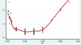
SUMMARY Objective. To describe the characteristics of acute ischemic stroke on conventional magnetic resonance imaging (MRI) and angiography using TOF 3D technique and to evaluate the time course of the apparent diffusion coefficient after cerebral infarction. Materials and Methods. We conducted a retrospective study in 221 patients with acute ischemic stroke who were performed MRI at the University Medical Center Hospital between January 2018 and December 2020. A radiologist who had more than 5 years of experience of MRI evaluates the changes of signal intensity on these sequences: T1- weighted, T2-weighted, FLAIR, DWI/ADC, Susceptibility weighted imaging (SWI) and TOF3D. Results : The study was composed of 221 patients (136 males, 85 females). The mean age of patients was 64.5 ± 13.7 years (range, 28- 96 years). 60 - 79 age group accounts for the majority with 105 people (47.5%). Anterior cerebral artery territory infarcts and cerebral peduncular infarction were the least common (1.1%). The rate of brain parenchymal signal abnormalities on the T1W, T2W, FLAIR, DWI / ADC sequences respectively was 85.5%, 87.3%, 90.0%, and 97.3%. The rate of a significant decrease in vascular signaling on TOF3D was 35.7%. The rate of susceptibility vessel sign and prominent vessel sign on SWI was 21.8% và 9.1% respectively. Immediately following a cerebral infarction, the relative ADC (rADC) value begins to decrease gradually and reaches its lowest level between day 2 and 4. Thereafter the rADC value increases gradually and reaches a pseudonormalization around day 7. Conclusion: Magnetic resonance imaging is of high value in the diagnosis of acute ischemic stroke. Diffusion imaging has the highest sensitivity in lesion detection. TOF3S and SWI were helpful in depiction the vascular signaling, the susceptibility vessel sign and prominent vessel sign. Immediately following a cerebral infarction, the relative ADC (rADC) value begins to decrease gradually and reaches its lowest level between day 2 and 4. Thereafter the rADC value increases gradually and reaches a pseudonormalization around day 7. Keywords : acute ischemic stroke, magnetic resonance imaging.
Vai trò của siêu âm trong chẩn đoán bệnh tim bẩm sinh trước sinh
06/05/2021 16:33:50 | 0 binh luận
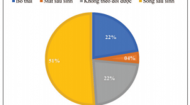
SUMMARY Purpose: Role of prenatal ultrasonographic in diagnostic of congenital heart defects Subjects and methods: Descriptive study on 98 fetuses diagnosed congenital heart defects (CHDs) from August 2018 to September 2020 at National Obstetric and Gynecology. 50 cases with live birth, 22 cases of pregnancy termination, 4 cases of perinatal mortality, 22 cases of losing follow-up. The patient was performed an prenatal fetal heart ultrasound and follow the outcome. Result: The mean maternal age was 28.5 ± 9.4 yrs old, the mean gestational age at the time of prenatal diagnosis of CHD was 28 ± 6,4 weeks. CHDs accounts for a high proportion on prenatal ultrasound: ventricular septal defect (VSD), atrioventricular septal defect (AVSD) and the double outlet right ventricle (DORV) account for 14-15% of CHDs. The mild CHD account for 27.6%, critical CHD was 72.4%. Accuracy of prenatal echocardiography diagnosis was 88%. The severity of CHDs prenatal ultrasound affects pregnancy outcome (p<0.05). Conclusion: Fetal echocardiography has high accuracy in prenatal diagnosis of CHDs. The severity of CHDs in prenatal ultrasound has an impact on pregnancy outcome. Key word: Fetal echocardiography, congenital heart defects.
Đánh giá giá trị chẩn đoán của các dấu hiệu nghi ngờ ác tính trên siêu âm vú
06/05/2021 16:41:52 | 0 binh luận
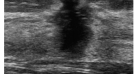
SUMMARY Objective: To evaluate the diagnostic value of suspected malignancy signs on breast ultrasound. Methods : patients who have the solid mass in breasts underwent ultrasound examination (they have at least one of eight signs of suspected malignancy on breast ultrasound) and performed the ultrasound guided needle biopsy of these mass at Bach Mai Hospital from July 2019 to July 2020 were investigated. Results: 268 patients have breast tumors with signs of suspected malignancy detected on ultrasound (mean age, 42,1 years; range, 12 to 75). There were 97 breast cancer patients accounted for 36,2 %, including 84.5% of patients over 40 years old. Cancer lesions are mainly at the upper - outer quadrant position, accounting for 58,8 %. The sensitivity, specificity, PPV and the OR value in regression analysis of suspected malignancies signs, respectively: spiculation and/or thick hyperechoic halo ( 67,01%; 97,07%, 92,87%; 67,44), irregular shape with angular margins (89,69% and 70,76%; 63,5%; 21,05), retrotumoral acoustic shadowing (18,56 % and 98,83 %; 90 %, 19,25), hypoechoic echo pattern (96.91% and 7.02%; 37.15%), orientation not parallel (includes round solid nodules with round shape) (57,73 % and 84.21%; 67,47 %; 7,29), duct changes - enlarged ducts within surrounding tissues (24,74 % and 95.91%; 77,42 %; 7,7), microlobulations (38,14 % and 90.06 %; 68,52 %; 5,59), microcalcifications within or outside a mass (49,48 % and 89,47 %; 72,73%; 8,33). Conclusion: The signs of suspected malignancy on breast ultrasound have high diagnostic value in diagnosing breast cancer, especially the signs of spiculation and/or thick hyperechoic halo, irregular shape with angular margins and retrotumoral acoustic shadowing. In which, spiculation and/or thick hyperechoic halo is the sign with the highest UTV diagnostic value, followed by a sign of irregular shape with angular margins and retrotumoral acoustic shadowing. Hypoechoic echo pattern sign have high Se but low Sp and PPV, have no meaning in the diagnosis of breast cancer.
Nhận xét mối tương quan giữa thể thông bào xương chũm và tình trạng thông khí của tai giữa trên cắt lớp vi tính ở tai xẹp nhĩ
06/05/2021 14:44:10 | 0 binh luận
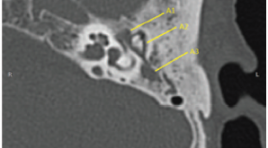
SUMMARY Objectives : Describe characteristic imagings and comment on the correlation between types of mastoid pneumatization and the aeration status of middler ear on computed tomography in atelectatic ears. Material and methods: The study describes 74 ears of 74 atelectatic patients who had 64-128 slice temporal bone CT, at Bach Mai Hospital and National Otorhinorarynology Hospital from 12/2018 to 3/ 2020. Results: Among atelectatic ears, condensed images of the middle ear on CT scanner contain: the anterior epitympanic recess (AER) in 35.1%, the inner epitympanum in 45.9%, the lateral epitympanum in 54.1%, the mesotympanum in 20.3%, the hypotympanum in 3.5%, the antrum in 52.7%. The mastoid pneumatizations on CT scanner include sclerotic mastoid in 44.6%, diploic mastoid accounts for 41.9%, the well pneumatized mastoid accounts for 13.5%, the difference has statistical significance with p = 0.001. There is a close significantly correlation between mastoid pneumatization and condensations in middle ear spaces (anterior epitympanic recess - attic - antrum) in atelectatic ears with p <0.0001, Cramer's V = 0.957. Conclusion: There is a close statistically significant correlation between aeration status of middle ear spaces and mastoid pneumatization on CT. Sclerotic mastoid or diploic mastoid are advantageous to the appearance and development of atelectatic ear. Keywords: atelectatic ear, mastoid pneumatization, the aeration status, attic, antrum, CT of temporal bone
Kết quả bước đầu nút thông động tĩnh mạch cửa trong điều trị bệnh nhân ung thư biểu mô tế bào gan
06/05/2021 14:32:25 | 0 binh luận
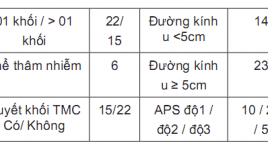
SUMMARY Advanced stage hepatocellular carcinoma with arterioportal shunts has worse prognosis and limited treatment. Transarterial chemoembolization (TACE) use embolic materials is a safe method and have efficacy in treatment. Aims: The aim of this study is to evaluate safety and efficacy of transarterial chemoembolization (TACE) with use embolic materials for the treatment of hepatocellular carcinoma (HCC) with arterioportal shunts (APS) Materials and Methods: From June 2019 to June 2020, 37 patients who had diagnosis of HCC with major portal vein thrombosis were perfomed TACE with diffrent embolic materials which depend on the level of APSs. The patients were followed after treatment 1 week for the clinical symptom and at 1-3 month after the initial intervention, contrast-enhannced multi slide computed tomography (MSCT) or magnetic resonance image (MRI) or DSA was performed to assess the APS’s treatment efficacy and tumor response by mRECIST and tumor markers (AFP or PIVKA-II). The primary safety endpoint was liver toxicity at 2-5 days and 1 month after intervention. Results: All interventional procedures were successful without any procedure relevant complications. The immediate APS improvement rate was 89.2% (33/37), and the APS improvement rate at first‑time follow‑up was 70.3% (26/37). Radiologically confirmed complete response (CR), partial response, stable disease, and progressive disease at 1 month after first chemoembolization were observed in 3 (8.1%), 17 (45.9%), 13 (35.1%) và 4 (10.9%) patients, respectively. Conclusions: Transarterial chemoembolization (TACE) use embolic materials is safety and have efficacy in treatmen of arterioportal shunt in patient with hepatocellular carcinoma. Key words: Arterioportal shunts (APS), Transarterial chemoembolization (TACE), Hepatocellular carcinoma (HCC).
Đánh giá hiệu quả nút mạch ngoài gan của ung thư biểu mô tế bào gan tại Trung tâm ĐIỆN QUANG Bệnh viện BẠCH Mai
06/05/2021 14:27:58 | 0 binh luận
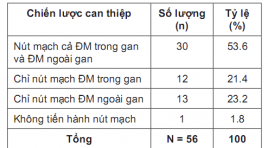
SUMMARY Purpose: Evaluation the effectiveness of the extrahepatic arterial chemoembolization on treatment of hepatocellular carcinoma. Subjects and methods: There is a non-controlled prospective intervention study in HCC patients with tumors which had blood supply from extrahepatic arteries from July 2019 to August 2020 in Radiology center of Bach Hospital. Results: . Our study was performed on 56 patients including 48 men (85.7%) and 8 women (14.3%) who were diagnosed with HCC. The mean age were 58.29 ± 10,849 years old. The rate of successful approaching to the extrahepatic vessel branches was 83.05%. There were 2 patients (3.58%) had acute complications during and immediately after intervention. Special complications related directly to the extra-hepatic embolization were observed in 14 patients (25%). After an average of 2.25 ± 0.919 months of follow-up, 1 patient (1.8%) re-examined due to rupture of liver tumor, the remaining 55 patients (98.2%) were reexamined by appointment. The results of tumor response according to mRECIST with complete response, partial response, stable disease and progressive disease were 3.6%, 52.7%, 25.5%, and 18.2% respectively. Conclusion : The results of the extrahepatic arterial chemoembolization in our study had high rates of the successful approaching to the extrahepatic vessel branches, the intervention safety and the post-intervention tumor response. Key words: TACE, Extrahepatic artery supply to HCC
Bạn Đọc Quan tâm
Sự kiện sắp diễn ra
Thông tin đào tạo
- Những cạm bẫy trong CĐHA vú và vai trò của trí tuệ nhân tạo
- Hội thảo trực tuyến "Cắt lớp vi tính đếm Photon: từ lý thuyết tới thực tiễn lâm sàng”
- CHƯƠNG TRÌNH ĐÀO TẠO LIÊN TỤC VỀ HÌNH ẢNH HỌC THẦN KINH: BÀI 3: U não trong trục
- Danh sách học viên đạt chứng chỉ CME khóa học "Cập nhật RSNA 2021: Công nghệ mới trong Kỷ nguyên mới"
- Danh sách học viên đạt chứng chỉ CME khóa học "Đánh giá chức năng thất phải trên siêu âm đánh dấu mô cơ tim"


