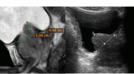
Nghiên cứu kết quả điều trị tăng sản lành tính tuyến tiền liệt thể lồi vào bàng quang bằng phương pháp nút mạch
06/05/2021 14:23:09 | 0 binh luận
SUMMARY Objective : To acess the results of prostatic artery embolization (PAE) in patients wwith significant intravesical prostatic protrusion (IPP). Material and methods: Prospective analysis of 45 consecutive patients with significant IPP undergoing PAE. We measured IPP on sagittal T2-weighted images before and after PAE. IPSS and clinical results were also evaluated before and after PAE. Results: PAE resulted in significant IPP reduction (16.9 ± 7.9mm before PAE and 13.2 ± 6.6mm after PAE, p < 0.001) (Fig. 1) with no complication. IPSS, quality of life (QoL), total prostate-specific antigen (PSA) level, and prostate volume (PV) showed significant reduction after PAE, and maximum urinary flow rate (Qmax) showed significant increase after PAE. A significant correlation was found between the IPP change and the IPSS change (r = 0.628, p = 0.001). Conclusion : Patients had significant IPP reduction as well as significant symptomatic improvement after PAE, and these improvements were positively correlated. Keywords: Benign prostate hyperplasia, Intravesical prostatic protrusion, Prostate artery embolization (PAE)
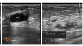
Nghiên cứu giá trị của phương pháp tiêm cồn tuyệt đối dưới hướng dẫn của siêu âm trong điều trị nang tuyến vú lành tính
06/05/2021 14:18:29 | 0 binh luận
SUMMARY Objective : To investigate the effectiveness of single- session ultrasound-guided percutaneous ethanol sclerotherapy in symptom breast cysts Methods: Breast cysts symptoms patients underwent ultrasound examination and treated by ethanol sclerotherapy at Bach Mai Hospital from July 2019 to February 2020 were investigated. All patients were aspirated using 20-22G needles refilled using 99% ethanol and reaspirated completely after 10 minutes of exposure under ultrasoundguidance. Follow-ups were done by ultrasound examination at one week and 3 months to 6 months by ultrasound examination. Results : 62 breast cysts (mean volume, 5.01 ± 4.8 ml; range, 0.8 to 22 ml) of 59 patients (mean age, 44.5 years) had symptoms were treated. 7 patients (11.2%) had painful symptoms, 17 patients (27,4%) had felling burn skin, and 15 patients (24.2%) felling uncomfortable while the ethanol sclerotherapy process and all symptoms disappeared after 5 minutes. 26 breast cysts (42%) were disappeared in sonography examinations and 36 breast cysts (58%) reduced in volume (mean 96,4% volume reduced) after one week. At 3 months to 6 months, 61 cysts (98,4%) completely responded therapy as undetectable on sonography, only one cyst (1.6%) was reduced 88,6% in volume. The success of this technique was 100 %, no patient had serve complication such as hemorrhage or abscess Conclusion: Ultrasound-guided ethanol sclerotherapy is a simple, fast, and safe method in the treatment of symptom breast cysts.
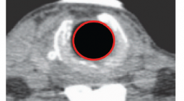
Khảo sát đặc điểm hình ảnh trên cắt lớp vi tính 128 dãy có dựng hình ba chiều trong bệnh lý sẹo hẹp khí quản tại Bệnh viện Bạch Mai
06/05/2021 12:36:00 | 0 binh luận
SUMMARY Objective: The study aimed to research causes, imaging features on CT scan and endoscopy, the correlation between CT scan and endoscopy in tracheal stenosis disease. Subjects and methods: Descriptive cross-sectional study, 14 patients was diagnosed tracheal stenosis at Bach Mai hospital from March 2019 to July 2020. Instruments of data collection included medical records, endoscopy and CT scan results. SPSS software was then employed for data analysis. Results: 14 patients in the study, 10 males (71,4%), 4 females (28,6%). Average age was 41.7±13.8. The cause of intubation and tracheostomy was diverse, but the main cause was traumatic brain injuries due to traffic accidents (4 patients). Prominent clinical symptom was difficult inspiration and stridor. Imaging features of tracheal stenosis on CT scan: The mean distance from vocal cord was 29mm. According to Cotton classification, grade III was highest proption ( 71,4%), the remaining was grade II (28,6%), no case was grade I and IV. The mean length of stenosis was 20mm. On endoscopy, the mean distance from vocal cord was 33mm. The correlation between three-dimensional 128-slice CT scanning and endoscopy was concluded: Similar evaluation of the distance from vocal cord to upper end of stenosis, no statistical significance (p>0.05) Key words: tracheal stenosis, intubation and tracheostomy, 128-slice CT scanning, the correlation between CT scan and endoscopy
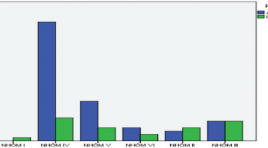
Nghiên cứu đặc điểm hình ảnh và giá trị siêu âm trong chẩn đoán hạch cổ ác tính
06/05/2021 14:14:26 | 0 binh luận
SUMMARY Background : The purpose of the study was to evaluate the characters and efficacy of B Mode and color Doppler ultrasound (CDUS) in diagnosis malignant cervical lymph nodes. Materials and Methods: In this cross-sectional prospective study, during a period of 12 months, performed on 85 patients including two groups: group I includes 63 patients with cervical lymphadenopathy with suspected lymph nodes on US and group II includes 22 patients with clinically suspected lymph nodes (not have any suspicious characteristics in US) were prospectively evaluated with B-mode and CDUS. Statistical analysis was carried out with histopathological or cytological diagnosis as gold standard. Results: We conducted ultrasound in 85 patients,. To compare with the pathology results of the disease, there are 56 metastatic lymph nodes, 04 lymphoma nodes, 01 plasmocyoma lymph node, 18 nonspecific inflammatory lymph nodes, 01 purulent lymph node and 05 granulomatous lymph nodes due to tuberculosis. The sensitivity, specificity, the accuracy of 2D ultrasound method combined with Dopper ultrasound are 95.08%, 79,2 %, 92%, 86% và 90,6%. Conclusions : Within the limitations of this study, B-mode and CDUS evaluations were found to be highly significant with a high sensitivity and specificity. B-mode and CDUS examinations provide a prospect to reduce the need for biopsy/fine needle aspiration cytology in reactive nodes. Keywords: B-mode ultrasound, Doppler color ultrasonography, histopathology, lymph node.
Giá trị chẩn đoán của các vi vôi hóa nghi ngờ ác tính trên X quang tuyến vú
06/05/2021 12:49:48 | 0 binh luận
SUMMARY Objective: Diagnostic value of some microcalcifications with suspected malignancy on mammograms. Methods: The study included 60 women with microcalcifications who underwent imaging-guided biopsy between July 2019 and July 2020 at Bach Mai Hospital. Digital mammograms procured before biopsy were analyzed independently by one breast imaging subspecialists blinded to biopsy results. Results : * Micro-calcification outside of a mass – 30 cases The overall positive predictive value of biopsies was 40%. The individual morphologic descriptors predicted the risk of malignancy as follows: fine linear/branching, 7 (87.5%) of 8 cases; fine pleomorphic, 4 (25%) of 16 cases; amorphous, 1 (16.7%) of 6 cases và coarse heterogeneous, 0 cases. Fisher’s Exact testing showed statistical significance among morphology descriptors (p < 0.01) * Microcalcifications in a mass – 30 cases The overall positive predictive value of biopsies was 96.7%. The individual morphologic descriptors predicted the risk of malignancy as follows: fine linear/branching, 16 (100%) of 16 cases; fine pleomorphic, 11 (92%) of 12 cases; amorphous 2 (100%) of 2 cases và coarse heterogeneous, 0 cases. * The positive predictive value for malignancy according to BI-RADS assessment categories were as follows: category 4B, 21,1%; category 4C, 66,7%; and category 5, 94.3%. Conclusion: Morphological description and distribution of microcalcifications on mammograms helps classify BI-RADS and assess the risk of malignancy for each case for diagnosis and treatment monitoring. The positive predictive values for breast cancer increased in order of amorphous, fine pleomophic, and fine linear/ branching microcalcification.
Vai trò của chụp cắt lớp vi tính mạch máu trong chẩn đoán hẹp tắc động mạch nội sọ ở bệnh nhân đột quỵ do thiếu máu não cấp
06/05/2021 12:16:52 | 0 binh luận
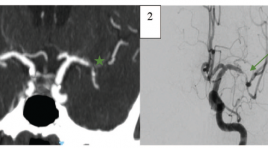
SUMMARY Background : Diagnosis of intracranial arterial stenooclusive disease and identification of intracranial atherosclerosis related occlusions (ICAS-O) in ischemic stroke patients is extremely important in order to plan a correct therapeutical approach. Few studies to date have examined the role of computed tomographic angiography (CTA) in diagnosing intracranial stenosis and predicting ICAS-related occlusions. Objective: To determine whether there is any correlation between CTA-determined truncal-type occlusion (TTO) and ICAS-related occlusions. To compare CTA to digital subtraction angiography (DSA) for detecting and measuring intracranial arterial stenoocclusive disease. Methods : We reviewed 129 ischemic stroke patients who underwent CTA and DSA. The occlusion and degree of stenosis of each intracranial arteries were calculated by WASID method. Occlusion type was classified as TTO or branching-site occlusion (BTO) on CTA. ICAS-O was detected by evaluating of underlying fixed focal stenosis (FFS) on DSA. Results: A total of 423 intracranial arteries were analyzed. CTA detected intracranial artery occlusion with sensitivity and specificity, and NPV 97,8%, 98,6% và 98,9% respectively. For detection of 50%-99% stenosis, CTA had 89,7% sensitivity and 98,2% specificity. TTO was more frequent in ICAS-O group than in the embolic group (78,1% versus 8,5%, p < 0,001). Conclusions: Compared to DSA, CTA has high sensitivity and specificity for diagnosing intracranial arterial stenooclusive disease. Preprocedural TTO on CTA is related to postprocedural ICAS-O in ischemic stroke patients.
Bước đầu đánh giá giá trị của kỹ thuật sinh thiết hút chân không trong chẩn đoán các tổn thương vi vôi hóa ở vú
06/05/2021 12:09:50 | 0 binh luận
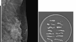
SUMMARY Objective: The purpose of this article is initial assessment the value of vaccum assited biopsy technique in diagnosis breast microcalcification lesions. Materials and Methods: T he prospective study included 17 women with 18 breast microcalcification lesions without mass that were classified BIRADS 4- 5.All lesions underwent imaging- vaccum assisted biopsy between August 2019 and July 2020 in Radiology Centre- Bach Mai Hospital. Results: The study involved 2 lesions with stereostatic vaccum assited biopsy- SVAB and 16 lesions with ultrasound guided vaccum assisted biopsy- USVAB. The mean duration of procedure was 53.22 ± 15.06 (minutes).The median number specimen was 10.9 ± 4.2. All lesions had microcalcifications in specimens. In terms of complications, the mild pain rate was 27.8% and 1/18 (5.6%) had hematoma without surgery. Biopsy results revealed 7 benign and 11 malignant lesions. The malignant rate in fine pleomorphic, amorphous and linear microcalcification were 87.5%, 33.3% and 100%, respectively.The malignant rate in regional, segmental and cluster distribution were 50%, 71.4% and 57.1%, respectively.9/11 malignant lesions had histopathologic results after surgery. The total upgrade rate at surgery was 22.2% (all for ductal carcinomas in situ- DCIS). Patients with benign lesions were followed up for a median period of 6 months, with no interval change. The sensitivity, specificity, positive predictive and negative predictive value of VAB in diagnostic breast microcalcification were 100%. Conclusion: Our first experiences: Image-guided vacuum-assisted breast biopsy is accurate, reliable and minimally invasive.It can be used as a safe approach for diagnosis in patients with breast microcalcifications. Keyword: VABB, breast microcalcification
Vai trò của cộng hưởng từ chức năng trong đánh giá vùng vận động bàn tay ở bệnh nhân u não
06/05/2021 12:02:47 | 0 binh luận
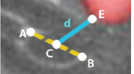
SUMMARY Background: Localizing the brain's functional cortex on functional magnetic resonance imaging (fMRI) plays an important role in brain tumor resection. Objective: To investigate imaging characteristics of the hand motor area on standardized MRI (sMRI) and fMRI in patients with brain tumors. To evaluate the correlation between lesion to motor cortex distance (LMD) on fMRI and preoperative motor deficit. Methods: Standardized and functional magnetic resonance images of 20 patients with rolandic brain tumors were included, all patients underwent tumor resection. Anatomic landmarks related to the hand motor area were interpreted on standardized MRI. Measure the distance between the hand motor area localized on fMRI and the hand motor area localized on standardized MRI. Compare the incidence of preoperative motor deficit of groups of patients with different LMDs Results: The rates of clearly defined anatomical landmarks related to the hand motor area on tumor-affected hemispheres are lower than those on unaffected hemispheres. The distance between the hand motor area localized on fMRI and the hand motor area localized on standardized MRI is 17.01 ± 3.63 mm on average and there are 6 cases where this distance is greater than 20 mm. The incidence of motor deficit in the “LMD<1cm” group, the “LMD from 1 to 2 cm” group and the “LMD>2 cm” group are 75%, 50% and 0% respectively. Conclusions: Standardized MRI should not be use to localize the hand motor area in patients with brain tumors. LMD is correlated with preoperative motor deficit. Keywords: Brain tumor, functional magnetic resonance imaging, hand motor area.
Bạn Đọc Quan tâm
Sự kiện sắp diễn ra
Thông tin đào tạo
- Những cạm bẫy trong CĐHA vú và vai trò của trí tuệ nhân tạo
- Hội thảo trực tuyến "Cắt lớp vi tính đếm Photon: từ lý thuyết tới thực tiễn lâm sàng”
- CHƯƠNG TRÌNH ĐÀO TẠO LIÊN TỤC VỀ HÌNH ẢNH HỌC THẦN KINH: BÀI 3: U não trong trục
- Danh sách học viên đạt chứng chỉ CME khóa học "Cập nhật RSNA 2021: Công nghệ mới trong Kỷ nguyên mới"
- Danh sách học viên đạt chứng chỉ CME khóa học "Đánh giá chức năng thất phải trên siêu âm đánh dấu mô cơ tim"


