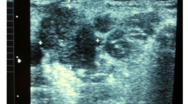
Nghiên cứu giá trị siêu âm và siêu âm hướng dẫn chọc hút tế bào bằng kim nhỏ hạch di căn trong ung thư thực quản
02/06/2020 17:26:05 | 0 binh luận
summary urpose : To survey ultrasonographic characteristics suggested benign and malignant cervical lymph node metastasized from EC methods (esophagus cancer). To find out the values of FNA in diagnosis of metastatic cervical lymph node from EC. Materials and Methods: 20 patients were diagnosed EC and ultrasound detected suspiciously malignant cervical lymph node, from 4/2012 to 6/2013. The patients were surveyed: Mode 2D, FNA, cytopathology in Hue Central Hospital. We divided the diagnosis into 2 groups: benign and malignant group. Results: In 20 patients with cytology, 8 cases are diagnosed malignant lymph node metastasized from EC. The remains, 12 patients are diagnosed with benign lymph node: lymphadenitis. In our research, the signs suggest malignant lymph node in sonography: hypoechonic, heterogeneous, loss of the fatty hyperechoic hilum, ill-defined border, size about 10mm in diameter (short axis), round-shaped, greater numbers. Se= 80%, Sp= 100%, PPV= 100%, no false negative cas. Conclusion: The combination of ultrasound and FNA is valuable in the diagnosis of suspected malignant lymph node in the neck, metastasized from EC. It is a safe and simple method for selected patients which allows diagnosis and staging in a single step.
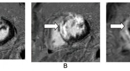
Đánh giá tổn thương cơ tim trên cộng hưởng từ ở bệnh nhân nhồi máu cơ tim cấp
06/06/2020 10:27:18 | 0 binh luận
Evaluation of myocardium injury on cardiac magnetic resonance imaging in patients with acute myocardial infarction SUMMARY Objective: To access the imaging characteristic of the myocardium injury on cardiacmagnetic resonance imaging (MRI) in reperfused acutemyocardial infarction (MI) after percutanous coronary revascularization. Materialand Methods: Cine sequence and Delayed Contrast- Enhanced MRIwere underwent in period of 9 days after percutanous coronary revascularization on 50 patients suffering from Acute Myocardial Infarction at Bach Mai Hospital. Left ventricular function was done on cine sequence and extent of infarction, infarct size was evaluated on delayed-enhancement images. Results : A total of 50 patients with acute MI were classified 90% as STEMI and 10% as NSTEMI. The sensitivity of delay-enhancement MRI for detecting MI reaching 98%. The accuracy of MRI for identifying MI location (compared with infarct-related artery perfusion territory) were 92%, kappa=0,842 with all the patients and were 97,8%, kappa=0,952 with STEMI. The infarcted areas in 49 patients were detected by use of cardiac delayedenhancement MRI. There was an excellent correlation between quantitative planimetry and scoring method for the hyperenhancement infarct size (r=0,976, p<0,0001). Infarct size on delayed-enhancement MRI showed a good correlation with left ventricular ejection fraction (r=-0,63,p<0,0001 with planimetry method; r=-0,602, p<0,0001 with scoring method). Conclusion : Cardiac MRI could evaluation of myocardium injury in patients with reperfused acute myocardial infarction. Key words: Cardiac Magnetic Resonace Imaging, delayed enhancement MRI, late gadolinium, Acute myocardial infarction, infarct size.
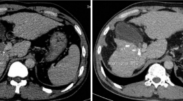
Đánh giá kết quả điều trị nút mạch hóa dầu kết hợp truyền CISPLATIN trong ung thư gan có huyết khối tĩnh mạch cửa
17/03/2020 16:39:48 | 0 binh luận
Evaluate treatment results of ctace combine with intratumoral injection of cisplatin in hepatocellular carcinoma with major portal vein thrombosis SUMMARY Advanced stage hepatocellular carcinoma with portal vein thombosis has worse prognosis and limited treatment. Transarterial chemoembolization use Lipiodol combined Cisplatin intratumoral is a safe method and have efficacy in treatment. Objective : To evaluate safety and efficay of cTACE combine with intratumoral injection of Cisplatin for hepatocellular carcinoma with major portal vein shunt treatment. Methods: From May 2018 to May 2019, 24 patients who had diagnosis of HCC with major portal vein thrombosis were perfomed cTACE (Famorubicin + Lipiodol) combined with intratumoral injection of Cisplatin. The patients were followed after treatment 1 week for the clinical symptom and after 1 month, used mRECIST and tumor markers (AFP or PIVKA-II) to aveluated treatment efficacy. The patients were perfomed consecutive courses of treatment and followed until died or until the end of study. Results: 24 patients (21 males, 3 females), age mean is 54,4 (from 32ys to 72ys, AFP mean 15600 ng/ml (from 3 to 121000), 19 patients (79%) has size of tumor ≥5cm. The classification of portal vein thrombosis: 6 patients Vp1 and Vp2; Vp3 18 patients. Total courses of treatment was 38 times. 15 patients (62,5%) had post embolization syndrome, 8 patients (33,3%) had decreasing of tumor marker. mRECIST: CR 3 patients (12,5%); PR 6 patients (25%); SD 4 patients (16,7%); PD 11 patients (45,8%). The mean survival time was 9,9 ± 1,1 months. The survival times is depend on the classification of PVTT (p=0.037 the mRECIST respondsibility (p=0.0001), the decreasing of tumor markers (p=0.01) and isn’t depend on the AFP before procedures. Conclusions : cTACE combine with intratumoral injection of Cisplatin is safety an have efficacy on treatment of HCC with major portal vein thrombosis. Keyword: Hepatocellular with portal vein thrombosis, cTACE, Cisplatin injection.
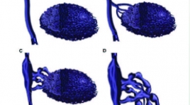
Đánh giá kết quả điều trị tiêm xơ dị dạng tĩnh mạch dưới hướng dẫn DSA
17/03/2020 16:57:22 | 0 binh luận
Evaluation the result of flouroscopy-guided sclerotherapy in treating superficial venous malformation SUMMARY Objective : Describe characteristics imaging of superficial venous malformation (VM) on flouroscopy and evaluate effectiveness of foam sclerotherapy. Methods: Prospective and retroprestive cohort from November 2015 till July 2019 on 17 patients with VM treated by flouroscopy-guided sclerotherapy with 21 lesions and 46 seasons sclerotherapy. Evualating results of treatment based on improvement in symtomps ( pain- Visual Analogue Score) and imaging ( MRI- repeat MRI after last season for 6 months . There are 4 grade in improvement in imaging: excellent ( reduction in size of lesion over 90 %), good ( reduce 50 -90 %), average ( 10-50 %) and no reponse ( less 10 %). Evualating recurrence based on increasing pain score (VAS) or size in MRI. Using SPSS 20.0 to analize and process data. Results : 17 patients (7 males and 11 females) with VM were involved in our study. Patients were a mean of 26.5± 12.9 years old ( range: from 6 to 59). Evualated by digital subtraction angiography, the lesion were categorized into 4 types according to the venous drainage features. Of the 21 lesion: 3/21 had type I (14.3 %), 12/21 had type II (57.1 %); 2/21 had type III (9.5 %) and 4/21 had type IV (19 %). Total seasons are 46. 8/21lesions (38.1 %) achieved excellent response, 9/21 (42.9 %) achieved good response, 3/21 (14.3 %) achieved average response and 1 patient (4.8%) no response in magnetic resonance imaging in magnetic resonance imaging assessments. Mean VAS scores after treatment for 1 month: 1.4; 3 months: 0.9; 6 months: 1.1, min: 0, max: 5. The long- term recurrence rates with follow-up time from 1 to 2 years is: 3/4 patients had recurrence (75 %) include of 1 patients had increasing imaging and pain score, 2 patients just had only increase imaging in MRI. Conclusion : Flouroscopy-guided sclerotherapy is a safe and effective procedure to reduce size and pain for patient with venous malformations that have symptoms. But the long – term recurrence rates is quite high. Keywords: venous malformation, sclerotherapy, flouroscopy-guided
Kết quả phương pháp tạo hình đốt sống ngực qua da ở những bệnh nhân xẹp cấp thân đốt sống do loãng xương
17/03/2020 16:51:05 | 0 binh luận
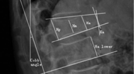
SUMMARY Objective : Report results of percutaneous vertebroplasty for treatment of osteoporotic compression fractures in the thoracic spine Methods: There is a retrospective combined prospective study in 40 patients who had fractured thoracic vertebrae due to osteoporosis were performed vertebroplasty at Bach Mai Radiology Center from January 2018 to March 2019. Patient’s pain status was rated with Visual Analogue Scale (VAS) score and Macnab system, pre- and post-operative vertebral heights and Cobb’s angles were measured and complications were noted 1 week, 1 month, 3 months after surgery. Results :40 patients with 56 fractured vertebraes show that there are no complications in the process of puncture needle and system complications, 12,5% cases of cement leakages into peri-vertebrae there is no case of cement cause pulmonary embolism, 5% case of pain post- operative. VAS scores were significantly reduced from 6.95 to 1.72, 86,95% has good and excellent, Cobb angle slightreduced from 14.4±9.1 to 13.2±8.5, the anteriorvertebral height slight increased from 18.5±4.3mm to 19.1±4.1mm, the middle vertebral height increases from 16.7±4.3mm to 17.4±4.3mmwith p<0.05, there is no significant changing in the post vertebral height after 3 months of intervention with p>0, 05. Conclusion: Percutaneous vertebroplasty thoracic spine is a safe and effective method in order to provide rapid pain relief, but is less significant in improving the Cobb angle and vertebral body height. Key words: Percutaneous vertebroplasty thoracic spine, treatment of Thoracic Fracture, pain reduction, change vertebral height.
Bước đầu đánh giá hiệu quả của phương pháp đốt sóng cao tần nhân lành tính tuyến giáp có triệu chứng
17/03/2020 16:32:47 | 0 binh luận
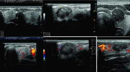
Evaluation of the early efficacy of radiofrequency ablation for benign thyroid nodules in symptomatic patients SUMMARY Objective: To evaluate the early efficacy of radiofrequency (RF) ablation for treatment of benign thyroid nodules, which causes symptoms in the Department Radiology- Bach Mai Hospital. Methods: We evaluated 51 benign thyroid nodules from 43 patients treated with RF ablation between 10/2016 and 4/2017. The procedure began with examining and diagnosing an benign thyroid nodules which causes symtoms by clinical physicians. The patients were diagnosed with a benign thyroid nodule according to the TIRADS classification combined with at least two appropriate results of cytology or biopsy by radiologists. The patients were then considered and performed radiofrequency ablation treatment for benign thyroid nodules. The follow-up examinations took place 1 month after RF ablation. Results: (1) 94% of the tumors needed only one time of RFA; (2) One treatment duration of RFA lasted an average of 21.8 minutes, (3) 76% of the tumors decreased by 30-50% volume, 11 % decreased by > 50% volume. (4) 100% of the treated individuals will reduce perfusion. (5) 96.4% of patients reduced or lost their symptoms. (6) There were no major complications during treatment, minor side effects (neck pain, bleeding,voice change …) restored after 2 weeks maximum. Conclusion: Radiofrequency ablation is a minimally-invasive method and an effective treatment for symptomatic thyroid nodules that are confirmed benign. Keywords: Thyroid nodule, benign thyroid nodule, symptomatic thyroid nodule, treatment for thyroid nodules, radiofrequency ablation.
Đánh giá hiệu quả điều trị trung hạn suy tĩnh mạch mạn tính bằng phương pháp can thiệp nội mạch
17/03/2020 16:18:24 | 0 binh luận
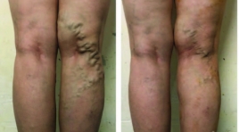
SUMMARY Objectives: The aim of this work was to evaluate the outcomes after the endovascular interventions for chronic venous insufficiency of the lower limbs. Methods: This study was conducted on 60 patients diagnosed with chronic venous insufficiency of the lower limbs at the Radiology Center, Bach Mai Hospital, 2018 – 2019. Convenience sampling technique was used for this study. Results : Endovenous laser ablation was indicated for 40 patients and endovenous radiofrequency (RF) ablation was for 20 patients, showing all patients were removed from the reflux line. For the patients with laser ablation, the mean Venous Clinical Severity Score (VCSS) were 3,67 ± 3,58 (0-11) before the treatment; 1,22 ± 2,03 (0-6) at 1 month; and 0,41 ± 1,01 (0-4) at 12 months. For the patients with RF ablation, the mean VCSS were 3,62 ± 3,45 (0-10) before the treatment; 0,15 ± 0,55 (0-2) at 1 month; and 0,08 ± 0,28 (0-1) at 12 months. 18 cases were recorded the complications after the intervention (30.00%), including dark skin, and paresthesia. 2 cases had the positions with varicose veins after 12 months (3.33%). There were no significant differences in the efficacy and complications amongst endovenous laser ablation and endovenous radiofrequency ablation (p >0.05). Conclusions : Endovascular intervention is minimally invasive safe effective method that improves long-term better symptoms.
Đánh giá kết quả điều trị dị dạng động - tĩnh mạch não dã vỡ ở trẻ em bằng nút mạch onyx
17/03/2020 16:14:01 | 0 binh luận
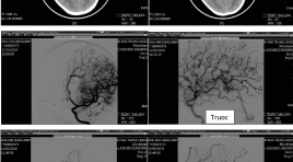
Evaluation of erlier outcome of treatment embolization for ruptured brain arterionous malformation in children with onyx SUMMARY Objective: The manifestation of clinical symptom, CT scanner, DSA imaging and evaluated earlier results of treatment embolization for ruptured brain arteriovenous malformations (bAVM) in children with Onyx. Methods: A prospective study was performed at National Children Hospital with treatment of ruptured bAVM by Onyx during the period from March 2017 to July 2019, including 45 patients. Results: Twenty-Five boys and Twenty Girls with a mean age of 8.7 years,. Result in on CT: cerebral hemorrhage on Supratentorial was 77.8%, infratentorial 11.1%, intraventricular hemorrhage 57.8%. DSA: according to Spetzler – Martin grading system: grade 1: 2.2%, grade 2 60%, grade 3 33.4%, grade 4 4.4%, 57 interventional sessions of embolization were performed, 1 to 3 sessions/patient with an average of 1.2 sessions/ patient, Complete obliteration of the AVM with Onyx was achieved in 26 of 45 patients (57,7%). Partial obliteration (>60%) was 17of 45 patients (37.7%), there were 8 patients with complications including hemorrhages and infarctions during embolization and good recovered, no died patient. Conclusion: Endovascular treatment of ruptured bAVMs with Onyx in children seems safe and effective with low complication rates. Key words : Arteriovenous malformation, Embolization, children
Bạn Đọc Quan tâm
Sự kiện sắp diễn ra
Thông tin đào tạo
- Những cạm bẫy trong CĐHA vú và vai trò của trí tuệ nhân tạo
- Hội thảo trực tuyến "Cắt lớp vi tính đếm Photon: từ lý thuyết tới thực tiễn lâm sàng”
- CHƯƠNG TRÌNH ĐÀO TẠO LIÊN TỤC VỀ HÌNH ẢNH HỌC THẦN KINH: BÀI 3: U não trong trục
- Danh sách học viên đạt chứng chỉ CME khóa học "Cập nhật RSNA 2021: Công nghệ mới trong Kỷ nguyên mới"
- Danh sách học viên đạt chứng chỉ CME khóa học "Đánh giá chức năng thất phải trên siêu âm đánh dấu mô cơ tim"


