Nghiên cứu giá trị chụp cắt lớp vi tính trong chẩn đoán viêm ruột thừa cấp
03/04/2020 09:04:58 | 0 binh luận
Value of CT- Scanner in the diagnosis of acute appendicitis SUMMARY: Acute appendicitis is most common in emergency surgical abdominal pathology. CT- Scanner is usually applied in atypical form of acute appendicitis which making difficult for diagnosis. Prompt CT-Scanner diagnosis can reduce the follow–up time, exclude the wrong so as justify the perforated one. Objective: Imaging description and assess the value of CTScanner in diagnosis of acute appendicitis. Subjects and Methods: Cross-section descriptions of 92 subjects were clinically diagnosed pre-operation. CT scanner was carried out also before surgery. The CT-Scanner results was confronted to results of surgery and anatomopathology. Study performed in the Department of Surgery of Bach Mai Hospital from December 2008 to July 2010. Results: The average diameter of the appendix inflammation was 10.32 ± 0.49 mm (min 5 mm, max 30 mm). Inflamed appendix diameter was more than 6 mm (83.5%) and wall thickness more than 2 mm (85.7%). Fat infiltration and fluid around the appendix was 84.6% and 50.5%, respectively. Appendix high density was compared with caecum (29.7%) and appendix enhancement was 89.0%. Thickness of caecum localized around the base of the appendix (26.4%). Stone of appendix, ganglion mesentericum and perforation complication were seen in 38.5%, 12.9% and 29.7%, respectively. Conclusion: The CT-Scanner was very effective in diagnsosis of appendicitis, especially in difficult cases. It confirmed the location, size and complications of appencitis. The value of this modality is higher than US. Key words: Acute Appendicitis, Acute abdominal pain, CT scanner.

Nghiên cứu giá trị của sinh thiết dưới hướng dẫn siêu âm qua đường trực tràng trong chẩn đoán ung thư tuyến tiền liệt
31/03/2020 21:29:51 | 0 binh luận
Researching the value of biopsy under guideline of transrectal ultrasound in the diagnosis of Prostate cancer SUMMARY: Purpose: To describe the image of prostate cancer in transrectal ultrasound and to define the value of biopsy under guideline of transrectal ultrasound Materials and method: Prospective study on 190 patients with prostate biopsy in Viet Duc hospital from Sep/ 2013 to Aug/ 2014, of those 68 cases are cancer. Describe the transrectal ultrasound image and study the sensitivity, the specificity, the accuracy of biopsy under guideline of transrectal ultrasound. Results : Prostate cancer on the transrectal ultrasound has the image of rough margin 73.5%; unclear between internal and external zone 54.4%; irregular hyper angiogenesis 61.8%; disappear the image of fat around prostate 52.9%; node with hypo-echo 100%, hyper angiogenesis 89.2%, rough margin 56.1% and in the external 67.6%. The 10 speciment prostate biopsy by transrectal ultrasound have the sensitivity 94.7%; the specificity 100.0%; the accuracy 98.0% and low complication with the rate of un-complication of 64.7%. Conclusion: The prostate biopsy under guideline of transcretal ultrasound have high value in early diagnosis the prostate cancer, safely and less complication. Keywords: prostate biopsy, transcretal ultrasound.

Bước đầu đánh giá kết quả điều trị lạc nội mạc tử cung trong cơ tử cung bằng phương pháp nút động mạch tử cung
02/04/2020 22:42:04 | 0 binh luận
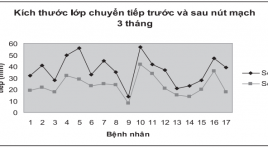
Preliminary evaluation of uterine artery embolization for treatment of adenomyosis SUMMARY: The uterine arterial embolization was carried out for 17 patients who suffered from adenomyosis, in which of them, 10 cases were followed-up by MRI at 1 and 3 months after embolization. The results showed that: free from abdominal pain were about 87.5 and 81.25% respectively and free from menorrhagia were 66.7% and 50% respectively. The thickness of transitional zone was reduced from 33 to 22.1mm. Average reducing of uterine volume was from 274.4 to 215.9cm3, corresponding to 19.5% of decreased volume. No contrast enhancement on MRI were found at 7 patients (7/10). 100% patients felt abdominal pain at the end of the procedure, especially 94,1% strongly pain. Hospitalization time was only 1-2 days in 94,1% of patients.

Chỉ định liều 131I trong điều trị bệnh nhân Basedow
02/04/2020 22:36:59 | 0 binh luận
Dose indication treated by iodine-131 for Grave’s patients SUMMARY: 543 Grave’s patients were treated by iodine-131. The treatment dose for patients were variable from 70 to 170 μCi/1g thyroid mass, depending on the value of thyroid volume and iodine uptake of thyroid. Follow up 6-24 months after treatment showed that: with appropriate dose indication had a significant reduction of the rate of hypothyroidism and hyperthyroidism.

Nghiên cứu đặc điểm tổn thương trên xạ hình SPECT tưới máu cơ tim ở bệnh nhân sau nhồi máu cơ tim
02/04/2020 22:32:58 | 0 binh luận
Characteristics of myocardial perfusion on SPECT at the post - infarct affection SUMMARY: Aims: the purpose of our study was to evaluate characteristics of myocardial perfusion defects in Tc99msestamibi gated SPECT myocardial perfusion imaging (MPI). Subjects and methods: 119 post-myocardial infarction (MI) patients were underwent gated SPECT in Nuclear Medicine Department, 108 Central Military Hosspital from March 2007 to May 2010. Results: in gated SPECT MPI, reversible, mixed and fixed perfusion defects were detected in 63.9%, 18.5% and 17.6%, respectively. In patient group with ESV ≥ 70 ml, SSS and SRS were significantly higher in group with ESV < 70 ml (18.63 ± 5.02 and 15.58 ± 4.99 vs 14.49 ± 4.83 and 11.15 ± 4.63 (p<0,001). There were significant correlations between SRS and SSS with WMS (r = 0.68, p<0.001 and r = 0.61, p<0.001). In patient group with EF ≤ 40%, SSS and SRS were significantly higher in patients with ESV>40% (19.83 ± 4.36 and 17.07 ± 4.58 vs 15.50 ± 5.2 và 12.13 ± 4.85; p<0.001). There were correlation between SRS and SSS with EF (r = - 0.47, p < 0.001) và SSS (r = -0.44, p<0.001). Conclusions: In post-MI patients, fixed, reversible and mixed defects are frequently detected in SPECT MPI. The extent and severity of perfusion defects are significantly correlated with wall motion, left ventricular volume and ejection fraction evaluated by gated SPECT MPI.

So sánh cộng hưởng từ và PET/CT trong chẩn đoán ung thư qua điểm y văn
02/04/2020 22:23:49 | 0 binh luận
Compare MRI and PET /CT in cancer diagnosis through literature SUMMARY: This paper aims to compare the ability of MRI and PET/CT in the diagnosis of cancer, assessment of tumor stages according to TNM classification, the advantages and disadvantages of each method through literature. The results showed that PET/CT has the advantages in the assessment of tumor stage (T) and the status of lymph nodes, especially with lung or head/neck primary tumors. However, the disadvantages of PET/CT are the high radiation exposure and the expensive cost. Whole body MRI has a high accuracy in the detection of distant metastases especially in bone, liver and central nervous system. MRI is considered as the first choice in the primary tumors which have bad FDG fixation such as renal cell and prostate carcinoma. Additionally, MRI is very good at the accurate evaluation of the entire skeleton condition and highly effective in the stage assessment of malignant bone marrow diseases.

Nghiên cứu ứng dụng kĩ thuật sinh thiết lõi qua thành ngực dưới hướng dẫn của siêu âm
02/04/2020 22:10:08 | 0 binh luận
Ultrasound-guided transthoracic core biopsy SUMMARY: Background: On the global scale, primary lung cancer is a major contributor to both cancer incidence and mortality. Definite diagnosis can be made from cytology or biopsy . A variety of techniques are available and helpful for the sampling include bronchoscopy, fine needle aspiration or image-guided biopsy, in which ultrasound-guided core biopsy of lung solid masses has been widely used as it is available, convenient and cost-effective. Material and method: A cross-sectional study was done on a sample of 77 individuals who had juxtapleural lung solid masses detected by Ultrasound or CT scan at Hue central hospital, from January-2010 to August-2011. All of these have undergone ultrasound-guided core biopsy. Objectives were to evaluate the efficacy, advantages and disadvantages of the technique. Results: The successful rate of single sampling was 89.6%. Five patients had twice sampling which raised the successful rate up to 94.8%, from which 89.6% were indicated malignant and the rest 5.2% were benign lesions. Advantages were multiple sampling (>2), supine and inclination position, skin surface-lesion distance less than 3 cm, pleura-lesion interface over 2 cm, lesion’s depth over 2 cm. Disadvantages include multiple sampling, lesion’s depth over 3 cm, FEV1/FVC < 0.7, needle size (<17G), prolonged procedure (>25 minutes). Rate of complication was 14.6%, in which pneumothorax was the most likely (4.9%), but no pleural drainage needed. Conclusion: ultrasound-guided core biopsy appeared safe, effective, non radiation exposure, cost-saving, simple and applicable in clinical setting. Key words: ultrasound-guided biopsy, lung solid mass.

Kết quả ban đầu điều trị can thiệp dò màng cứng vùng xoang hang đường tĩnh mạch bằng Coils qua 8 trường hợp
02/04/2020 21:57:18 | 0 binh luận
Endovascular treatment of cavernous sinus dural arteriovenous fistula using coils through venous approach. Report of 8 cases SUMMARY: Purpose: To analyze technical indication and result of endovascular treatment of cavernous sinus dural arteriovenous fistulae using coils through venous approach Material and methods: Eight patients were included in the retrospective study during the interval from 2/2012 to 7/2012. All of them were diagnosed of complex cavernous sinus dural arteriovenous fistulae and embolized by coiling through venous approach. The degree’s occlusion was evaluated at the end of procedure. The clinical symptoms of pre and post embolization were compared. Results: All the patient have cavernous dural arterio-venous fistula (Barrow type D and Cognard type IIIc), in which three cases were approached through facial vein, one case through superficial vein, and four cases through inferior or superior petrous sinus. DSA at the end of procedure showed complete occlusion all of them. Clinical symptoms, such as titinuos, exophthalmoses, and conjunctive edema were disappeared post embolization. One patient has internal strabismus as complication. Conclusion: Using coils via venous approach for cavernous sinus DAVF’s treatment is safe and effective.
Bạn Đọc Quan tâm
Sự kiện sắp diễn ra
Thông tin đào tạo
- Những cạm bẫy trong CĐHA vú và vai trò của trí tuệ nhân tạo
- Hội thảo trực tuyến "Cắt lớp vi tính đếm Photon: từ lý thuyết tới thực tiễn lâm sàng”
- CHƯƠNG TRÌNH ĐÀO TẠO LIÊN TỤC VỀ HÌNH ẢNH HỌC THẦN KINH: BÀI 3: U não trong trục
- Danh sách học viên đạt chứng chỉ CME khóa học "Cập nhật RSNA 2021: Công nghệ mới trong Kỷ nguyên mới"
- Danh sách học viên đạt chứng chỉ CME khóa học "Đánh giá chức năng thất phải trên siêu âm đánh dấu mô cơ tim"


