
Can thiệp nút mạch điều trị tổn thương động mạch vùng đầu tụy tá tràng
02/04/2020 21:51:09 | 0 binh luận
Endovascular embolisation methode in treatment of pancreatico duodenal arterial injuries SUMMARY: Purpose: Study about the endovascular embolisation methode in treatment of pancreatico duodenal arterial injuries. Materials and menthod: between 11/2011 and 4/2012, four patients with pancreatico duodenal vascular injuries on CT-Scanner were treated by endovascular embolisation. Result: All had hematoma and arterial injuries in pancrea ticoduodenal area on CT-Scanner,no indication of surgical intervention. On angiogra phy, two patients had an injury of anterior superior pancreaticoduodenal artery, one had gastroduodenal artery injury and one had injuries of posterior superior pancreatico duodenal and inferior pancreaticoduodenal artery. The patients had successfully embolized by n-BCA glue. No complication was noted. Conclusion: endovascular embolization is a safe, effective and suitable methode for pancreaticoduodenal arterial injury. The technique can develop in many healthcare centre in order to reducing the ratio of surgical intervention. Key word: endovascular embolisation, arterial injuries, CTScanner, angiography.
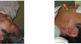
Tác dụng của phương pháp tiêm cồn tuyệt đối diệt hạch dây V qua da dưới hướng dẫn của màn tăng sáng
02/04/2020 21:32:15 | 0 binh luận
Effect of absolute alcohol infection in Gasserian ganglion neurolyse under Fluoroscopic guidance SUMMARY: Objective: By 3 cases of Gasserian ganglion neurolysis with absolute alcohol injected percutaneously through the foramen ovale under fluoroscopic guidance this article is aimed to present this technique and to evaluate the short-term efficacy of this procedure. Results: The efficacy on pain is immediately. The advantage is low cost. The disadvantage is the numbness and paralysis of the muscles of mastication however the patient accepted those effects than the pain they’ve had. Conclusion: The technique was found to be safe, cheap and effective in treating trigeminal neuralgia
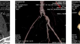
Kết quả ban đầu can thiệp nội mạch trong tái thông hẹp tắc mạn tính động mạch chậu
02/04/2020 21:28:25 | 0 binh luận
The short-term primary patency of endovascular intervention in recanalrization of chronic iliac arterial occluded diseases SUMMARY: Purpose: to evaluate the short-term primary patency of endovascular intervention in recanalrization of chronic iliac arterial occluded diseases. Method and materials: prospective study, group of 21 patients with diagnosis of chronic occlusion of iliac arteries in Bach Mai hospital from 9/2011 to 6/2012. The patients were indicated to do revascularization by endovascular intervention. Result: 21 patients with 28 iliac arteries which were treated by endovascular intervention including percutaneous angioplasty alone and Stenting. Arterial access were performed at common femoral arteries bilateral (retrograde) in 100%, including one failed case that required second intervention by brachial arterial access. No complication relates arterial access. There are 89,3% procedures of stenting with angioplasty post-Stenting and 100% completed deployment of stents. No complication of angioplasty and stent deployment such as arterial rupture or perforation. The success rate of recanalrization is 96% with 96% successful rate in the first approach to cross the over the occlusion. No required 2nd reintervention in duration of follow up from 1 to 6 months, 100% of case with improved intermittent claudication and ABI. Conclusion: the initial results confirm that the endovascular intervention is a safe, effective method in the goal of recanalrization of chronic occluded iliac arteries.
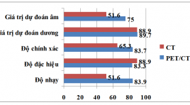
Gía trị của 18FDG PET/CT và CT ở bệnh nhân ung thư tuyến giáp biệt hóa có nồng độ Thyroglobulin huyết thanh cao và xạ hình toàn thân với 131I âm tính
02/04/2020 21:23:58 | 0 binh luận
Value of PET /CT and diagnostic CT scan in differentiated thyroid carcinoma patients with high serum thyroglobulin and negative 131I whole body scan SUMMARY: Objective: to determine value of FDG-PET/CT and diagnostic CT scan in post-surgical differentiated thyroid carcinoma patients with high serum thyroglobulin and negative 131I whole body scan. Patients and method: We performed FDG-PET/CT and diagnostic CT scan in 49 post-surgical differentiated thyroid carcinoma patients with high serum thyroglobulin and negative 131I whole body scan who already underwent 131I therapy in Department of Nuclear Medicine, 108 Central Military Hospital. Results: 41 lesions detected in 29/49 patients (59.2%) with positive PET/CT scan compared to only 20 lesions 18/49 patients (36.7%) detected on positive CT scan. The Sn, Ac and NPV of FDG-PET/CT were 83.9%, 83.7% and 75% higher than those of CT (51.6%, 65.3% and 51.6%, respectively). Sp and PPV of FDG-PET/ CT (83.3% and 89.7%, respectively) were similar to those of CT (88.9%). SUVmax thresholds with good diagnostic value were 4.5 ( Sn 84.6%, Sp 66.7%) and 5.0 ( Sn 65.3%, Sp 100%). FDG-PET/CT had altered treatment plan in 25/49 patients (51.2%). Conclusion: FDG-PET/CT is superior than CT in detecting reccurent/metastatic lesions in post-surgical differentiated thyroid carcinoma patients with high serum thyroglobulin and negative 131I whole body scan. Key words: FDG PET/CT, CT, differentiated thyroid carcinoma
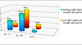
Nghiên cứu giá trị tiên lượng của xạ hình SPECT tưới máu cơ tim ở bệnh nhân sau nhồi máu cơ tim
02/04/2020 21:13:45 | 0 binh luận
Evaluation the prognostic of SPECT in post myocardial infarct SUMMARY: Aims: The purpose of our study was to evaluate the prognostic value of characteristics of defects in Tc99m-sestamibi gated SPECT myocardial perfusion imaging (MPI) in post-MI patients. Subjects and methods: 116 post-myocardial infarction (MI) patients were underwent gated SPECT in Nuclear Medicine Department, 108 Central Military Hospital from March 2007 to May 2010. Mean follow-up time was 23.27 ± 9,9 months. Results: The reversible perfusion defects had higher rate of patients with unstable angina (92.1% vs 76.9%, p<0.05) and revascularization (95.8% vs 72.1%, p<0.01), respectively. The fixed defect had higher rate of patients with congestive heart failure compared to patient group without congestive heart failure (47.2% vs 6.1%, p<0.001). In patients with unstable angina (UA) and/or recurrent MI, severe and large defects were significant highger than that of patients without UA and/or recurrent MI (p<0.05). In congestive heart failure (CHF) patients, severe and large defects were 97.1% and 76.8% in comparison with 88.2% and 70.7% respectively (p < 0.05). In fatal patient group, 100% had large defects compared to 72% in non-fatal MI patients. SSS and SRS were higher in post-MI patient with coronary events compared to those without coronary events (18.17 ± 5.3 vs 13.5 ± 3.65, p<0.001 and 14.77 ± 5.46 vs 11.04 ± 3.58, p < 0.01). Conclusions: the characteristics of myocardial perfusion defect on MPI have prognostic value in post-MI patients.
Gía trị chẩn đoán lệch bội nhiễm sắc thể trên thai kỳ nguy cơ cao của nhưng dấu hiệu siêu âm mới ở ba tháng đầu
02/04/2020 21:09:04 | 0 binh luận
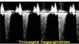
Chromosomal defects’ evaluation with new ultrasound markers in first trimester having high genetic risk pregnancies SUMMARY: Objective: To determine sensitivity (Se), specificity (Sp), false positive rate (FPR), positive predictive value (PPV) of new ultrasound markers on high genetic risk pregnancies between weeks 11 and 14. Before CVS procedure, the new ultrasound markers nasal bone(NB), DV and TR were assessed. We determined diagnostic evaluation of new ultrasound markers based on ultrasound findings and genetic results from CVS – gold standard. Results: NB marker: Se: 60%, Sp: 97.1%, FPR: 2.9% for T21. Se: 60%, Sp: 94.5%, FPR: 5.5% for T18. Se: 50%, Sp: 93.6%, FPR: 6,4% for T13. Se: 100%, Sp: 92.9%, FPR::7,1% forTurner syndrome. DV marker: Se: 70%, Sp: 96.2,1%, FPR: 3.8% for T21. Sens: 60%, Spec: 92.7%, FPR: 7.3% for T18. Se: 50%, Sp: 91.8%, FPR: 8.2% forT13. Se: 100%, Sp: 91.1%, FPR: 8.9% forTurner syndrome. TR marker: Sens: 60%, Sp 96.1%, FPR: 3.9% for T21. Se: 40%, Sp: 92.7%, FPR: 73%, for T18. Se: 50%, Sp: 92.7%, FPR: 7.3% for T13. Se: 100%, Sp: 92%, FPR: 8.0% for Turner syndrome. Combine 3 new markers for detecting chromosome defects: Increase Se, Sp and decrease FPR.Se: 60%, Sp: 97,1%, FPR: 2,9%. Reducing 73% placental biopsies… Conclusion: New ultrasound markers in the first trimester improved diagnostic evaluation of chromosome defects and avoiding unnecessary placental biopsy. Key words: Chorionic villus sampling, nasal bone, ductus venosus, tricuspid flow.
Bước đầu nghiên cứu siêu âm đàn hồi mô tuyến giáp ở người bình thường bằng phương pháp tạo hình và đo vận tốc sóng biến dạng qua kĩ thuật ARFI
02/04/2020 20:59:31 | 0 binh luận
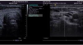
Preliminary studies on ultrasonic elastic tissue in the normal thyroid with the method of measuring shape and value velocity of shear wave by using a acoustic radiation force impulse image technology (ARFI) SUMMARY: Objectives: Measure the reference value of average velocity of shear wave in healthy healthy thyroid tissue by using acoustic radiation force impulse imaging. Methods: Evaluate 49 healthy volunteers with normal thyroid: 27 women and 22 men without thyroid pathology, thyroiditis, localized lesions,calcium metabolic disease, not taking these drugs affect thyroid. Identify Region Of Interest (ROI ) for thyroid tissue depth of 1.2 cm, for respectively stenocleidomastoid muscle depth of 0,6 cm.The result measured by two observers with different levels of experience performed independently and blindly. Results: Average velocity of shear wave in the healthy’s thyroid tissue is 1,47 ± 0,41m/s. There is no statistically significant difference in shear wave velocity between two gender and age (p > 0,05). Average velocity of shear wave in the healthy’s stenocleidomastoid muscle is 1,42 ± 0,32m/s. There is no statistically significant difference in shear wave velocity between three age groups but there is statistically significant difference in shear wave velocity between two gender (p < 0,05).In term of interobserver results, no statistically significant difference in shear wave velocity obtained by two observers with different levels of experiences (P > 0,05). Conclusions : The results of this study show that shear wave velocity measurement in healthy’s thyroid of women (1,50 ± 0,41 m/s) was significantly higher than in men but there is no statistically significant (p > 0,05). In contrary, average velocity of shear wave in the healthy’s stenocleidomastoid muscle of men (1,54 ± 0,29 m/s) was higher than in women (1,33 ± 0,32 m/s) statistically significant ( p < 0,05). Thyroid tissue elastogram shows the structure of the tissue is quite soft, homogeneous and smaller B-mode image. ARFI can be performed in the thyroid tissue with reliable results.

Xác định kích thước hố yên người Việt Nam bằng chụp cắt lớp điện toán
02/04/2020 20:52:36 | 0 binh luận
Normal sizes of the sella turcica of VietNamese on computed tomography SUMMARY: Objectives: To determine the normal sizes of the sella turcica of Vietnamese from 1 to 30 y.o. on computed tomography. Methods: cross-sectional description of retrospective study. The length, depth, width and volume of sella turcica were measured on computed tomography of 705 patients at the No.1 Children’s Hospital and the Trưng Vương Emergency Hospital, HCM city, Vietnam , from January to June, 2011. Results: The mean size values of the sella turcica were divided into six age groups:1 - 5, 6 - 10, 11 - 15, 16 - 20, 21 - 25, 26 - 30 with length (7.16 ± 1.43 mm, 8.11 ± 1.09 mm, 8.48 ± 1.14 mm, 9.99 ± 1.47 mm, 10.20 ± 1.16 mm, 10.39 ± 1.17 mm); depth (5.74 ± 1.16 mm, 6.74 ± 1.10 mm, 11.35 ± 1.42 mm, 12.67 ± 1.86 mm, 12.73 ± 1.5 2mm, 12.72 ± 1.41 mm); width (9.14 ± 1.67 mm, 10.44 ± 1.41 mm, 11.35 ± 1.42 mm, 12.67 ± 1.86 mm, 12.73 ± 1.52 mm, 12.72±1.41mm) and volume (196.69 ± 85.28 mm3, 286.33 ± 77.18 mm3, 336.19 ± 90.90 mm3, 491.18 ± 146.68 mm3, 500.05 ± 130.09 mm3, 523.80 ± 119.58 mm3). Conclusions: The sizes of sella turcica increase from 1 to 20 years old; no significant increasing from 21 to 30 y.o. Differences in the sizes of sella turcica between males and females are not statistically significant. Keywords: Sella turcica
Bạn Đọc Quan tâm
Sự kiện sắp diễn ra
Thông tin đào tạo
- Những cạm bẫy trong CĐHA vú và vai trò của trí tuệ nhân tạo
- Hội thảo trực tuyến "Cắt lớp vi tính đếm Photon: từ lý thuyết tới thực tiễn lâm sàng”
- CHƯƠNG TRÌNH ĐÀO TẠO LIÊN TỤC VỀ HÌNH ẢNH HỌC THẦN KINH: BÀI 3: U não trong trục
- Danh sách học viên đạt chứng chỉ CME khóa học "Cập nhật RSNA 2021: Công nghệ mới trong Kỷ nguyên mới"
- Danh sách học viên đạt chứng chỉ CME khóa học "Đánh giá chức năng thất phải trên siêu âm đánh dấu mô cơ tim"


