Xạ hình 99mTc-MDP phát hiện di căn xương ở bệnh nhân ung thư
02/04/2020 14:21:28 | 0 binh luận
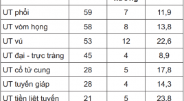
Detection of bone metastases by spect 99m Tc-MDP of cancer patients SUMMARY: 99mTc-MDP bone scan for 425 patients with different stage cancer. Bone metastases were detected on 52 patients (12.2%). High rate of bone metastases had origine from prostate, breast, uterin cervix cancers. Almost were multifoci asymmetric lesions with increased uptake of radiopharmatical activity. The most common site of bone metastases are spine, pelvis (hip) and ribs. Key words: Bone metastases, Bone scan 99mTc-MDP.

So sánh giá trị của siêu âm đường bụng và đường âm đạo trong chẩn đoán thai ngoài tử cung
02/04/2020 14:17:53 | 0 binh luận
Evaluated comparison of transvaginal and abdominal ultrasound in diagnosis of ectopic pregnancy SUMMARY: Purpose: To compare the value of abdominal and transvaginal ultrasound in diagnosis of ectopic pregnancy. Materials and methods: Cross-section and descriptive study was underwent on 140 patients who were suspected of ectopic pregnacies, from June 2009 to June 2010. All of them were done both abdominal and transvaginal ultrasound. The value of each modality was analysed base on gold standard of histopathology. Results: 140 cases of suspected ectopic pregnancy in which 110 were ectopic pregnancies, 10 viable intrauterine pregnancies, 19 abortions and 1 hydatidiform mole. The suggestive abdominal ultrasound diagnosis of ectopic pregnancy has Sn 71%, Sp 86%, PPV 95% and NPV 44%, those of transvaginal ultrasound has Sn 92.7%, Sp 96.6%, PPV 98% and NPV 96,6%. Conclusion: Transvaginal ultrasound has a primary role in the diagnosis of ectopic pregnancy

Nghiên cứu đặc điểm hình ảnh và giá trị chẩn đoán của cộng hưởng từ đối với u nguyên bào thần kinh đệm hai bán cầu
02/04/2020 14:10:20 | 0 binh luận
Study of imaging characteristic and value of MRI in diagnosis of hemisphere glioblastoma SUMMARY: Purpose: Describing imaging characteristic and assessing the MRI value in the diagnosis of hemisphere glioblastoma. Materials and methods: 2505 patients of brain tumors were selected in retrospective describe study, diagnosed, operated and having histopathological result from January 2009 to March 2011 in Viet Duc Hospital, in which 45 patients were histologically confirmed of glioblastoma. Based on gold standard of histopathology, the value of MRI in diagnosis of hemisphere glioblastoma was assessed. Results: Mean age 51±16. Gender: M/F 1.3/1. Location at frontal lobe (26.4%), fronto -temporal lobe (20%), less frequently at occipital lobe. Average diameter more than 5cm (60.4%). Hyposignal on T1W (66%), hypersignal on T2W (70%), marked and heterogenous enhancement (43.4%), rim enhancement (47.3), necrosis (95.6%), cyst component (52.8%), hemorrhage (28.4%); surrounding edema of grade II and III (90%), meningeal and copus callosum invasion (47.2% and 39.6%, respectively). Accurate diagnosis of tumor location was 100% comparing to surgery. Se, Sp and Acc respectively 81.1%, 97.6%, and 98%. Conclusion: MRI had high value in diagnosis of hemisphere glioblastoma.

Bước đầu đánh giá vai trò của cộng hưởng từ tim trong chẩn đoán tứ chứng Fallot
02/04/2020 14:07:17 | 0 binh luận
Initial evaluation of cardiac magnetic resonance in for diagnosis of Fallot tetralogy SUMMARY: Purposes: To apply cardiac magnetic resonance (CMR) for diagnosis preoperative Tetralogy of Fallot patients and to compare the findings on echocardiography with CMR. Methods and materials: 32 patients were included in a prospective study during the interval from June 2008 to August 2009 at Bach Mai hospital. Both echocardiography and CMR had been perrformed to evaluate the variables: Ejection Fraction (EF), the right ventricular outflow tract, main pulmonary artery (MPA), left and right pulmonary artery (LPA & RPA), major aortopulmonary collateral arteries (MAPCAs)… The correlation between the findings on CMR and echocardiography was compared by using Pearson statistics. Results: 32 patients (14 males) with average age was 19.94 ± 8.20 (range: 8 -53) y. o. 100% success CMR with average time: 80.93 ± 50.41 minutes. There was a close correlation between the findings on CMR and on echocardiography with Kappa statistics r = 0.63-0.70 (p < 0.05). CMR detect more MAPCAs and additional anomalies than echocardiography. Conclusions: The results of our study indicate the usefulness of CMR for evaluation pre-operative patients with Tetralogy of Fallot. CMR has more advantages than echocardiography for detection MAPCAs and additional anomalies Key words: Tetralogy of Fallot; Cardiac magnetic resonance imaging -CMR; Echocardiography

Nghiên cứu đặc điểm hình ảnh cộng hưởng từ chảy máu nhu mô não không do chấn thương
02/04/2020 14:01:57 | 0 binh luận
The study of characteristic imaging of non-traumatic cerebral parenchymal hemorhage on MRI SUMMARY: Objective: This study aims to describe the characteristic and find out the causes of non-traumatic cerebral parenchymal hemorhage on MRI. Materials and methods: Across descriptive study was underwent on 45 patients suffering from cerebral parenchymal hemorhage in BachMai hospital, from January to Sseptember, 2010. Results: Super-acute stade: The hematoma is isointense on T1, central hyperintense and peripheric hypointense on T2, Flair, T2* (5/5 cases). Acute stade: isointense on T1; hypointense in the centre, hyperintense in peripheric on T2, Flair (2/2 cases). Early subacute stade: homogeneous hyperintense on T1W (6/8 cases). Later subacute stade: homogeneous hyperintense on T1W (84.6%), peripheric hypointese on T2W (88.5%). Chronic stade: isointense on T1, Central hyperintense on T2, Flair, T2* (2/2 cases). Hypertension is the most common causes (48.5%) and leads to central noyal hematoma. Meanwhile lobular hematoma is due to vascular malformation (35.3%). The hyperintense is more frequently in over 50 years old (73.1%) and vascular malformation is seen in the under 50 years old (47%). Conclusion: MRI is effective method to evaluate the characteristic of cerebral parenchymal hematoma and has important role in searching the causes.

Bước đầu nghiên cứu giá trị của chụp cộng hưởng từ toàn thân trong đánh giá giai đoạn của ung thư phổi
02/04/2020 13:58:16 | 0 binh luận
Initial study of whole-body MRI value in assessement of lung cancer staging SUMARY: Purpose: This study aims to describe the characteristic of the primary and mestastasis lesions of pulmonary cancer on wholebody MRI and to study the value of whole-body MRI in assessement of pulmonary tumor staging. Materials and methods: Prospective and describe study included 46 patients, confirmed of lung cancer base on histopathology, from January to October 2011 in Bach Mai Hospital. All of them were done whole-body MRI and 21 patients in which were taken FDG-PET/CT. The imaging characteristic of original tumor and metastasis lesions were describe on whole-body MRI. Assessement of lung cancer staging on whole-body MRI was compared with FDG-PET/CT. Results: The mean diameter of original tumor is about 44.4±22.15mm. The average restriction of lung cancer is about 4.02±0.83 points. 67.5% of lung cancer presented mediastinal and pleural invasion. The metastasis rate of lymph node, bone, brain, liver and adrenal gland were 54.3%; 28.3%; 26.1%; 15.2% and 8.6% respectively. Assessement of lung cancer staging according to T, N, M and TNM between 2 methods of whole-body MRI and FDG-PET/CT with Kappa coefficence were 0.601; 0.516; 0.904 and 0.846 respectively, p < 0.05. Conclusion: High field whole-body MRI were considered as an alternative modality to assess the lung cancer staging.

Đánh giá hiệu quả bước đầu phương pháp lấy huyết khối cơ học bằng stent Solitaire trong điều trị nhồi máu não tối cấp
31/03/2020 15:29:03 | 0 binh luận
Đột quỵ não là bệnh lý thường gặp ở người lớn tuổi, phổ biến nhất là những người có tiền sử bệnh lý về tiểu đường, tim mạch, huyết áp, hay tăng cholesterol máu... trong đó 90% các trường hợp là thiếu máu - nhồi máu não. Đây là nguyên nhân hàng đầu gây tàn tật và gây tử vong đứng hàng thứ 3 tại Hoa Kỳ [1]. Tỉ lệ đột quỵ não được dự báo sẽ tăng lên hàng năm và sẽ đạt 1,2 triệu người mỗi năm vào năm 2025 [2]. Giảm tốc độ dòng máu và thiếu máu kéo dài sẽ chuyển thành nhồi máu thực sự nếu không được điều trị kịp thời và để lại nhiều di chứng nặng nề. Vì vậy, mục tiêu của điều trị can thiệp nội mạch trong nhồi máu não cấp là giảm thiểu tác hại đến nhu mô não bằng việc khối phục dòng máu đến vùng đang thiếu máu càng sớm càng tốt với độ an toàn đảm bảo. Điều trị lấy huyết khối bằng phương pháp cơ học, sử dụng stent Solitaire là phương pháp hiện đại, mới được áp dụng lần đầu ở Việt Nam tại Khoa Chẩn đoán hình ảnh Bệnh viện Bạch Mai vào tháng 5 năm 2012 [3]. Chúng tôi nghiên cứu đề tài này nhằm mục đích đánh giá hiệu quả bước đầu phương pháp lấy huyết khối cơ học bằng stent Solitaire ở các bệnh nhân nhồi máu não cấp.

Cập nhật nhanh và xử lý sớm sốc phản vệ do thuốc cản quang
03/04/2020 09:48:01 | 0 binh luận
Phản vệ được định nghĩa là một phản ứng dị ứng nghiêm trọng mà xuất hiện và có thể dẫn đến tử vong một cách nhanh chóng [2, 3]. Phản vệ có thể xuất hiện sau vài phút (thường là trong vòng những giờ phút đầu tiên sau khi tiêm (tiếp xúc với dị nguyên) [1]. Thuốc cản quang được xem như là một trong những dị nguyên gây phản vệ nghiêm trọng nhất trong bệnh viện. Hàng năm, toàn thế giới có hơn 70 triệu thăm dò chẩn đoán hình ảnh có sử dụng thuốc cản quang, riêng ở Mỹ có ít nhất 10 triệu người [4]. Các thăm dò chẩn đoán hình ảnh bao gồm chụp ống tủy, chụp mạch (động - tĩnh mạch, chụp UIV, chụp đường mật ngược dòng - ERCP, chụp khớp gối, chụp CLVT…). Sốc phản vệ xảy ra chủ yếu khi dùng thuốc cản quang dạng tiêm đường tĩnh mạch. Phát hiện sớm và xử trí kịp thời sốc phản vệ liên quan đến thuốc cản quang theo phác đồ là yêu cầu bắt buộc với các nhân viên y tế nói chung, đặc biệt là bác sĩ, kĩ thuật viên tại khoa chẩn đoán hình ảnh. Adrenaline là thuốc cấp cứu cơ bản trong sốc phản vệ do bất cứ dị nguyên nào. Không có chống chỉ định tuyệt đối giành cho Adrenalin, song thuốc này vẫn chưa được sử dụng đúng cách và triệt để trong cấp cứu sốc phản vệ. Một số nghiên cứu cho thấy 70% bệnh nhân sốc phản vệ có các triệu chứng rầm rộ cần ít nhất hai lần tiêm adrenaline [1]
Bạn Đọc Quan tâm
Sự kiện sắp diễn ra
Thông tin đào tạo
- Những cạm bẫy trong CĐHA vú và vai trò của trí tuệ nhân tạo
- Hội thảo trực tuyến "Cắt lớp vi tính đếm Photon: từ lý thuyết tới thực tiễn lâm sàng”
- CHƯƠNG TRÌNH ĐÀO TẠO LIÊN TỤC VỀ HÌNH ẢNH HỌC THẦN KINH: BÀI 3: U não trong trục
- Danh sách học viên đạt chứng chỉ CME khóa học "Cập nhật RSNA 2021: Công nghệ mới trong Kỷ nguyên mới"
- Danh sách học viên đạt chứng chỉ CME khóa học "Đánh giá chức năng thất phải trên siêu âm đánh dấu mô cơ tim"


