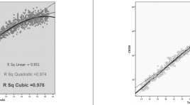
Xây dựng và ứng dụng biểu đồ phát triển thai nhi cho người Việt Nam ở bệnh viện Từ Dũ
03/04/2020 09:39:42 | 0 binh luận
Establish a fetal growth development chart for vietnamese in Tu Du Hospital SUMMARY: Background and objective: Ultrasound has been used extensively in obstetrics in recent years. Among many of its applications, gestational assessment and monitoring of fetal growth are the most important ones. Fetal growth problems such as IURG or macrosomnia fetus, are diagnosed base on reliable fetal growth development chart. Aim: modeling fetal development chart from 14 to 20 weeks of gestation by fetal biparietal diameter, head and abdomen circumference, femur length. Methods: cross-sectional study conducted from 3/2009 to 8/2010, 1843 recruited pregnancies, using randomized selection. Select each regression equation parameters, a correlation coefficient R2 highest gestation after testing the suitability of the regression model. Result and conclusion: may be our table percentage of ultrasound parameters according to gestational age brings characteristic and different from other authors. Keywords: ultrasound parameters, regression model
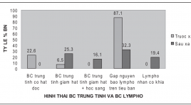
Nghiên cứu ảnh hưởng của I-131 liều 100 MCI tới tủy xương trong điều trị ung thư tuyến giáp thể biệt hóa đã phẩu thuật
03/04/2020 09:22:13 | 0 binh luận
Research effects of 100 mCi dose Iodine- 131 on bone marrowin therapy for operated differentiated thyroid carcinoma SUMMARY: Researching into the effects of 100 mCi dose I-131 on bone marrow of 31 operated differentiated thyroid carcinoma patients, after 03 months of I-131 therapy, through the characteristics of peripheral blood cells and hematopoietic precursors of bone marrow, comparing with prior to I-131 therapy, the results showed that: On the peripheral blood circulation: Lymphocyte and thrombocyte counts were decreased obviously (p< 0.05). However, the mean values of lymphocyte and thrombocyte counts still belong to normal reference values; Erythrocyte lineage has not yet had affected signs of I-131 at dose of 100 mCi, that causes the decresae in erythrocyte count and hemoglobin concentration; The increase of patient percentage with unusual cytoplasm of Neutrophils (hypogranular neutrophil or the presence of vacuoles in cytoplasm) (p= 0.01); The increase of patient percentage with large thrombocyte in dimension (p= 0.038). On bone marrow: bone marrow cell counts were decreased (p= 0.0001). However, the mean value of bone marrow cell counts still belong to normal reference value; The decrease of patient percentage found lymphocytic precursors and Basophilic meakaryocytes (p < 0.001); The increase of patient percentage found lymphocytes with cleaved nucleus (p= 0.01); The increase of patient percentage with unusual cytoplasm of granulocytic precursors (decreased azurophilic granules or the presence of vacuoles in cytoplasms) (p < 0.05). Key words: Eeffects of 100 mCi dose Iodine- 131 on bone marrow.
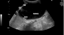
Đặc điểm hình ảnh nang ống mật chủ ở trẻ em trên siêu âm và cộng hưởng từ 1.5T
31/03/2020 21:43:09 | 0 binh luận
Imaging characteristics of choledochal cysts in children on ultrasound and MRI 1.5T SUMMARY: Purposes: The description of Imaging characteristics of choledochal cysts in children on Ultrasound and MRI 1.5T Material and methods: 44 patients were diagnosed choledochal cysts on Ultrasound and/or had Ultrasound and MRCP from 7/2012 to 9/2013 in National Hospital of Pediatrics. 16 patients with choledochal cysts underwent MRCP using a half –Fourier acquisition single-shot turbo spin-echo sequence. MRCP findings were compared with intraoperative cholangiography. Result: In 44 patients choledochal cysts including 34 girls and 10 boys:age range, 2 months -16 years; mean age 3.4 years. Clinical presentation: abdominal pain is the most common symptom (72.72%), vomiting (63.18%), fever (15.90%), jaundice (13.63%). The type of choledochal cysts (according to Todani): type I (93%), type IVa (7%); Type of dilatation: cystic dilatation (77.27%), fusiform dilatation (22.73%); mean measurement: 39.47mm; stone in cyst (29.5%); intrahepatic duct dilatation (43.18%); gallstone (6.8%). MRCP findings (n=16): with the most common form being typeI 87.5% (14/16), typeIVa 12.5% (2/16); cystic dialatation (93.7%), fusiform dilatation 6.3%. Cystolithiasis (75%); intrahepatic duct dilatation (56,25%). Kappa value was good agreement (k: 0.717-0.738) when compared Ultrasound and MRCP. The presence of the anomalous junction of pancreaticobiliary duct was revealed by MRCP in only 10 cases of 13 cases choledochal cysts with Kappa value was good agreement (k=0.612) when compared with intraoperative cholangiography. Conclusion: Ultrasound and MRI showed overall good accuracy in the detection and the classification of choledochal cysts and revealed associated complications. MR cholangiopancreatography provides information about anomalous pancreaticobiliary ductal union in children with choledochal cyst. Key words : Choledochal cyst, Ultrasound. Magnetic resonance cholangiopancreatography (MRCP).
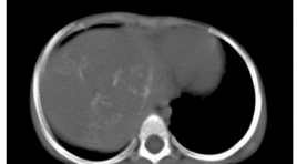
Đặc điểm hình ảnh của u nguyên bào gan trẻ em trên phim chụp cắt lớp vi tính hai dãy đầu thu
31/03/2020 21:48:55 | 0 binh luận
Characteristic of pediatric hepatoblastoma on 2 detector computed tomography SUMMARY: Purpose: to describe the characteristics of hepatoblastoma (HB) on 2 detector computed tomography (CT) images Materials and method: 49 under 15 year-old patiens with pathological results of HB were undergone 2 detector computed tomography from 2010 to 2014. Result: 100% was solid tumor, the average diameter was 8.48mm, most of them were single tumor, located at the right lobe, lobulated and defined margin, heterogeneous structure, 31.5% had calcification, strong contrast enhancement in the arterial phase, less than liver parenchyma in the portal phase. 63% enhanced less than normal liver parenchyma in both aterial and portal phase. Pretext II and III is 81.7%, lung metastasis in 4 cases, portal vein thrombosis in 2 cases, 4 cases infiltrated to extra-hepatic spaces, 1 tumor was ruptured, 2 caseshad hepatic umbilical nodes. Conclusion: HB appears on CT images as a solid tumor, heterogeneous, irregular margin mass, 31.5% had calcification. After injecting the material contrast most of tumors enhance trongly during the arterial phase and less density than the surrounding liver parenchyma in the portal phase. The most common is PRETEXT II and III. It may metastase to lung, lead to portal vein thrombosis, hepatic hilar lymph node. Keywords: hepatoblastoma, liver tumors in children, liver mass in children, hepatoblastoma imaging.
Đặc điểm hình ảnh và các giá trị cắt lớp vi tính trong bệnh lý u nguyên bào thần kinh sau phúc mạc
03/04/2020 09:17:28 | 0 binh luận
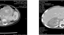
Radiographic features and accuracy of CT in diagnosis and evaluation of the dissemination of retroperitoneal neuroblastoma in children SUMMARY: Purpose: This study aims to characterize radiographic features on CT of retroperitoneal neuroblastoma. Evaluate Se., Sp., PPV., NPV.,Ac., of CT in diagnosis of retroperitoneal neuroblastoma and dissemination of the disease. Materials and Method: 111 children with 112 retroperitoneal tumors were enrolled in a prospective study. Abdominal CT findings of all the patients were analyzed, correlated with clinical, laboratory data, results of biopsy and surgical findings in order to assess sensitivity, specificity and accuracy values of CT in diagnosis and evaluation of tumor extent Result: Radiographic features of retroperitoneal neuroblastoma on CT: lobulated, ill-defined, uncapsulated, calcified mass with mild or moderate heterogeneous enhancement and vessel encasement. Among these signs, “lobulated, ill-defined and vessel-encasement” are the most valuable (Ac. 72-81%). The other signs have low to medium Se., Sp., Ac in diagnostic value (52%-96%) and should be combined with the former. CT is good at evaluating the tumor extent: renal invasion (>94%), vessel encasement (100%), abdominalorgan invasion (>75%), retroperitoneal lymphadenopathy (>85%) except for detecting peritoneal fluid (Se. 36%) Conclusion: Radiographic features of retroperitoneal neuroblastoma on CT: lobulated, ill-defined, uncapsulated, calcified, with mild or moderate heterogeneous enhancement and vessel-encased. Among them, “lobulated, ill-defined vesselencasement” are the most valuable signs. We should combine signs to increase accuracy of diagnosis. For evaluation of tumor extent, CT have high sensitivity, specificity and accuracy except for detecting peritoneal fluid. Abbreviation: CT: Computed Tomography, Se: Sensitive, Sp: Specificity, PPV: Positive Predictive Value, NPV: Negative Predictive Value, Ac: Accuracy.

Điều trị can thiệp nội mạch các tổn thương mạch trong chấn thương tạng đặc
03/04/2020 09:14:24 | 0 binh luận
Interventional endoluminal treatment for vascular lesion of solid organ post traumatism SUMMARY: Pupose: Estimation the efficacy of embolization in abdomen injury. Material and method: 37 injury patients were underwent angiography and emboli zation in Viet Duc hospital from 2008 to 2012 with 25 cases hepatic trauma, 10 renal trauma, 2 splenic trauma. Result: All of patients undergone embolization hadn’t extravasation in angiography (100%), no on going hemorrhage required laparotomy. Conclusion: Embolization in abdomen trauma is an efficacy therapeutic method should be widely applied in clinical application.
Gía trị của chụp cắt lớp vi tính đa dãy trong chẩn đoán u mạc treo
31/03/2020 21:35:54 | 0 binh luận
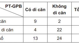
The value of MSCT in diagnosing of mesenteric tumors SUMMARY: Objectives: Evaluating the value of MSCT in diagnosing of mesenteric tumors. Subjects and Methods: including 55 patients from October 2010 to June 2014 with MSCT and with the sugery result and pathology of mesenteric tumor, Gist (Gastrointestinal stroma tumor), retroperitoneal tumor. Result: Determining diagnosic: Se:86.5%, Sp: 92.9%, PPV: 64%. Local fat invasion: Se: 86.6%, Sp: 86.3%, ACC: 86.4%. Evaluating vasculary invasion: Se: 66.6%, Sp: 92.8%, ACC: 86.4%. Evaluating gastrotestinal invasion: Se: 69.2%, Sp: 91.6%, ACC: 83.7%. Evaluating exact location of tumor: 35.1%. Evaluating peritoneal invasion: Se:71.4%, Sp: 90%, ACC: 86.4%. Conclusion: MSCT has hight Se and Sp in determining diagnosis, evaluating the invasion level of mesenteric tumor. MSCT has average PPV and low value in evaluating exact location of mesenteric tumor Key words : MSCT, Gist, mesenteric tumor , retroperitoneal tumor.
Nhận xét đặc điểm hình ảnh và kết quả chẩn đoán cắt lớp vi tính so với phẫu thuật trong di dạng teo thực quản, rò khí thực quản bẩm sinh
03/04/2020 09:10:39 | 0 binh luận
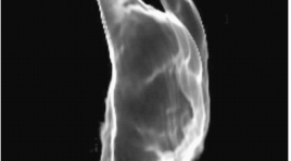
CT imaging in congenital esophageal atresia, tracheo-esophageal fistula (TEF ) comparing with surgical results SUMMARY: Purpose: To review some CT signs in congenital esophageal atresia, tracheo-esophageal fistula (TEF) and compare the CT findings with surgical results. Materials and Methods: Images, CT diagnostic results were collected and compared with the surgical descriptions in 7 cases TEF from 12/2007-11/2010 at Binh Dinh hospital. Results: Direct signs: atresis image of esophageal was visualized in 100% cases. The image of fistula between proximal esophagus-trachea: true positive is 2/3 cases, true negative is 3/4. The image of fistula distal esophagus-trachea: true positive is 3/6 cases, true negative are 1/1. Indirect signs: pneumonia and esophageal dilatation were visualized in 100% cases. Gaseous distention of the stomach and bowel was visualized in 3/4 cases which exist only lower fistula and 2/2 cases which exist both upper and lower fistula. Conclusion: Between CT with surgery is concordantly in 5/7 cases, nonconcordantly in 2/7. Key words: esophageal atresia, tracheoesophageal fistula, congenital tracheoesophagus
Bạn Đọc Quan tâm
Sự kiện sắp diễn ra
Thông tin đào tạo
- Những cạm bẫy trong CĐHA vú và vai trò của trí tuệ nhân tạo
- Hội thảo trực tuyến "Cắt lớp vi tính đếm Photon: từ lý thuyết tới thực tiễn lâm sàng”
- CHƯƠNG TRÌNH ĐÀO TẠO LIÊN TỤC VỀ HÌNH ẢNH HỌC THẦN KINH: BÀI 3: U não trong trục
- Danh sách học viên đạt chứng chỉ CME khóa học "Cập nhật RSNA 2021: Công nghệ mới trong Kỷ nguyên mới"
- Danh sách học viên đạt chứng chỉ CME khóa học "Đánh giá chức năng thất phải trên siêu âm đánh dấu mô cơ tim"


