Đánh giá kết quả siêu âm doppler xuyên sọ trong chẩn đoán chết não tại bệnh viện Trung Ương Huế
23/05/2020 11:07:39 | 0 binh luận
SUMMARY Background : Although the clinical examination has the most important role and documentation of the clinical signs of brain death are very uniform, it is necessary to use technical confirmatory tests to corroborate the clinical signs such as Transcranial Doppler ultrasonography (TCD) and Electroencephalography (EEG). The current study examined (1) Hemodynamic of cerebral arteries in 16 death brain cases (2) the role of TCD in confirmation of brain death. Subjects and methods :16 patients clinical brain deathwere included in the study. The following TCD findings were accepted as confirmatory of brain death when they were found at least one of two within the same examination: (1) brief systolic forward flow or systolic spikes and diastolic reverse flow or no diastolic flow, (2) Vmax <10cm/s or no demonstrable flow in a patient in whom flow had been clearly documented in a previous TCD examination. Results : All of cases were confirmed as brain death on TCD. 14/16 patients (87.5%) performed the waveform abnormality, the rest was found decreasing velocity or missing the blood flow on TCD. Conclusion: The sensitivity of TCD is increased with repeat examinations and should be repeated in cases in which systolodiastolic forward flow is demonstrated after the first TCD. TCD may prolong or shorten the time to declaration of brain death. The necessity of demonstrating cerebral circulatory arrest in patients with clinical brain death is debatable. Key words: brain death, doppler ultrasonography.
Nghiên cứu đặc điểm siêu âm lỗ dò động mạch quay - tĩnh mạch đầu có giảm lưu lượng ở bệnh nhân lọc máu chu kỳ
23/05/2020 11:02:13 | 0 binh luận
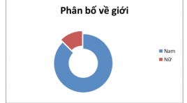
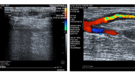
SUMMARY Objectives : Assessing the characteristics of radio - cephalic fistulas with insufficient flow volume in dialysis patients by Dupplex sonography. Subjects and methods : A cross - sectional study from 20 dialysis patients who have radio - cephalic fistulas with insufficient flow volume. Results: In 20 patients (13 males and 7 females), average age (43.7 ±1.18 years of age). Diameter of juxta-anastomotic orifice is 3.1± 0.9 mm in males and 2.7 ± 1.2 mm in females. Diameter of cephalic vein is 3.6 ± 0.9 mm in males and 2.8 ± 1.2 mm in females. Some hemodynamic indexs of fistular: PSV (89.55 ±101.61cm/s), ESV (51.80 ± 45.50 cm/s), RI 0.76 ± 0.23) and flow volume (130.95 ± 110.04 ml/minute). Some causes of insufficient flow volume: thrombosis 9/20 (45%), accessory veins (3/20 (15%), intimal hyperplasia 2/20 (10%), juxta-anastomotic stenosis due to thrombosis 1/20 (5%) and no cause found 5/20 (25%). Conclusions : Dupplex sonography can assess the flow volume of fistula and causes of insufficient flow volume. It is a useful tool for clinicians in following up the AV fistulas.
Bước đầu đánh giá hiệu quả của can thiệp nội mạch trong tổn thương động mach vùng hàm mặt do chấn thương
23/05/2020 10:33:48 | 0 binh luận
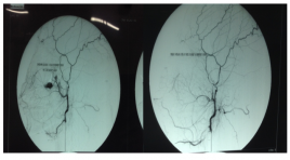
Initial evaluation of Transcatheter Arterial Embolization in the Treatment of Maxillofacial Trauma SUMMARY Purpose: To evaluate the results of Transcatheter Arterial Embolization in the Treatment of Maxillofacial Trauma. Methods and Materiels: Prospective description study on 18 patients admitted for maxillofacial injuries with life-threatening hemorrhaging and hemodynamic instability from January 2011 to December 2013, at Viet Duc Hospital. Results: Maxillofacial trauma was caused by traffic injuries (89%). 16/18 (89%) patients exhibited documented Le Fort III fractures. Internal maxillary artery often the most artery injury (77.8%). Transcatheter arterial embolization (TAE) was successful in all patients. No complications was found post-TAE. Three patients deaths (16.5%) occurred from severe traumatic brain injury. Conclusion: Transcatheter Arterial Embolization in the Treatment of Maxillofacial Trauma was safe and effective to control acute bleeding. Key Words: Maxillofacial trauma, Hemorrhage, Transcatheter arterial embolization (TAE), Facial fractures, Le Fort fractures.
Chẩn đoán chứng tự kỷ với cộng hưởng từ
23/05/2020 12:32:24 | 0 binh luận
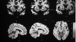
MRI in Autism SUMMARY The diagnosis of Autism based chiefly on clinical manifestations which are polymorphous. Some years recently owing to the fast development of diagnosis imaging particularly MRI, several articles had demonstrated the morphologic changing in autism. Rely on high spatial resolution, it can find the abnormality in the gray, white matter based on diffusion sequence and displaying neural tractus in the predilection area of autism such as callosus corpus, frontal, temporal, amydal, ROI-based Volumetry, Voxel-based Morphometry, Surface-based Morphometry, Tensorbased morphology, DTI... The autistic pathologic changing can be found by decreasing of fractional anisotrope FA, increasing of voxel-volume, thinning of the grey matter particular the neural fiber deficit on tractography. The equipement must be high MRI unit 3.0T and specific soft ware, however MRI can display the quality variation of the affection.
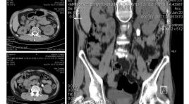
Chiến lược xử lý cơn đau quặn thận
23/05/2020 12:30:25 | 0 binh luận
Management of renal colic SUMMARY Renal colic is a frequently situation seen in emergency. The pathophysiological mechanisms of pain in acute renal colic is due to increased intrapyelic pressure by ureteral obstruction with renal synthesis of prostaglandin E2. Intrarenal hemodynamic changes by increase the blood’s flow. The pharmacological treatment base on these pathophysiologicals mechanisms. The diagnostic and radiological mission are relatively well codified in all emergency center. For the simple renal colic, the radiograph of the abdomen without preparation can identify the calcifications of stones on urinary tract. The ultrasound can detect the presence of stones in the kidney and in the urinary tract. In somes cases, ultrasound find only indirect sign obstruction of which suspect the migration of the renal stones into urinary tract. For complex colic, considering the sensitivity, the dose of radiation, the cost and quality of the information obtained, the scanner is the test of choice of the initial diagnostic tool. Keywords: Renal colic, ureteral obstruction, simple renal colic, complex colic, radiograph, ultrasound, scanner.
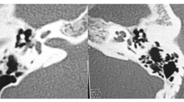
Nghiên cứu đặc điểm hình ảnh và giá trị chụp cắt lớp đa dãy trong chẩn đoán xốp xơ tai
22/03/2020 20:34:46 | 0 binh luận
The imaging and the role of multislice computed tomography of otosclerosis SUMMARY Objective : To describe the imaging characteristics of otosclerosis and to assess the role of multislice computed tomograph in diagnosis of this disease. Methods : About 45 patients (31 females, 14 males), clinically diagnosed as otosclerosis or hearing loss with unknown causes, underwent MSCT in Radiology Department between November 2013 and September 2014. All patients were surgically confirmed in ENT Department of Bach Mai Hospital or National ENT Hospital. Assessmentis based on the classification of Veillon and Portmann, comparing the differences of hearing threshold, bone and ABG between lesion-type groups and comparing with surgery records. Results: Mixed hearing loss accounts for the highest proportion. Regarding the affected sites on CT, the antefenestrum was affected in 33 ears, the other sites are rare. The otosclerotic foci that greater than 1mm in diameter and not spreading into the membrane in the cochleaare dominate. According to the classification of Veillon, type Ib and II account for the highest percentage. When comparing the average air or bone conduction threshold and ABG between differents groups, there are no statistically significant difference. The diagnostic value of CT scan in otosclerosis is up to 91.5% and its sensitivity and specificity are more than 95%. Conclusion : MSCT is an useful method in the diagnosis of otosclerosis and in the differential diagnosis with others causes of hearing loss. Keywords: Otosclerosis, the role of MSCT in diagnosis of otosclerosis.
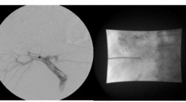
Đánh giá tính an toàn và hiệu quả của kỹ thuật nút tắc tĩnh mạch cửa phải bằng dù kim loại gây phì đại gan trước phẫu thuật
23/05/2020 12:27:11 | 0 binh luận
The assessement of the safety and efficacy of preoperative portal vein embolization using an Amplatzer vascular plug (AVP ) SUMMARY Purpose : To determine the safety and efficacy related to the use of Amplatzer Vascular Plugs (AVP) for preoperative portal vein embolization. Subjects and methods : Between 7/2011-7/2012, a total 16 AVP were embolized into the portal vein of 12 patient (HCC) prior to extended hepatic resection where the residual liver volume (RLV) was deemed sufficient (RLV < 30% in patient with normal liver, RLV <40% in patient with liver cirrhosis. AVP were used combined with gel foam in 5 patients and histoacryl in 7 patient. Result : The procedure was technically successful in 100% of cases. The rate of RLV growth was from 30 to 233 cm3 (mean at 133cm3). The rate surgical was 75% (3 patients were excluded: one insufficient growth of RVL, two metastasis). There were no major complication and minor complication in two patients: abdominal pain. Conclusion : AVP appear to be safety an effective for the preoperative embolization of portal vein, with low morbidity and sufficient growth of RLV. Key word: Amplatzer Vascular Plugs, portal vein embolization.
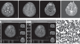
Giá trị của CHT khuếch tán trong chẩn đoán phân biệt áp xe não và u não hoại tử hoặc u não dạng nang
23/05/2020 12:23:51 | 0 binh luận
Role of diffusion weighted mr imaging in the differentiation between brain abscess and cystic/ necrotic brain tumors SUMMARY Objective: To evaluate the role of diffusion-weighted MR Imaging in the differentiation between brain abscess and cystic/necrotic brain tumors. Material and method: A cross-sectional study was conducted on 53 individuals presenting with rim enhancement intra-axial mass on brain MR image at Da Nang Hospital from August 2012 to August 2013. Diffusion-weighted image were also acquired with b value were 0, 50, 1000 using 1.5T Phillp Achieva MR system. Results: 28 patients had brain abscesses and 23 were diagnosed with brain tumors pathologically. The sensitivity and specificity of MRI in the diagnosis of brain abscess were 96.4% and 96%, respectively. PPV was 96.4% and NPV was 96%. There was a statistically significant difference in mean central ADC value between abscess and tumor, as 0.71 ± 0.24 x 10⁻³ mm2/s and 2.23 ± 0.44 x 10⁻³ mm2/s (p = 0,004) respectively. Mean peripheral ADC value showed no difference between 2 groups, as 0.78±0.26 x 10⁻³ mm2/s for abscess and 0.82±0.28 x 10⁻³ mm2/s for tumor (p = 0,132). At cut-off point of ADC ≤ 0.86 x 10⁻³ mm2/s, ROC curve for ADC showed 94.6% sensitivity and 100% specificity. Conclusion: Diffusion-weighted MR image had high sensitivity and specificity in differentiation between brain abscess and cystic/necrotic tumor. Keywords: Diffusion-weighted MR image, brain abscess, brain tumor, ADC value.
Bạn Đọc Quan tâm
Sự kiện sắp diễn ra
Thông tin đào tạo
- Những cạm bẫy trong CĐHA vú và vai trò của trí tuệ nhân tạo
- Hội thảo trực tuyến "Cắt lớp vi tính đếm Photon: từ lý thuyết tới thực tiễn lâm sàng”
- CHƯƠNG TRÌNH ĐÀO TẠO LIÊN TỤC VỀ HÌNH ẢNH HỌC THẦN KINH: BÀI 3: U não trong trục
- Danh sách học viên đạt chứng chỉ CME khóa học "Cập nhật RSNA 2021: Công nghệ mới trong Kỷ nguyên mới"
- Danh sách học viên đạt chứng chỉ CME khóa học "Đánh giá chức năng thất phải trên siêu âm đánh dấu mô cơ tim"


