


Vai trò của cộng hưởng từ trong chẩn đoán và theo dõi điều trị lao não, màng não
06/05/2021 15:55:20 | 0 binh luận
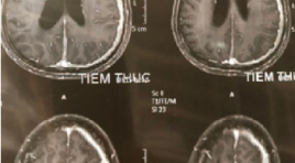
SUMMARY Purpose: Characteristic description image and evaluate the role of CHT in diagnosis and follow up and treatment of meningitis tuberculosis. Subjects and methods: Prospective and retrospective description 45 patients meningitis tuberculosis with evidence of TB bacteria in the cerebrospinal fluid undergoing MRI before and after treatment from July 2019 to September 2020. Comparison of lesions on MRI before and after tuberculoma, tuberculosis meningitis treatment. Result: In a total of 45 study patients, the average age is 28.8, male / female = 1.5, the rate of patients developing lesions on MRI before treatment (88.9%), The most common signs of damage in patients with tuberculosis of the brain, the common meningitis before treatment include: sign of meningitis enhancement (84.4%), basal meningeal enhancement (66.7%), Sylvian fissures (6.7%) , meninges of supersellar cistern (17.8%), tuberculoma (44.4%), hydrocephalus (31.1%), cerebral infarction (13.3%). Following 40 patients with meningeal tuberculosis lesions on CHT after treatment, the rate of detecting damage after treatment is 75%, of which: signs of meningitis enhancement (72.5%), basal meningeal enhencement (37.5%), sylvian fissures (2.5%), meninges of supersellar cistern (7.5%), tuberculoma (42.7%), hydrocephalus (20%), infarction cerebral (2.5%). Keyword: tubercoulosis meningitis, tuberculoma, MRI
Vai trò của chụp cắt lớp vi tính mạch máu trong chẩn đoán hẹp tắc động mạch nội sọ ở bệnh nhân đột quỵ do thiếu máu não cấp
06/05/2021 12:16:52 | 0 binh luận
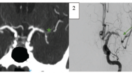
SUMMARY Background : Diagnosis of intracranial arterial stenooclusive disease and identification of intracranial atherosclerosis related occlusions (ICAS-O) in ischemic stroke patients is extremely important in order to plan a correct therapeutical approach. Few studies to date have examined the role of computed tomographic angiography (CTA) in diagnosing intracranial stenosis and predicting ICAS-related occlusions. Objective: To determine whether there is any correlation between CTA-determined truncal-type occlusion (TTO) and ICAS-related occlusions. To compare CTA to digital subtraction angiography (DSA) for detecting and measuring intracranial arterial stenoocclusive disease. Methods : We reviewed 129 ischemic stroke patients who underwent CTA and DSA. The occlusion and degree of stenosis of each intracranial arteries were calculated by WASID method. Occlusion type was classified as TTO or branching-site occlusion (BTO) on CTA. ICAS-O was detected by evaluating of underlying fixed focal stenosis (FFS) on DSA. Results: A total of 423 intracranial arteries were analyzed. CTA detected intracranial artery occlusion with sensitivity and specificity, and NPV 97,8%, 98,6% và 98,9% respectively. For detection of 50%-99% stenosis, CTA had 89,7% sensitivity and 98,2% specificity. TTO was more frequent in ICAS-O group than in the embolic group (78,1% versus 8,5%, p < 0,001). Conclusions: Compared to DSA, CTA has high sensitivity and specificity for diagnosing intracranial arterial stenooclusive disease. Preprocedural TTO on CTA is related to postprocedural ICAS-O in ischemic stroke patients.
Vai trò của cộng hưởng từ chức năng trong đánh giá vùng vận động bàn tay ở bệnh nhân u não
06/05/2021 12:02:47 | 0 binh luận
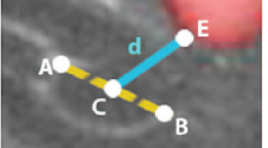
SUMMARY Background: Localizing the brain's functional cortex on functional magnetic resonance imaging (fMRI) plays an important role in brain tumor resection. Objective: To investigate imaging characteristics of the hand motor area on standardized MRI (sMRI) and fMRI in patients with brain tumors. To evaluate the correlation between lesion to motor cortex distance (LMD) on fMRI and preoperative motor deficit. Methods: Standardized and functional magnetic resonance images of 20 patients with rolandic brain tumors were included, all patients underwent tumor resection. Anatomic landmarks related to the hand motor area were interpreted on standardized MRI. Measure the distance between the hand motor area localized on fMRI and the hand motor area localized on standardized MRI. Compare the incidence of preoperative motor deficit of groups of patients with different LMDs Results: The rates of clearly defined anatomical landmarks related to the hand motor area on tumor-affected hemispheres are lower than those on unaffected hemispheres. The distance between the hand motor area localized on fMRI and the hand motor area localized on standardized MRI is 17.01 ± 3.63 mm on average and there are 6 cases where this distance is greater than 20 mm. The incidence of motor deficit in the “LMD<1cm” group, the “LMD from 1 to 2 cm” group and the “LMD>2 cm” group are 75%, 50% and 0% respectively. Conclusions: Standardized MRI should not be use to localize the hand motor area in patients with brain tumors. LMD is correlated with preoperative motor deficit. Keywords: Brain tumor, functional magnetic resonance imaging, hand motor area.
Nghiên cứu giải phẩu chức năng của tiểu não trong trí nhớ làm việc ở trẻ em sau điều trị u nguyên bào tủy
01/04/2020 12:00:28 | 0 binh luận
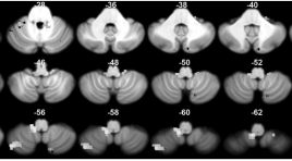
Anatomo-functional study of the cerebellum in working memory in children treated for medulloblastoma SUMMARY Introduction and Purpose: Medulloblastoma is the most common malignant cerebral tumor during childhood, arising in the posterior fossa. Children treated for medulloblastoma often experience working memory (WM) deficits, affecting their quality of life and school performance. The aim of the present study undertaken to describe the cerebellar involvement in WM deficits observed in these children. Method: Healthy children and children treated for medulloblastoma were included into study. All subjects performed a detailed neuropsychological examination, an anatomical and functional MRI. Stimuli were presented to the participants with alternating sensory modality and nature of communication in a block design during functional magnetic resonance imaging acquisitions. A Mann- Whitney U test was used for analyzing neuropsychological and behavioral data. The SPM8 and the SUIT (Spatially Unbiased Atlas Template) were utilized for anatomical and functional MRI data. Results : The patients had cerebellar lesions locating principally in the left posterior lobe. These patients were significantly reduced intelligence quotient, central executive and visuospatial WM. In healthy children group, fMRI showed robust activations for nonverbal or visuospatial WM in the left posterior cerebellar lobe. Conclusion: This study provides further evidence that the cerebellum plays a role in WM. Lesions of the left posterior cerebellar lobe may lead to nonverbal WM impairment in children. These finding contribute to treatment planning and to rehabilitation for improving the quality of life of children treated for cerebellar medulloblastoma. Keywords: Medulloblastoma, cerebellum function
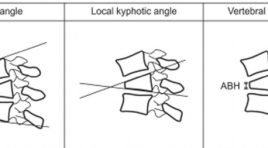
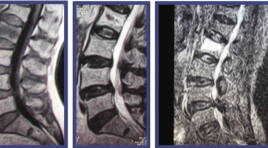
Nghiên cứu hiệu quả điều trị của phương pháp tạo hình đốt sống qua da phối hợp chỉnh hình bằng tư thế
01/04/2020 09:03:16 | 0 binh luận
Eficacy evaluation of percutaneous vertebroplasty combined with preprocedure orthopaedic positioning SUMMARY Purpose: To evaluate the effectiveness of Percutaneous ertebroplasty combined preprocedure orthopaedic positioning in treating fresh vertebral compression fractures. Methods : From January 2012 to May 2014, the data of 31 patients (23 females, 8 males; mean age, 72 years) with new vertebral compression fractures were prospectively and retrospectively analyzed. At least 6h before vertebroplasty produce, the patients were positioned to straighten the vertebral column. The radiographies of spinal column is face and lateral view before and post produce were analyzed to evaluate the vertebral body height, as well as scoliosis. Effectiveness of pain-relief was evaluated based on Visual Analog Scale (VAS). Results: The body height vertebral of compression fractures in these patients was improved by a mean of 56.2%. We achieved a mean improvement of the wedge angle 5.9o and the cobb angle 4,90 (p < .05). The VAS score is significantly improved (mean 7,8 before and 1,6 after procedure, p < 0,05). Conclusions : The combination between pre-procedure positioning and vertebroplasty brought good results in pain relief and height of vertebral body with low price. Keywords : Percutaneous vertebroplasty, Kyphoplasty, Vertebroplasty versus Kyphoplasty, Vertebral body height in vertebropasty.
Đánh giá hiệu quả bước đầu phương pháp lấy huyết khối cơ học bằng stent Solitaire trong điều trị nhồi máu não tối cấp
31/03/2020 15:16:34 | 0 binh luận
Evaluating the initial results of the thrombectomy using Stent Solitaire in patients with acute ischemic stroke summary Objective : Evaluating the initial results of the thrombectomy using stent Solitaire in patients with super acute ischemic stroke. Method and result: From May 2012 to August 2013, 14 patients suffered from hyper acute ischemic stroke underwent the thrombectomy treatment by using stent Solitaire. There are 5 males and 9 females with mean age 58.2 ± 7.9. Mean interventional time in DSA (digital subtraction angiography) room was 70.7 ± 40.2 mins. Ratio of revascularization was 80%. After 3 months, there are 9 patients with good recovery (64.3%), 3 patients with slow recovery (21.4%), and 2 mortalities (14.3%). Conclusion: Using stent solitaire in thrombectomy for the super acute ischemic stroke patients is a new and potential treatment including revascularization and clinical recovery results.
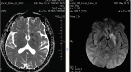
Ảnh hưởng của sự tái thông lòng mạch sớm tới tiến triển và tiên lượng ngồi máu não cấp
31/03/2020 15:09:23 | 0 binh luận
The impact of early recanalization on the growth and the prognosis of brain acute ischemic stroke summary Purpose : to study the impact of early recanalization on the growth and the outcomes of the brain acute ischemia. Method: Cerebral MRI was performed in 53 stroke ischemia patients (from 1/2010 to 6/2013) with our stroke protocols (T2*, FLAIR, Diffusion, perfusion and MR angiography) before 6h from onset, using 1.5tesla Siemens system. We ruled out the patients who had no arterial occlusion on TOF sequence. These patients were treated at Bach mai Hospital with IV thrombolysis, IA thrombectomy (solitaire stent) or without specific treatment. A second MRI was performed at 24h after onset. The patients were divided in to 2 groups: recanalization and non-recanalization. We compared the mean infarcted volume before and after treatment of the 2 groups and between the initial and the second MRI of the each group. We compared also the rate of hemorrhage transformation and the clinical outcomes (modified Rankin Scale) at 3 months of the two groups to assess the impact of early racanalization on ischemic stroke Results : 53 patients (31 recanilized, 22 non- racanilized). No difference about the age, time from onset, NIHSS, initial volume infracted between 2 groups. The final volume of infaction was 54cm3 versus 141cm3 for the recanalized and non- recanalized patients (p=0.029). An increased volume of the infarction for nonrecanalized patient group (54 versus 141cm3) with significant difference (p=0.0009). There was no difference of symptomatic hemorrhage rate (9% versus 9.6%, p=0.99) between the nonrecanalized and recanalized group. Good clinical outcomes (mRs ≤ 2) was 64.5% versus 22% for the recanalized and non- recanalazed group, p=0.13. Among the patient without recanalization, there were 4 patients died (18%) and 3 patients with mRs 5 at 3 months. Conclusion: Early recanalazation reduces the growth of ischemic lesion and it is strongly associated with improved functional outcomes.
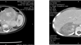
Đặc điểm hình ảnh và các giá trị cắt lớp vi tính trong bệnh lý u nguyên bào thần kinh sau phúc mạc
03/04/2020 09:17:28 | 0 binh luận
Radiographic features and accuracy of CT in diagnosis and evaluation of the dissemination of retroperitoneal neuroblastoma in children SUMMARY: Purpose: This study aims to characterize radiographic features on CT of retroperitoneal neuroblastoma. Evaluate Se., Sp., PPV., NPV.,Ac., of CT in diagnosis of retroperitoneal neuroblastoma and dissemination of the disease. Materials and Method: 111 children with 112 retroperitoneal tumors were enrolled in a prospective study. Abdominal CT findings of all the patients were analyzed, correlated with clinical, laboratory data, results of biopsy and surgical findings in order to assess sensitivity, specificity and accuracy values of CT in diagnosis and evaluation of tumor extent Result: Radiographic features of retroperitoneal neuroblastoma on CT: lobulated, ill-defined, uncapsulated, calcified mass with mild or moderate heterogeneous enhancement and vessel encasement. Among these signs, “lobulated, ill-defined and vessel-encasement” are the most valuable (Ac. 72-81%). The other signs have low to medium Se., Sp., Ac in diagnostic value (52%-96%) and should be combined with the former. CT is good at evaluating the tumor extent: renal invasion (>94%), vessel encasement (100%), abdominalorgan invasion (>75%), retroperitoneal lymphadenopathy (>85%) except for detecting peritoneal fluid (Se. 36%) Conclusion: Radiographic features of retroperitoneal neuroblastoma on CT: lobulated, ill-defined, uncapsulated, calcified, with mild or moderate heterogeneous enhancement and vessel-encased. Among them, “lobulated, ill-defined vesselencasement” are the most valuable signs. We should combine signs to increase accuracy of diagnosis. For evaluation of tumor extent, CT have high sensitivity, specificity and accuracy except for detecting peritoneal fluid. Abbreviation: CT: Computed Tomography, Se: Sensitive, Sp: Specificity, PPV: Positive Predictive Value, NPV: Negative Predictive Value, Ac: Accuracy.
Bạn Đọc Quan tâm
Sự kiện sắp diễn ra

THÔNG BÁO SINH HOẠT KHOA HỌC 19/01/2024: CẬP NHẬT CHẨN ĐOÁN VÀ ĐIỀU TRỊ UNG THƯ TRỰC TRÀNG
19-01-2024 09:47:01 0 bình luận

Thông tin đào tạo
- Những cạm bẫy trong CĐHA vú và vai trò của trí tuệ nhân tạo
- Hội thảo trực tuyến "Cắt lớp vi tính đếm Photon: từ lý thuyết tới thực tiễn lâm sàng”
- CHƯƠNG TRÌNH ĐÀO TẠO LIÊN TỤC VỀ HÌNH ẢNH HỌC THẦN KINH: BÀI 3: U não trong trục
- Danh sách học viên đạt chứng chỉ CME khóa học "Cập nhật RSNA 2021: Công nghệ mới trong Kỷ nguyên mới"
- Danh sách học viên đạt chứng chỉ CME khóa học "Đánh giá chức năng thất phải trên siêu âm đánh dấu mô cơ tim"


Đơn vị hợp tác

