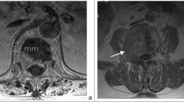
Nghiên cứu các dấu hiệu cộng hưởng từ thường quy trong chẩn đoán phân biệt xẹp đốt sống do loãng xương và xẹp đốt sống do nguyên nhân ác tính người cao tuổi
01/04/2020 08:35:48 | 0 binh luận
The conventional MRI features in different benign and malignant vertebral collapse SUMMARY Purpose: To points out the MRI important features in different benign and malignant vertebral collapse. Method: Retrospective research bases on the conventional MRI with enhancement of 40 patients who were more than 50 year old (20 patients with osteoporosis vertebral collapse and 20 patients with malignant collapse) in Ajou university hospital, Suwon, Korea from 01-01-2010 to 01-01-2012. Using Chi-square test to evaluation the significant different in frequency of MRI features of two group. Result: Lesion with wedge symmetric sharp, posterior wall intact or concave, no or less of paravertebral soft tissue, isointence on T1W, isointence or heterogenous on T2W, no enhance after contrast were favored as benign collapse; lesion with crush asymmetric collapse, big paravertebral mass, include posterior arch lesion, posterior wall convex were favored as malignant collapse. Conclusion: Conventional MRI enables differentiate between osteoporosis vertebral collapse and malignant collapse. Key word: osteoporosis vertebral collapse, malignant vertebral collapse.
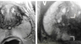
Phân loại PI-RADS của cộng hưởng từ( CHT) tuyến tiền liệt
31/03/2020 19:50:16 | 0 binh luận
Classification of Prostatic cancer with PI-RADS
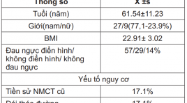
Bước đầu áp dụng cộng hưởng từ tim trong chẩn đoán bệnh tim thiếu máu cục bộ mãn tính
31/03/2020 19:35:01 | 0 binh luận
Cardiac Magnetic Resonance Imaging in diagnose Ischemia Heart Disease SUMMARY Objective : Accuracy of Cardiac Magnetic Resonance in diagnose ischemia heart disease with patients suspected coronary artery disease (CAD) in comparison to invasive angiography. Material and Methods: Thirty-five patients (61.54±11.23 years, 27 men, 71.4% CAD) underwent CMR including cine, short axis to evaluate EF, EDV, ESV, stress PERF (adenosine 140 μg/ min/kg), rest PERF (SSFP, 3 short axis, 1 saturation prepulse per slice) and LGE (3D inversion recovery technique) using Gd- BOPTA. Images were analyzed visually. Stenosis >50% in invasive angiography was considered significant. Results : Mean study time was: 41.37±11.04 minutes, EF: 48.95±18.55%, Hypokinesia: 57.1%, Akinesia: 17.1%. Sensitivity for PERF, LGE and the combination of PERF/LGE was 100%, 82.4%, 100%, respectively and specificity 80%, 80%, 80%, respectively. PPV: 94.4%, 93.3%, 94.4%, NPV: 100%, 57.1%, 100%. A good relation (p<0.01) between deficit perfusion state, the levels of myocardial delayed enhancement correlation with coronary stenosis of LAD, RCA, LCx. Conclusion: In patients with CAD, the combination of stress PERF, LGE is feasible. A combined perfusion and infarction CMR examination with can diagnose CAD in the clinical setting. The combination is superior to perfusion-CMR alone. Key words : Cardiac Magnetic Resonance(CMR), diagnose, ischemia heart disease (IHD), coronary artery, late gadolinium, perfusion.
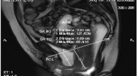
Khảo sát sự tương quan giữa sa trực tràng dạng túi với các bệnh lý sàn chậu thường gặp khác
31/03/2020 19:27:58 | 0 binh luận
Investigation of the relationship between rectocele and other pelvic floor disorders SUMMARY Background - Objectives: Pelvic floor dysfunction and prolapse are common condition of women past the middle age, with nonspecific clinical symptoms. Many cases of rectocele occurs combine with other pelvic floor disorders. Failure to recognize the complex set of pelvic floor defects leads to most therapy failures. The aim of this study is order to evaluate the correlations between age, number of birth and rectocele, and to define the role of dynamic magnetic resonance defecography in diagnosis. Methods : Cross-study description. Patients with pelvic floor dysfunction had done clinical examinations and they were indicated dynamic MR Defecography at University Medical Center, HCM City by urologist or gynecologist and proctologist. Results: MR Defecography of 1683 patients was evaluated from 01/2008 to 6/2012. Most patients are about 40 to 50 years old with 2 to 3 parity. 1218 patients with incontinence; 1311 patients (77.9%) has rectocele. Prolapse of the posterior compartment is the most common type of prolapse. Rectocele combines with more than one pelvic organ prolapse 77.4%; 64.2% of the patients with anismus had rectocele. There are statistically significant in the correlation of age, the number of birth with rectocele or pelvic organ prolapse (OR # 1.04-2.67 and p<0.005). Risk of rectocele was higher in the patients with pelvic organ prolapse than in the patients without pelvic organ prolapse (p<0.005). Conclusions: Complex pelvic floor disorders often involve several compartments. Dynamic MR Defecography allows evaluating the both morphological and functional assessment of the pelvic floor, selecting the most appropriate treatment.

Đánh giá hiệu quả ban đầu của can thiệp mạch trong điều trị dị dạng mạch máu tủy
31/03/2020 19:20:03 | 0 binh luận
The preliminary result of endovascular treatment in spinal vascular malformation SUMMARY Objective : To evaluate the preliminary result of endovascular treatment in spinal vascular malformation. Material and method: We prospectively studied patients with spinal arteriovenous shunt who were diagnosed and endovascular treatement with spinal arteriovenous shunt at Bach Mai hospital from 2012 to 2013. Clinical features were analyzed before and after treatment by Aminoff-Logue disability scale. MR imaging characteristics were evaluated. Result: 9 patients were treated by endovascular embolization, 44.4% were spinal arteriovenous malformation, 55.6% were spinal dural arteriovenous fistulae. MRI studies showed intramedullary increased T2 signal and dilated venous drainage in all patients. The rate of complete angiographic obliteration was 55.5% and nearly occluded in 45.5 . After follow up of 3 months, clinically significance improvement was achieved in 66.7%, partial recovery in 22.2%. Conclusion: n-BCA glue embolization for spinal arteriovenous shunt should be considered the treatment of choice with satisfactory outcomes. Large studies with longer follow-up are required to determinate the safety and efficacy of endovascular treatment.
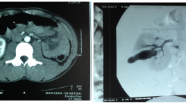
Hiệu quả điều trị can thiệp nội mạch tổn thương động mạch thận do chấn thương
31/03/2020 19:14:13 | 0 binh luận
The efficacy embolization in treatment of renal arterial trauma SUMMARY Pupose: to apply and estimate the efficacy of embolization in renal injury. Material and method: 26 patients were undergone renal angiography and embolization at Viet Duc hospital from 1/2010 to 8/2013. Among those patients, 23 cases showed pseudoaneurysm, 2 cases showed extravasation and 1 case showed AVM on CT scan. Result: All patients who underwent embolization did not show extravasation (100%) and ongoing hemorrhage required laparotomy on angiography postoperative. Conclusion : In hemodynamically stable and controlled patients, selective and superselective embolization is a safe and effective method for the management of renal vascular injury. Keyword: : Embolization- Aneurysm- Arterial injuries- Arteriography- Renal trauma- Arteriovenous fistula- Angiography- Extravasation- Arterial bleeding.
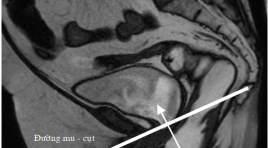
Đánh giá đặc điểm sa trực tràng kiểu túi ở bệnh nhân rối loạn chức năng sàn chậu bằng cộng hưởng từ động
31/03/2020 19:09:28 | 0 binh luận
Evaluation of rectocele in patients with pelvic floor dysfunction by dynamic magnetic resonance imaging SUMMARY Objectives : Rectocele is a bulge or a prolapse of the anterior rectal wall into the posterior vaginal wall. It is relatively common with diversified and nonspecific symptoms. Clinical examination is easily confused and/or sometimes omitted the prolapse of other pelvic organs. Imaging to assess the pelvic floor dysfunction is an important and useful diagnostic test, especially dynamic MR. Methods : Our study was cross-sectional descriptive. Patients with pelvic floor dysfunction were undergone clinical examinations and then were indicated to have dynamic MR scanning at Ho Chi Minh City Medical University by anorectic doctor, urologist and gynecologist. Results: 1.863 patients were evaluated from January 2008 to June 2012. Most of them are women, middle-aged and used to give birth. The rate of rectocele with its depth from 2 to 4 cm was 77.9%, mainly with the shape of “finger”. The depth of more than 2 cm with the shape of “bag” has the high risk of stagnancy. The factor of age and being used to give birth have a significant relation with rectocele (p<0.001). The rate of rectocele in the group of anismus was 64.2%. The combination of the prolapse of more than one pelvic chamber accounted for 77.4% (p<0.001). Conclusions : Dynamic MR of the pelvic floor helps to diagnose in details the characteristics of the rectocele and other pelvic organ prolapse, helping clinicians to choose the appropriate treatments.
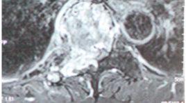
Mô tả đặc điểm hình ảnh Xquang và cộng hưởng từ trong chẩn đoán ung thư di căn đôt sống
31/03/2020 19:04:57 | 0 binh luận
Conventional Radiography and MRI for the diagnosis of vertebral metastases SUMMARY Purpose : Describe the imaging characteristics and evaluate the value of conventional radiography and MRI for the diagnosis of vertebral metastases. Material and methods : 62 patients with vertebral lesions suspected metastates were taken biopsy at radiology department in Bach Mai Hospital from November 2011 to August 2013. Result : A total of 42 patients with vertebral metastases,male / female 26/16, the mean age is 57.43 ± 13.29. The percentage of lesions detected on X-ray were 52.4% with the sensitivity of signs of vertebral collapse,condensation of vertebral body, pedicle destruction were 31.8%; 18.7%; 21.4%. MRI: the sensitivity, specificity, accuracy were 97.6%; 75%; 90.3%. Conclusion: Although X-ray is the first method but less valuable in diagnosing vertebral metastases. Magnetic resonance imaging has great value in the diagnosis of vertebral metastases with many special signs.
Bạn Đọc Quan tâm
Sự kiện sắp diễn ra
Thông tin đào tạo
- Những cạm bẫy trong CĐHA vú và vai trò của trí tuệ nhân tạo
- Hội thảo trực tuyến "Cắt lớp vi tính đếm Photon: từ lý thuyết tới thực tiễn lâm sàng”
- CHƯƠNG TRÌNH ĐÀO TẠO LIÊN TỤC VỀ HÌNH ẢNH HỌC THẦN KINH: BÀI 3: U não trong trục
- Danh sách học viên đạt chứng chỉ CME khóa học "Cập nhật RSNA 2021: Công nghệ mới trong Kỷ nguyên mới"
- Danh sách học viên đạt chứng chỉ CME khóa học "Đánh giá chức năng thất phải trên siêu âm đánh dấu mô cơ tim"


