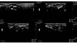
Nghiên cứu giá trị chẩn đoán ung thư tuyến giáp của phân độ EU - tirads 2017
04/12/2019 21:35:27 | 0 binh luận
Research into the value in the diagnosis of thyroid cancerof the EU-TIRADS 2017 classification SUMMARY A diagnostic test study was conducted at Bạch Mai hospital to evaluate the efficacy of Ultrasound andthe EU-TIRADS 2017classsification of thyroid nodules. Result: 170 patients with thyroid noduleswere prospectively evaluated by B-mode ultrasound and the EU-TIRAD 2017 classsification, followed by the fine needle aspiration (FNA) biopsy. The averae age is 46,7 ± 11,5 years old and female/male = 5,5. The sensitivity, specificity, positive predictive value, negative predictive value, accuracy for the EUTIRADS 2017 were 98,2%; 34,5%; 74,3%; 90,9%; 76,7%.TIRADS 5 is the highest (64,7%). 4 features of high suspicion are irregular margins, microcalfifcations, marked hypoechogenicity, “taller – then -wide” shape; the sensitivity, specificity 70% and 93%; 35% and 91%; 50% and 79%; 58% and 82%. Conclusion: TIRADS 5 is the highest and the EU-TIRADS 2017 classification and pathology indicates strong evidence. Key Words: The EU-TIRADS 2017, thyroid nodules on ultrasound, thyroidcancer.

Nghiên cứu giá trị chẩn đoán ung thư tuyến giáp của phân độ EU - TIRADS 2017
16/04/2020 23:13:02 | 0 binh luận
Research into the value in the diagnosis of thyroid cancerof the EU-TIRADS 2017 classification SUMMARY A diagnostic test study was conducted at Bạch Mai hospital to evaluate the efficacy of Ultrasound andthe EU-TIRADS 2017classsification of thyroid nodules. Result : 170 patients with thyroid noduleswere prospectively evaluated by B-mode ultrasound and the EU-TIRAD 2017 classsification, followed by the fine needle aspiration (FNA) biopsy. The averae age is 46,7 ± 11,5 years old and female/male = 5,5. The sensitivity, specificity, positive predictive value, negative predictive value, accuracy for the EUTIRADS 2017 were 98,2%; 34,5%; 74,3%; 90,9%; 76,7%.TIRADS 5 is the highest (64,7%). 4 features of high suspicion are irregular margins, microcalfifcations, marked hypoechogenicity, “taller – then -wide” shape; the sensitivity, specificity 70% and 93%; 35% and 91%; 50% and 79%; 58% and 82%. Conclusion: TIRADS 5 is the highest and the EU-TIRADS 2017 classification and pathology indicates strong evidence. Key Words : The EU-TIRADS 2017, thyroid nodules on ultrasound, thyroidcancer.
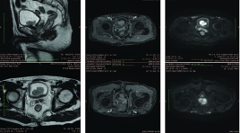
Nghiên cứu đặc điểm hình ảnh và giá trị CHT chẩn đoán phân độ giai đoạn T của ung thư bàng quang
04/12/2019 21:29:46 | 0 binh luận
Researching imaging characteristics and assessing values of MRI in the diagnosis of bladder cancer at T-stage SUMMARY Purpose: Describing imaging characteristics and assessing values of MRI in the diagnosis of bladder cancer at T-stage. Subjects and methods : 43 patients with bladder tumors were selected to be in adescriptive study (38 of whom had tumors from tumor histopathology and 5 patients had histology from other organs or benign paraganglioma bladder tumor), in which they got diagnosed, operated (transurethral resection or radical cystectomy) and had pathology results from May 2017 to June 2018 at K hospital in Tan Trieu. All MRI films were evaluated preoperatively and compared with histopathology postoperativelydistinguishing superficial tumors (T1 or lower) and invasive tumors (T2 or higher). Results : Among 38 patients being studied, the mean age is 56 ± 13.24 andthe gender ratio is M/F ≈ 7/1. Out of 61 tumors, the most common tumor location is bilateral bladder 30.7%, mostly one tumor (26/38). Featured images: mean size 23.47 ± 14.09 mm, max size 68mm, min size 7 mm; most frequently found polype-shaped tumor 25/38 patients (65.8%). According to the assessment of MRI results with T2W, DCE and DWI distinguishing T staging, tumors in T1 or lower 30/38 patients (78.9%) and T2 or higher 8/38 patients (21,1%, in which 4 patients in T2, one in T3 and 3 in T4). There is no correlation between the number of tumors and T staging (p>0.05). There is a strong correlation between the shape of tumors and T staging (p<0.001). When combining T2W with DCE, the sensitivity, specificity and overall accuracy of two observers for differentiating Tis to T1 tumor from T2 to T4 tumors were 79.3 %, 100% and 84.2% respectively. When using all three image types together (T2W, DCE and DWI) to assess T staging, the sensitivity rose up to 96.5%, specificity 66.7%, overall accuracy 89.5% and PPV 90.3%. The Cohen’s Kappa score of 0.685 had a good correlation between MRI results and histopathology in distinguishing T-staging of bladder cancer (p<0.001). In addition, a total 36 patients had urothelial carcinoma, ADC values of 29/36 patients at T1 or lower were 1.138 ×10⁻³mm2/s and 7/36 patients at T2 or higher were 0.79 × 10⁻³mm2/s, and this difference of ADC was significant between Grade 1 and Grade 3. The total of 29/38 patients (76.3%) underwent TUR and deep muscle biopsy was performed at the base tumor, 9/38 patients underwent radical cysectomy or chemotherapy before surgery. Key words : Bladder Cancer, MRI of Bladder cancer.
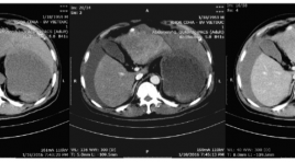
Đánh giá hình ảnh và kết quả nút mạch cầm máu cấp cứu ung thư biểu mô tế bào gan vỡ
25/03/2020 23:29:45 | 0 binh luận
Evaluate the imaging characteristics of ruptured hepatocellular carcinoma and the effectiveness of embolization for controlling hemorrhage SUMMARY Objects : Evaluate the effectivenes of transcatheter arterial embolization for controlling arterial hemorrhage due to spontaneous ruptured hepatocellular carcinoma (HCC). Methods: analyze retrospectively the outcomes of 22 patients who underwent abdominal CTscanner and urgent transarterial embolization for spontaneous ruptured HCC during the period from 01/2014 to 06/2016 in Viet Duc hospital. Results: Mean tumor size: 83.95mm (longest diameter). 7/22 patients (31.8%) exhibited contrast extravasation on angiography, 2/22 patients (9.1%) exhibited pseudoaneurysm, one patient (4.6%) showed arterioportal shunt, 12/22 (54.5%) showed no vascular injury. The embolization materials we used mostly was Spongel in 19/22 patients (86.4%), histoacryl 3/22 (14.6%). The success rate of embolization on angiography is 22/22. The average volume of blood tranfusion was 969ml. 1 patient die in one months after the procedure due to liver failure. 6/9 (66.7%) patients with thrombosis of portal vein die in less than 6 months after procedure. Conclusion: Transarterial embolization is a safe and effective method for controlling spontaneous rupture of HCC. Key words: angiography, embolization, hepatocellular carcinoma, spontaneous rupture.
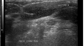
Di căn hạch của u mô đệm dạ dày ruột (GIST). Tổng quan tài liệu và nhân một trường hợp
30/03/2020 15:44:08 | 0 binh luận
Lymph node metastasis in GIST. Literature overview and case report SUMMARY GIST is considerd as rare tumor coming from Cajal cell. Due to poor clinical manifestations or vague symptoms, the disease is seen at late phase, some can be earlier by hazardous examination. The late comming with great dimension and existing metastatic lymph node generally having bad prognose. Lymph node metastatic is rare and not to be concentrated as liver and peritoneal metastasis. Absence of lymph node pre- post operation, intervention combining with adjuvant therapy is also difficult to prognose but generally can have 5 year survival. A case presenting in this paper with good prognose after treatment though lymph node metastasis after 4 years.
U mô đệm dạ dày ruột ( GIST) từ siêu âm đến cộng hưởng từ
30/03/2020 15:26:06 | 0 binh luận
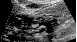
Gastrointestinal Stromal Tumor: from ultrasound to MRI SUMMARY Gastrointestinal Stromal Tumor: From ultrasound to MRI. Former GIST was considered as leiomyoma, shwannoma, epithelioma. Histoimmuno logy determined with Protein KIT CD117 and the presence of interstitial cell Cajal. GIST is not rare but not be early diagnosed. Conventional X Ray, USG, CT scanner, MRI and PET can find. Though almost discovered hazardly during screening examination, we use USG for detection then CT Scanner detail study. Comment and Results : 6 cases was noted, 4 hazardly, operation and histology affirmed. Sex M/F 2/6, 4 cases under 3 cm as dimension, unique cas 30x40 cm. Metastase not yet found. All are healthy after 3 years. Conclusion: The affection is hazardly found. All imaging modalities can detected. 5 year survival depend on tumor dimension. Metastasis often to liver, mesentery but late. USG and CT is used also to follow up after interventional and target therapy. Early diagnosis rely on screening control and community care. Key words : Gastrointestinal stromal tumor, GIST, Ultrasound, CT scanner, MRI.
Nghiên cứu đặc điểm hình ảnh cắt lớp vi tính đa dãy đầu thu và phân loại LUNG-RADS các nốt mờ phổi
30/03/2020 14:36:19 | 0 binh luận
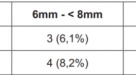
MDCT images and The ACR Lung Imaging Reporting and Data System (Lung-RADS™) of Pulmonary nodules - Research in Hue National Hospital and Hue Medic Clinic SUMMARY Objective: Solitary pulmonary nodule may be benign or malignant. The purposes of this study is to illustrate the clinical characteristics, describe the images characteristics of chest X-ray and MSCT, classification of lung nodules by The ACR Lung Imaging Reporting and Data System (Lung-RADS™) then offer management and monitor strategies for this disease at Hue National Hospital and Hue MEDIC clinic. Methods: The study design was cross-sectional descriptive. Describe the clinical characteristics and images of lung nodules on chest X-ray and MSCT with LungCAD software to determine the nodular lesions during 1 year (5 / 2015-5 / 2016), in 2 centers (Hue National Hospital and Hue MEDIC clinic). Results : In our study, there was 49 patients with pulmonary nodules. Male was 38/49 (77.6%), more than female. Mean age was 57 ± 2 years old. Smallest nodule is 5mm, average size is 18.7 ± 9mm. There was 31/49 (63.3%) patients with lung size 15-30mm. MSCT has higher sensitivity than X-ray in detecting nodules <6mm and ground glass nodule. Base on The ACR Lung RADS classification, Lung - RADS 4B was seen most with 19/49 (38,8%) patients. Number of patients with Lung - RADS 4X was 8 (16.3%), including 5 patients who underwent surgeries, 3 of them had malignant pulmonary nodules. Conclusions : Patients with pulmonary nodules should be evaluated by estimating the probability of malignancy, be performed imaging tests to characterize the lesions better, be assessed the risks and benefits of different management strategies (biopsy, surgery and observation with serial imaging tests). Lung - RADS classification is simple, easy to apply and make appropriate recommendations for the management of solitary pulmonary nodules. Keywords : Solitary pulmonary nodule. Lung - RADS.
Biến thể giải phẩu động mạch gan trên 300 trường hợp chụp mạch máu số hóa xóa nền
30/03/2020 14:11:17 | 0 binh luận
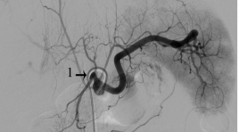
Variant Hepatic arterial anatomy in 300 patients underwent digital subtraction angiography SUMMARY Objective: to evaluate and describe the prevalence of hepatic arterial variants seen at digital subtraction angiography. Materials and methods : data were collected at Interventional Unit in Diagnostic Imaging department of Viet Duc hospital from May 2015 to May 2016. Results: 300 cases underwent at least one visceral angiographic examination during the study period. 232 (77.3%) cases had standard hepatic arterial anatomy. 14 (4.7%) cases had replaced left hepatic artery. 11 (3.7%) cases had replaced right hepatic artery. 2 (0.7%) cases had variant anatomy involving replacement of both left hepatic artery and right hepatic artery. 16 (5.3%) cases had accessory left hepatic artery. 3 (1%) cases had accessory right hepatic artery. 1 (0.3%) case had both left and right accessory hepatic artery. 12 (4%) cases had replaced common hepatic artery. 3 (1%) cases had double hepatic artery. 1 case had replaced common hepatic artery and accessory left hepatic artery at the same time. 1 case had right hepatic artery arises from aorta and left hepatic artery arises from left gastric artery. Conclusion : in this study the percent of standard hepatic artery is larger than previous published study while the percent of cases had other variants of hepatic artery is smaller. In 2 cases, we saw uncommon variants which weren’t described in previous study. However we didn’t see some other uncommon variants which were described in other study. Keyword : Hepatic artery, anatomy, variant, DSA.
Bạn Đọc Quan tâm
Sự kiện sắp diễn ra
Thông tin đào tạo
- Những cạm bẫy trong CĐHA vú và vai trò của trí tuệ nhân tạo
- Hội thảo trực tuyến "Cắt lớp vi tính đếm Photon: từ lý thuyết tới thực tiễn lâm sàng”
- CHƯƠNG TRÌNH ĐÀO TẠO LIÊN TỤC VỀ HÌNH ẢNH HỌC THẦN KINH: BÀI 3: U não trong trục
- Danh sách học viên đạt chứng chỉ CME khóa học "Cập nhật RSNA 2021: Công nghệ mới trong Kỷ nguyên mới"
- Danh sách học viên đạt chứng chỉ CME khóa học "Đánh giá chức năng thất phải trên siêu âm đánh dấu mô cơ tim"


