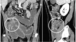
Giá trị cắt lớp vi tính trong chẩn đoán phân biệt các u nguyên phát thường gặp ở ruột non
04/12/2019 20:32:12 | 1 binh luận
SUMMARY Objectives: The purpose of the this study was to analyze imaging roles to assessthe diagnostic capacity for differentiating the common primary small bowel tumors. Methods: We performed a retrospective study from the medical database from January 2015 to May 2018 at University medical center and Cho Ray hospital. The inclusion criteria were as follows: pathologically proven primary small bowel neoplasms andpatients were performed MDCT with intravenous contrast media. Radiologist were blinded to the pathological information, reviewed the image findings according to the data collection paper. Radiologist collects the characteristics of neoplasm such as anatomical distribution, growth, enhancement, wall thickening patterns, size, hyperplasia vascular on tumor surfaces and lymph node characteristics. Then, comparing each findings to pathology report to access specificity, sensitivity and positive predictive value (PPV) of them. Results : A total of 98 patients met the criteria for analysis in thepresent retrospective study, 31 adenocarcinomas, 22 lymphomas, 30 GISTs anf 15 others. The extramural growth pattern isreliable prediction of GIST, with PPV of 82.3%. All of GISTs show moderate to avid enhancement. Tumor density of greater than or equal to 110 HU is likely to be GIST, with PPV of84.9%. Proliferation of blood vessels on tumor surfaces can help discriminate GIST from the others, with PPV of92%. Bowel wall thickening is the common patternof adenocarcinoma and lymphoma. Apple-core-like, shoulder defect and focal involvement are probably findings of adenocarcinoma, with PPV of 81.8%, 71.4% and 76.9%, respectively. Aneurysmal dilatation of the lumen and marked thickening wall bowel equal or greater than 25mm can strongly suggest lymphoma, with PPV of 87.5% and 72.7%, respectively. Enlarged lymph node with shorter axis greater than 20mmor multiple lymph nodes fused together forming a bulky massare likely to be lymphoma, with specificity of 100%. Conclusion : MDCT findings could potentially be useful to differentiate the common primary small bowel neoplasms based on analyzing specific imaging characteristics of each tumor after classifying by growth pattern lesion. Keywords : Small bowel neoplasm, differentiate

Đặc điểm lâm sàng, giải phẩu bệnh, siêu âm bệnh nhân u tuyến thượng thận đã phẩu thuật tại bệnh viện chợ rẫy năm 2014 - 2015
13/04/2020 15:25:10 | 0 binh luận
Clinical, pathology, ultrasound in patients with adrenal tumors, had surgery in Choray Hospita SUMMARY Objectives : Clinical, pathology, ultrasound in patients with adrenal tumors. Methods: Retrospective - Described a case series Results : 1/2014-6/2015, 84 patients. Pathology: 34 (40.5%) adrenocortical adenomas and 23 (27.4%) pheochromocytoma,. Clinical: Group of patients without clinical symptoms 57 (67.9% Utrasound : adrenal tumor in the left/right # 1, tumors 50.52 ± 27,19mm mix-echo 41.7% 47.6% hypoechoic, clearly limited tumor (casings unknown) 91.7%. Ultrasound correctly identified 97.6% of adrenal tumors. Conclusions: Ultrasonography correctly identify and detect adrenal tumors accidental high percentage. Key words :Adrenal tumor, Ultrasound.
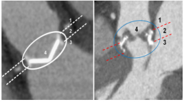
MSCT - 640 trong đánh giá van tim nhân tạo: Bước đầu khảo sát 36 vạn
14/04/2020 22:48:29 | 0 binh luận
Prosthetic heart valve assessment with 640-Slice MSCT: Initial Experience with 36 Prosthetic heart valv es SUMMARY Objectives: Multislice CT(MSCT) has shown potential for prosthetic heart valve (PHV) assessment. We assessed the image quality of different PHV types to determine which PHV are suitable for MSCT evaluation and accuracy of 640-Slice MSCT for PHV dysfunction assessment. Subjects and methods: Cardiac 640- Slice MSCT examinations performed at the Medic medical center since 6/2013 to12/2016 were reviewed for the presence of PHVs. Image quality of the supravalvular, perivalvular, subvalvular and valvular regions was scored on a four-point scale (1=non-diagnostic, 2= moderate, 3=good and 4=excellent). Causes of PHV dysfunction were confirmed by surgery. Results : 28 patients with a total of 36 PHVs (4 monoleaflets, 25 bileaflets and 7 biological PHVs) in the aortic(n=22), mitral(n=14) position were included. Median image quality scores for the supra-,peri-and subvalvular regions and valvular detail were 4, 3.7, 3.7 and 3.5, respectively for bileaflet PHVs; 3, 2.6, 2.5 and 1.6, respectively for monoleaflet PHVs and 4, 3.8, 4.0 and 3.7 respectively for biological PHVs. In 3/4(75%) monoleaflet valves with severe artefacts and non - assessment. In 22(17 bileaflets and 5 biological PHVs ) of the 32 PHVs(68,75%) detect PHV dysfunction. In the PHV dysfunction group, the mechanism of dysfunction (pannus, thrombosis , patient prosthesis mismatch , paravalvular leakage, endocarditis and degenerate) was correctly identified by surgery in 100% of the cases. Conclusion : Implanted bileaflet and biological PHVs have good image quality on 640-Slice MSCT and are suitable for 640-Slice MSCT evaluation. Causes of PHV dysfunction were correctly evaluated by 640-slice MSCT in all PHVs except for monoleaflet PHVs.
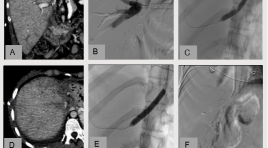
Điều trị can thiệp nội mạch hẹp tĩnh mạch gan sau ghép gan từ người cho sống ở trẻ em: Nhân một trường hợp
15/04/2020 20:54:05 | 0 binh luận
Endovascular treatment of hepatic vein stenosis following liver transplant in a child: a case report SUMMARY Liver transplantation is the major therapeutic option for end-stage liver disease as recent improvement in surgical technique, immune-suppressant contribute to better post-transplant outcome. However, significant graft failure as a result of vascular complications is still noted, especially in partial liver transplant with living donor graft and complications is higher risk in children compared with adults. The incidence of hepatic vein stenosis in pediatric liver transplant is 6% with living donor graft. A first case of pediatric liver transplant with living donor graft which performed in Viet Duc hospital have complication of hepatic vein stenosis and success with balloon angioplasty and stent placement. Key word: Living donor transplants, hepatic vein stenosis.
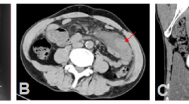
Tụy lạc chỗ tại ruột non với biến chứng viêm hoại tử ruột: Báo cáo một trường hợp hiếm và tổng kết trên y văn
15/04/2020 20:42:50 | 0 binh luận
Ectopic pancreas in the wall of intestine complicated with necrotic and inflamed intestine: A case report and review literature SUMMARY Background: Ectopic pancreas is a rare congenital condition characterized by pancreatic tissues located outside normal of confines of pancreas and lacking any anatomic or vascular connection with main pancreas. It can occur anywhere in the gastrointestinal tract but rarely are found in small intestine. Its preoperative diagnosis is difficult because the clinical symptoms are often nonspecific. We introduce a case of ectopic pancreas in the intestinal wall complicated with necrotic and inflamed intestine, which received a treatment by resection. Case presentation : A 44 years old man attended to Bach Mai hospital due to acute abdominal pain in epigastrium as result of gastrointestinal perforation. Contrast enhanced computed tomography (CT) of abdomen showed a mesenteric mass surrounded by inflamed fat in the left lower quadrant abdomen. In addition, CT images also suggested necrosis of the bowel wall next to the mass caused by twisting the mesentery and mesenteric vessels (whirlpool sign). The patient underwent local surgical resection and following histology revealed ectopic pancreatic tissues in the wall of intestine and necrosis of intestine. Conclusion : Although ectopic pancreas is rare, it should be considered in the differential diagnosis of a mesenteric or intestinal mass surrounded by necrotic and inflamed intestine. Keyword: Ectopic pancreas, mesenteric mass, intestinal mass, whirlpool sign.
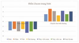
Bước đầu nghiên cứu đặc điểm hình ảnh chuyển hóa 18-FDG PET/CT não ở bệnh nhi tự kỷ
15/04/2020 20:33:18 | 0 binh luận
Preliminary evaluation of F-18 FDG PET findings in pediatric patients with autism SUMMARY Objectives: the purpose of this study was to evaluate F-18 FDG Positron Emission Tomography (PET) findings in pediatric patients with autism. Subjects and methods: This study includes 15 autism pediatric patients who were diagnosed at the Vinmec international hospital from January 2017 to May 2017 and five oncology patients without neurology’s diseases underwent to whole body PET scan (from vertex to midthigh) were selected as control group. All patients underwent PET/CT brain examination in Nuclear Medicine Department, 108 Central Military Hospital. Results: Mean patient age (7.1 ± 0.24). On the PET scans of the 15 patients with autism, 13 (86%) had significantly decreased metabolic activity of both hemispheres. All of the patients had decreased metabolism in temporal, parietal and cingulate gyrus. Mean of Z-scores of autism group showed significantly decreased activity in comparison with Z-score of the control group. There were relations between clinical and grade of hypometabolism of autism. Conclusions : hypometabolism in the brain of autism pediatric patients may present a role in diagnosis and prognosis. Key words : autism pediatric patients, hypometabolism in the brain
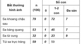
Đặc điểm hình ảnh cộng hưởng từ động học tổng phân ở nhóm bệnh nhân nữ rối loạn chức năng sàn chậu trên 60 tuổi
14/04/2020 23:27:30 | 0 binh luận
Magnetic Resonance Defecography in female patients with pelvic floor dysfunction age from 60 SUMMARY Objective: We describe characteristics dynamic MR defecography in female patients with pelvic floor dysfunction, age from 60. Methods Describing cross-study. 106 patients were indicated magnetic resonance defecography by coloproctologist from 09/2016 to 04/2017, at Thong Nhat Hospital, Ho Chi Minh City. Results Defecatory dysfunction is the most common symptom (83,1%). The prevalence of urinary incontinence and pain is 34% and 59,4%, respectively. There is a significant difference in the ratio of pelvic floor descent between the groups who have and no have children. The ratio of pelvic floor descent of the group who have 1-2 children is significantly greater than the group have more than 3 children. The correlation between age and the degree of pelvic floor descent is weak. The combination of pelvic organ prolapses usually occurs. If there is only one pelvic compartment prolapse, it is the posterior compartment prolapse, which counts for only 5,6%. The degree of all the rectoceles is from the second degree and 83,1% of this still contain ultrasound gel after defecation phase. The prevalence of Anismus is only 3/106 and none of this combine with retocele. Conclusions Age, menopause and childbirth all have influence on the weakness of pelvic floor. In the older group, the combination of pelvic floor organ prolapses usually occurs. Rectocele is also common while Anismus is a uncommon condition. Keywords : MR defecography, pelvic floor dysfunction, female age from 60, rectocele, Anismus.
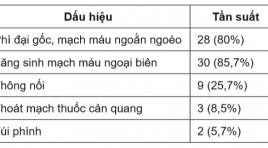
Vai trò can thiệp nội mạch trong điều trị ho ra máu nặng
14/04/2020 23:19:28 | 0 binh luận
The role of endovascular treatment for massive hemoptysis SUMMARY Objectives: To assess the safety and effectiveness of arterial embolization in patients with massive hemoptysis. Subjects and Method: All patients were diagnosed massive hemoptysis and treated endovascular intervention in Cho Ray Hospital from January 2016 to March 2017. Some variants were asscessed: etiologies, important angiographics findings, the clinical success, complications and follow-up outcomes within 1 month. Results: 35 patients were treated by endovascular intervention. Massive hemoptysis was caused by bronchiectasis (37,1%), pulmonary tuberculosis (20%), pulmonary aspergilloma 14.3%). A total of 69 bleeding arteries were found, an average of four arteries per patient. Important angiographics findings were: vascular hypertrophy and tortuosity (80,0%), neovascularity and hypervascularity (85,7%), shunting (25,7%), aneurysm formation (8,5%) and active extravasation (5,7%). Immediate clinical success achieved was 97,1% (34/35 patients) and 11,7% of patients had recurrent over 1 month; aspergilloma and shunting were asociated with early recurrent (p<0,05). No serve complications were reported and the most common complication was trasient chest pain (28,6%). Conclusion: Endovascular treament is an effective and safe procedure in the management of massive hemoptysis. Keywords: massive hemoptysis, embolized arteries, clinical success, early recurrent.
Bạn Đọc Quan tâm
Sự kiện sắp diễn ra
Thông tin đào tạo
- Những cạm bẫy trong CĐHA vú và vai trò của trí tuệ nhân tạo
- Hội thảo trực tuyến "Cắt lớp vi tính đếm Photon: từ lý thuyết tới thực tiễn lâm sàng”
- CHƯƠNG TRÌNH ĐÀO TẠO LIÊN TỤC VỀ HÌNH ẢNH HỌC THẦN KINH: BÀI 3: U não trong trục
- Danh sách học viên đạt chứng chỉ CME khóa học "Cập nhật RSNA 2021: Công nghệ mới trong Kỷ nguyên mới"
- Danh sách học viên đạt chứng chỉ CME khóa học "Đánh giá chức năng thất phải trên siêu âm đánh dấu mô cơ tim"


