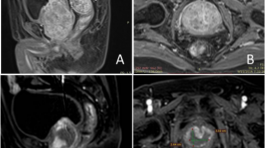
Phương pháp nút động mạch điều trị tăng sinh lành tính tuyến tiền liệt có thể tích lớn: kết quả trên 32 trường hợp (>80 GAM)
12/04/2020 22:28:28 | 0 binh luận
Prostatic arterial embolization for the treatment of benign prostatic hyperplasia due to large: result in 32 case (>80 gam) SUMMARY Background : Currently, large prostate size (>80 g) of benign prostatic hyperplasia still pose technical challenges for surgical treatment with complication such as: hemorrhage, endoscopy syndrome,… Objective : to explore the safety and efficacy of prostatic arterial embolization as an alternative treatment for patients with lower urinary tract symptoms due to large benign prostatic hyperplasia. Methods : A total of 32 patients with prostates >80 g were included in the study; all were failure of medical treatment and unsuited for surgery. Prostatic arterial embolization was performed using combination of 250 μm and 400μm particles in size, under local anaesthesia by a unilateral femoral approach. Clinical follow-up was performed using the international prostate symptoms score (IPSS), quality of life (QoL), peak urinary flow (Qmax), post-void residual volume (PVR), international index of erectile function short form (IIEF-5), prostatic specific antigen (PSA) at 1, 3, 6 month and prostatic volume measured by magnetic resonance imaging at 3 month after intervention. Results : Prostatic arterial embolization was technically successful in 32 patients (100%). Follow- up data were available for the those patients with a mean follow-up of 6 months. The clinical improvements in IPSS, QoL, Qmax, PVR, and PV at 6 month was 74.1 %, 152%, 68.7%, 92.6 %, and 35.5% (3 months), respectively. The mean IPSS (pre PAE vs post PAE 27.5 vs 7.1; P < 0.01), the mean QoL (4.7 vs 1.7; P < 0.01 ), the mean Qmax (7.5 vs 18.9; P < 0.01), the mean PVR (65 vs 20.3; P < 0.01), and PV (98.0 vs 65.0, with a mean reduction of 33.6 %; P < 0.01 ) at 3 month after PAE were significantly different with respect to baseline. The mean IIEF-5 was not statistically different from baseline. No major complications were noted. Conclusions : Prostatic arterial embolization is a safe and effective treatment method for patients with with lower urinary tract symptoms due to large volume. Prostatic arterial embolization may play an important role in patients in whom medical therapy has failed, who are not candidates for any surgical treatment. Keywords : Benign prostatic hyperplasia (BPH), Prostatic artery embolization (PAE), large prostate size
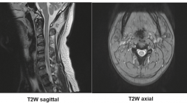
Đặc điểm hình ảnh tổn thương tủy sống gây bởi ngộ độc gây bởi khí Nitrous Oxide - nhân 2 trường hợp
07/04/2020 21:30:59 | 0 binh luận
SUMMARY Nitrous oxide gas (N2O), known as laughing gas, is widely used in medicine for the purpose of pain relief. However, nowadaysN2O are abused for recreational purposes and causes several harmful effects, especially neurological damages. In two cases of patients with nitrous oxide toxicity, we would like to make some remarks about the characteristic features of myeloneuropathy caused by N2O abuse Key word : metabolic, N2O, nitrous oxide, toxic

Thực quản đôi: Một trường hợp hiếm gặp tại bệnh viện Bạch Mai
07/04/2020 21:22:50 | 0 binh luận
Incomplete duplication of the esophagus: a case report SUMMARY Double lumen esophagus is a very rare disease. Approximately 20 cases have been reported in the past. Dysphagia and odynophagia are common symptoms. Symptomatic management is the mainstay of treatment.We report a extremely rare case of 57-year-old woman with a incomplete duplication of the esophagus. Patient‘s symptoms are dysphagia with solid food, regurgitation of great amount of the liquid and chest pain. The exact diagnosis is make by X-ray films of the chest with a water soluble contrast esophagogram, esophagogastroscopyand computed tomography (CT) of the thorax . For its rarity, this case is reported and review about literature of double lumen esophagus. Keywords : esophagus, incomplete esophageal duplication.
Đánh giá đặc điểm hình ảnh của chấn thương cột sống ngực - thắt lưng theo phân loại TLICS
07/04/2020 21:16:42 | 0 binh luận
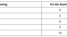
Evaluating the imaging characteristics of thoracic - lumbar spine injury according to the tlics classification SUMMARY The study was carried out with the purpose of evaluating the visual characteristics of the thoracolumbar spine injury according to the TLICS classification (The Thoracolumbar Injury Classification and Severity Score). From September 2016 to July 2017, at Viet Duc hospital, 80 patients with thoracolumbar spinal injury had been diagnosed using CT scanner and MRI, as well as receiving treatment at the hospital. The result was that 43 patients received preservative treatment, and 37 patients were operated upon, 14 of whom had TLICS < 4 point. With the use of MRI for evaluating PLC (posterior ligamentous complex), in comparision with post-operative results, the aforementioned methods showedthe sensitivity level of 96% and the specificity level of 100%. Keywords: thoracolumbar Injury, TLICS Classification, ligamentous complex.
Chẩn đoán không xâm lấn mức độ ác tính của u thần kinh đệm sử dụng cộng hưởng từ tưới máu và cộng hưởng từ phổ đa thể tích
07/04/2020 21:11:36 | 0 binh luận
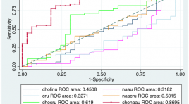
Noninvasive evaluation of glioma grade using perfusion and multivoxel 3d proton mr spectroscopy SUMMARY The purpose of this study was to clarify the usefulness of perfusion and multivoxel 3D proton MR spectroscopy in glioma grading. Study included 85 patients underwent preoperative conventional MR Imaging, multivoxel proton MR spectroscopy and histopathologically determined gliomas after stereotactic biopsy or partial resection. Receiver operative characteristic (ROC) curve analyses were performed to asscess the cutoff of rCBV and metaboite parameters of which the sensitivity and specificity are highest.A cut-off value of 2,56 for rCBV resulted in sensitivity, specificity of 86,54%, 75,76%, respectively. The cut-off value was taken as 2,76 in the Cho/NAA ratio provided sensitivity, specificity of 89,66%; 88%, respectively. The combination of rCBV and Cho/NAA ratio provided sensitivity, specificity of 71,15%; 78,79%, respectively. rCBV and Cho/NAA can increase the ability of glioma grading preoperatively. Key word : glioma, perfusion MR, spectroscopy MR

Hình ảnh cắt lớp vi tính và cộng hưởng từ bất thường dây thần kinh ốc tai trên 22 bệnh nhân điếc tiếp nhận bẩm sinh
11/04/2020 22:57:13 | 0 binh luận
CT scanner and MRI imagingof cochlear nerve deficiency in 22 patients with bilateral congenital sensorineural hearing loss SUMMARY Objective: To describe CT scanner and MRI imagingof cochlear nerve deficiency (CND) and cochleovestibular nerve abnormality in association with cochlear aperture, internal auditory canal (IAC) and labyrinthine malformations. Material and Methods : 22 patients with CNDin 43 ears. Aplasia or hypoplasia of the cochlear branch was evaluated on high resolution 3D gradient-echo MRI. Cochlear aperture, IAC and bony labyrinthine malformations was evaluated on high resolutionCT scanner. Results : 22 patients with CNDin 43 ears. Cochlear nerve aplasia in 20 ears (46,5%), cochlear nerve hypoplasia in 2 ears (4,7%), presence of vestibulocochlear nerve with no cochlear branch in 21 ears (48,8%). Labyrinthine malformation in 25 ears (58,1%).The mean IAC diameter 3,03 ± 1,03mm.Cochlear aperture stenosis and atresia 76,7%. Conclusion :CND frequently associated with labyrinthine malformations, cochlear aperture stenosis or atresia and IAC stenosis. Key words : Cochlear nerve deficiency, CT scanner and MRI.

Đánh giá kết quả bước đầu về cộng hưởng từ đàn hồi gan tại trung tâm y khoa Medic
11/04/2020 14:58:33 | 0 binh luận
Evaluate the initial result of liver mr elastography at the medic medical center SUMMARY Purpose: To evaluate the initial result of liver magnetic resonance elastography was performed in the patients and to compared with fibroscan. Methods and Materials: We retrospectively 210 patients was performed MR elastography, with 80 patients was performed MRE and Fibriscan at Medic medical center from July 2016 to June 2017, age range, 31-71 years. All the patients executed MRE on the GE signa explorer 1,5T MRI, and on Fibroscan of Echosens. Results: I n 210 patients was executed MRE, No. of patients no Fibrosis (F0): 29; Light fibrosis (F1): 39; Significant fibrosis (F2): 36; progressive fibrosis (F3): 41; Cirrhossis (F4): 65. Had detected 12 tumors (5 HCC, 1 FNH, 6 Hemangioma) and 14 cysts in liver. In 80 patients was executed MRE and Fibroscan: 9 patients F0 with MRE, the same 3F0 and 6F1 with Fibroscan; 12 patients F1 with MRE, same 5F1 and 7F2 with Fibroscan; 20 patients F2 with MRE, same 16F2 and 4F3 with Fibroscan; 15 patients F3 with MRE, same 13F3 and 2F4 with fibroscan; 24 patients F4 with MRE, same 24F4 with Fibroscan. Conclusion: MRE is a reliable non-invasive technique, safer, less expensive for evaluating hepatic fibrosis. Can be carried out at the same time as MRI examination with a search for hepatic tumor, and performed in most patients with liver disease including those with ascites or obesity. Can replace the method of liver biopsy unnecessary, risk of complications.
Nghiên cứu đặc điểm hình ảnh cộng hưởng từ sọ não trong bệnh bại não của trẻ em
07/04/2020 20:44:15 | 0 binh luận
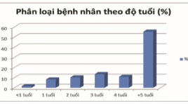
The imaging of cerebral palsy on MRI in children SUMMARY Objective : Description the image on MRI 3T of cerebral palsy and the correlation between imaging and clinical findings. Methods: From 01/2015 to 12/2016, 496 patients were diagnosed as cerebral palsy, have been done cerebral MRI in Diagnostic Imaging service. Results: 496 patients with age average 6.04 ± 4.08, youngest was under 1 year old and oldest was 15 years old. MRI shows the periventricular leukomalacia (white matter lesion) is the most popular position in CP which found in hypoxie in prenatal or perinatal, especially postnatal. The patients who had long term - hyperbilirubin have some lesion in the globus palidus and some basal ganglias. Abnormal formation of the brain, including cortical dysplasia, polymicrogyria, lissencephaly, pachygyria, schizencephaly, polymicrogyria and agenesis of the corpus callosum, have a significant rate 7.7 %. Normal MRI finding was noticed with 17.14%. Conclusion: Cerebral MRI is useful tool in confirming the degree of cerebral palsy and provides some correlation between imaging and clinical. Keywords : cerebral palsy, imaging of cerebral palsy.
Bạn Đọc Quan tâm
Sự kiện sắp diễn ra
Thông tin đào tạo
- Những cạm bẫy trong CĐHA vú và vai trò của trí tuệ nhân tạo
- Hội thảo trực tuyến "Cắt lớp vi tính đếm Photon: từ lý thuyết tới thực tiễn lâm sàng”
- CHƯƠNG TRÌNH ĐÀO TẠO LIÊN TỤC VỀ HÌNH ẢNH HỌC THẦN KINH: BÀI 3: U não trong trục
- Danh sách học viên đạt chứng chỉ CME khóa học "Cập nhật RSNA 2021: Công nghệ mới trong Kỷ nguyên mới"
- Danh sách học viên đạt chứng chỉ CME khóa học "Đánh giá chức năng thất phải trên siêu âm đánh dấu mô cơ tim"


