


Bước đầu đánh giá tái thông túi phình và vai trò chụp mạch cộng hưởng từ 1,5 Tesla trong theo dõi phình mạch não sau điều trị can thiệp nội mach
31/03/2020 15:44:12 | 0 binh luận
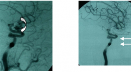
summar y The assessment of aneurysms recanalization and the role of MR angiography 1,5 Tesla in follow-up of intracranial aneurysms embolization in comparison with digital subtraction angiography. Purpose : The evaluation of aneurysmal recurrence and the role of three-dimensional time of flight MR angiography in follow-up of intracranial aneurysms embolization Material and methods: 66 patients harbored 68 selectivetreated intracranial aneurysms, in which 30 patients were both underwent three-dimensional time of flight MR angiography (MRA) and DSA, 33 patients were done only one method MRA, 0 patient were done only one method DSA. Results: The recanalization was observed in 68 selectivetreated intracranial aneurysms (39.7%), including major recanalization in 11 patients (16.1%). Compared with DSA, the overall sensitivity and specificity of MRA were 100% and 93.75%. MRI found out the ischemie lession concerning aneurysmal embolization about 8.8% and the hydrocephalus about 9.1%. Conclusion: The problem of aneurysmal recurrence shoud be considered. MRA was non-invasive method and was of very high sensitivity and specificity in follow-up of intracranial aneurysms embolization.
Ứng dụng lâm sàng của chuổi xung CISS 3D trong bệnh lý thần kinh
31/03/2020 14:05:56 | 0 binh luận
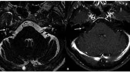
Clinical application of CISS 3D sequence in neurological diseases SUMMARY CISS 3D is a fully refocused steady-state gradient-echo sequence. This sequence is now available in almost every MRI scanners and is frequently used to evaluate several pathologies when routine MRI sequences do not provide desired anatomic information. Aplications in nerves include evaluation of cranial and spinal nerves and pathologies, which include neurovascular compression, tumor, traumatic nerve root injuries and compression by herniation disc. The cisternography aplications include CSF rhinorhea, postoprative dural tears.
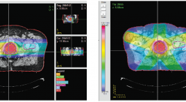
Đánh giá dòng chảy dịch não tủy ở cống não bằng kỉ thuật cộng hưởng từ
31/03/2020 13:51:45 | 0 binh luận
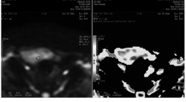
Evaluation of cerebrospinal flow at cerebral aqueduct by using magnetic resonance imaging technique SUMMARY Background : To quantify the parameters of cerebrospinal flow at the cerebral aqueduct by using cine phase - contrast magnetic resonance imaging. Methods: Twenty patients with normal ventricule were performed with 1.5T magnetic resonance system (Avanto, Siemens). Using cine phase - contrast magnetic resonance imaging, put perpendicular at the cerebral aqueduct level. Phase, rephase and magnitude images were acquired. The parameters peak velocity, diastole volume, systole volume, net volume, average area were studied. Results : The average speak speed, diastolic volume, systolic volume, average volume, and average area were 4.32cm/s, 0.05ml, 0.014ml, 0.036ml, 0397cm2 respectively. There was a statistically significant difference in peak velocity between the age groups. There were no statistically significant differences in cranial and caudal volume, net volume and average area flow parameters among different age groups. Statistically significant differences were not detected in flow parameters between sexes. Conclusion: MRI is a non-invasive method to quatify the flow of cerebrospinal fluid. Key words: cerebrospinal flow, cerebral aqueduct, cine phase - contrast magnetic resonance imaging, peak velocity, diastole volume, systole volume, net volume, average area.
So sánh giá trị của cộng hưởng từ khuếch tán và chọc hút kim nhỏ trong chẩn đoán xác định ung thư tuyến giáp
31/03/2020 13:24:49 | 0 binh luận
Compare between DWI and fnac in dignostis thyroid cancer summa ry Retrospective study on 21 patients (29 thyroid nodules: 21 benign nodules, 7 malignant nodules) who were acquired the diffusion-weighted magnetic resonance imaging (DW-MRI) with 200, 300, 400, 500, 600, 700, 800 b values and ADC maps were calculated, fine needle aspiration cytology (FNAC) were performed in 21 nodules. Diagnosis confirmed by postsurgical histopathologic examinations. Results: ADC values of nodules provide useful data about the nature of a thyroid nodule, with b200 DW-MRI, the mean ADC values of thyroid nodules were 1.45±0,30x10⁻³mm2/s in the malignant group and 2.26±0.33x 10⁻³mm2/s in the benign group, with significantly different (P< 0.001). According to an ADC cut-off value of 1.98x10⁻³mm2/s, the sensitivity, specificity, positive predictive value, negative predictive value, and accuracy in differentiating benign from malignant thyroid nodules are calculated as 100%; 81%; 66.7%; 100% and 86.2%, respectively. The DW-MRI has the higher values than FNAC for diagnosis of thyroid cancer. We can applied DW-MRI for diagnosis of thyroid nodule in the
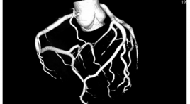
Gía trị của chụp cắt lớp vi tính hai nguồn năng lượng trong đánh giá bệnh lý hẹp động mạch vành không sử dụng thuốc kiểm soát nhịp tim
31/03/2020 13:07:13 | 0 binh luận
Value of dual-source computed tomography for evaluation coronary arterial stenosis without heart rate control medicine summa ry Objective : Accuracy of dual-source computed tomography (DSCT) for evaluation of coronary arterial stenosis without heart rate control. Material and methods : During 1 year (between January 2012 and December 2012), 108 patients were examined by second-generation dual-source computed tomographycoronary angiography without heart rate control at Radiology Departement of Bach Mai hospital and underwent conventional coronary angiography. All the data of DSCT were compared with the result of CCA to assess the sensitivity, specificity and accuracy for evaluation of coronary artery stenoses. Resul t: We enrolled 108 patients (65 male, 43 female, mean age 62,9 years). Mean heart rate was 77.4 bpm, radiation dose was 3.66 mSv. Eighty-four patients (77.8%) had good image quality. Ninety-seven patients (89.8%) were identified as having significant coronary stenoses. 11 patients (11.2%) having no significant coronary stenoses. Sensitivity, specificity and accuracy were 93.9%, 93.6% and 93.7%. Conclusion: DSCT provides a high diagnostic accuracy for evaluation coronary arterial stenosis.
Lập kế hoạch xạ trị điều biến cường độ chùm proton bằng hệ thống lập kế hoạch CERR
03/04/2020 13:26:53 | 0 binh luận
Planning impt treatment by using cerr program SUMMARY Up to now, beside two conventional methods are using electron beam and photon beam, radiotherapy using high energy proton beam has been attracted more and more scientists and hospitals. In this work, we using CERR program to make a treatment planning. The simulation results show that the treatment plan is better compared with by photon beam. The risk organs surrounding the tumour received negligible low dose. So the side effect decreases significantly. We also make a comparison the treatment planning by proton beam with by photon beam. Keywords: cancer, treatment planning, high energy proton beam therapy.
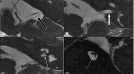
Vai trò MRI trong điếc bẩm sinh
03/04/2020 13:23:09 | 0 binh luận
The Role of MRI in the congenital hearing SUMMARY In the number of cochlear implants being performed and, consequently, there has also been a steady increase in the imaging, done as a part of the preoperative workup of these patients. High-resolution CT scan (HRCT) and MRI are routinely performed prior to cochlear implant surgery. These modalities help assess the status of the inner ear structures. A few patients have significant anomalies, which need to be assessed and understood in detail. MRI is now increasingly being used to study the membranous labyrinth and the cranial nerves; it provides accurate information and exquisite anatomical detail. This paper is a pictorial essay on the various congenital temporal bone anomalies seen in patients being investigated prior to cochlear implant surgery. There are several complex congenital anomalies that are encountered while imaging such patients, needs to follow a clinically oriented classification of these anomalies, which helps the implant surgeon plan the correct management strategy. Keywords: Congenital hearing loss, Cochlear implant, MRI – CT in congenital temporal bone.
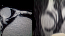
Hình ảnh siêu âm và chụp cắt lớp vi tính nang ống Nuck nhân một trường hợp
03/04/2020 12:52:34 | 0 binh luận
Ultrasound and CT image of the Nuck canal hydrocele. Case report SUMMARY Hydrocele (or cyst) of the Nuck canal is a rare condition in female resulting from incomplete obliteration of the canal and entrapment of fluid in the inguinal canal. Ultrasonography play an important role to diagnostic and differentiate from the other conditions presenting with inguinal swelling. MRI and CT can help identify an inguinal cystic mass when sonographic findings are not clearly. A case of hydrocele of the Nuck’ canal diagnosed withUSG and CT scan, comparison with surgery and pathology result at the E hospital. Keywords: Hydrocele, Cyst, Canal of Nuck, Ultrasonography.
Bạn Đọc Quan tâm
Sự kiện sắp diễn ra

THÔNG BÁO SINH HOẠT KHOA HỌC 19/01/2024: CẬP NHẬT CHẨN ĐOÁN VÀ ĐIỀU TRỊ UNG THƯ TRỰC TRÀNG
19-01-2024 09:47:01 0 bình luận

Thông tin đào tạo
- Những cạm bẫy trong CĐHA vú và vai trò của trí tuệ nhân tạo
- Hội thảo trực tuyến "Cắt lớp vi tính đếm Photon: từ lý thuyết tới thực tiễn lâm sàng”
- CHƯƠNG TRÌNH ĐÀO TẠO LIÊN TỤC VỀ HÌNH ẢNH HỌC THẦN KINH: BÀI 3: U não trong trục
- Danh sách học viên đạt chứng chỉ CME khóa học "Cập nhật RSNA 2021: Công nghệ mới trong Kỷ nguyên mới"
- Danh sách học viên đạt chứng chỉ CME khóa học "Đánh giá chức năng thất phải trên siêu âm đánh dấu mô cơ tim"


Đơn vị hợp tác

