


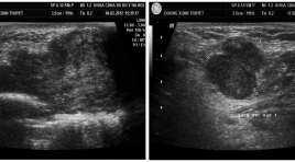
Ung thư tuyến mồ hôi nhân một trường hợp
02/04/2020 15:16:11 | 0 binh luận
Sweat gland carcinoma study case SUMMARY: Sweat gland carcinomas are rare maglignant tumors derived from sweat glands of the skin and occur in elderly persons. The disease grows slowly and often diagnosed in late stage. Dfinitive tumor diagnosis based on histopathology. Radiology plays important role in tumor determining, assessing enlargement and staging. We introduce a case study with diagnosis sweat gland carcinoma in the cheek and discuss features of tumor that are helpful in early diagnosis and treatment. Key words: sweat gland carcinoma features, ultrasound, MRI, CT Scanner.
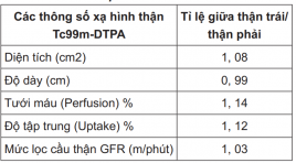
Nghiên cứu một số thông số xạ hình thận TC99- DTPA ở người bình thường
02/04/2020 15:09:14 | 0 binh luận
Quantitative evaluation of Tc99m – DT PA Renoscintigraphy parameters in healthy people SUMMARY: Aims: The purpose of our study was to determinate quantitative parameters in Tc99m-DTPA Renoscintigraphy in healthy people. Materials and Methods: Tc99m-DTPA renoscintigraphy was performed in 44 consecutive healthy people, mostly potential live kidney donors in Nuclear Medicne Department, 108 central military hospital. Quantitative parameters including Glomerular Filtration Rate (GFR) were determined by gamma camera using the dedicated software. Results: There were no significant differences of kidney areas, depths and quantitative parameters such as perfusion %, uptake % between the two kidneys. There was no diffirence between mean GFR ± SD of the left and right kidney (52.8 ± 13.4 vs 51.07 ± 12. 65 ml per minute, respectively) and total GFRs were in wide range from 78 to 155 ml per minute. Conclusions: Tc99m-DTPA renoscintigraphy quantitative parameters in the healthy people could be used as reference to research and clinical practice.
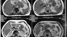
Nhân một trường hợp chẩn đoán và điều trị tụy đôi, ống tụy phụ có sỏi, giản dạng nang
02/04/2020 15:05:43 | 0 binh luận
Diagnosis and treatment pancreas divisum together calculi and cystic dilatation of the accessory duct SAMMARY: A patient, 46 year old, female, with abdominal pain, was admitted to Hospital of Hanoi medical university. Divisum pancreas was diagnosed in MR image and in operation, the accessory pancreatic duct was largely dilated which formed a cyst in the pancreatic head with some calculi inside. Divisum pancreas is a common congenital abnormaly of the pancreatic ducts. The human embryo starts life with two ducts in the pancreas; the ventral duct and the dorsal duct. In more than 90% of the embryos, the dorsal and the ventral ducts will fuse to form one main pancreatic duct. The main pancreatic duct will join the commom bile duct and drains into the duodenum through the major papilla. In approximately 10% of embryos, the dorsal and the ventral ducts fail to fuse. Failure of the ventral and the dorsal pancreatic ducts to fuse is called pancreas divisum. In pancreas divisum, the ventral duct drains into the major papilla, while the dorsal duct drains into a separate minor papilla. In patients with divisum pancreas, the majority of the pancreatic secretions pass through dorsal duct and the minor papilla (instead of the major papilla) that can result in inadequate drainage and pain caused by obstruction. And this obstruction may be the cause of acute pancreatitis, chronic pancreatitis and pancreatic lithiasis
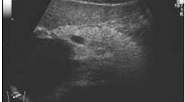
SARCOMA cơ vân của tuyến tiền liệt hình ảnh cộng hưởng từ
21/11/2019 08:46:55 | 0 binh luận
Rhabdomyosarcoma of the prostate: Mr findings SUMMARY: Prostatic rhabdomyosarcoma is a rare tumor, especially in the elderly, and has extremely poor prognosis. We report a case of rhabdomyosarcoma of the prostate on a 64 year-old patient who was admitted to hospital, complaining of dysuria, and pain in anus region. Rectal digital examination found a large mass compressing the rectum, which was suspected tumor of the prostate. Ultrasound: Prostate was enlarged and heterogenous. Perineal biopsy revealed rhabdomyosarcoma. MR examination demonstrated the tumor arises from peripheral gland zone of the right lobe, developing outward, encasing the left lobe, seminal vesicles, compressing the rectum. The tumor was lobulated, seperated by fibrous septa, heterogenous hyperintensity on T2W, with many hemorrhage foci due to recent biopsy. The tumor enhanced heterogenously after contrast administration. Key worlds: MRI, prostate, sarcoma
Nút mạch điều trị u máu gan khổng lồ báo cáo 2 ca
02/04/2020 14:24:59 | 0 binh luận
Arterial embolization in the treatment of giant hepatic hemangiomas Report 2 cases SUMMARY: Objective: Present 2 cases giant hemangiomas treated by selected arterial embolization. Materiel and method: 2 hepatic hemangiomas, 16 x 17cm and 6 x 9cm were diagnosed with CT scanner and embolized with PVA particles. Result and Discussion: No severe complication is noted exempt fever and pain covered by medicaments. Short and long time follow-up, 4 years, prove the pain was removed and reducing the tumor dimension. Many authors have determined the effective of this method in the literature. Conclusion: Arterial embolization can be carried out in conservative treatment of giant hepatic hemangioma against the pain and reducing the tumor volume, together it can be used in preoperative for reducing the volume tumor to avoid hemorrhage and give safety for operation.
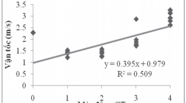
Gía trị vận tốc sóng biến dạng trong mô gan xơ qua kỉ thuật xung áp lực nén siêu âm
03/04/2020 09:33:44 | 0 binh luận
The value of shear wave average velocity in healthy’s liver by using acoustic radiation force impulse imaging SUMMARY: Objectives: The purposes of this study were to measure the reference value of average velocity of shear wave in healthy’s liver by using acoustic radiation force impulse imaging. Methods: Two hundred and of healthy volunteers with normal liver function test values were selected for the study. Shear wave velocity measurements, expressed in meters per second, were taken in liver segment 7 or 8 at depth from 3 to 4cm below the body surface portion of. Among these volunteers, we chose fifty ones to whom two observers with different levels of experience performed the measurements independently and blindly. Results: Total the measurements are taken 2.532 times. The value of average velocity of shear wave in the health’s liver is 1.05 ± 0.12m/s. There is no statistically significant difference in shear wave velocity between two genders. In term of interobserver results, there is no statistically significant difference in shear wave velocity obtained by two observers with different levels of experiences (P < .005). Conclusions: The results of this study show that shear wave velocity measurement in health’s liver by using the acoustic radiation force impulse technique is about 1.05 ± 0.12m/s and this technique is reproducible and independent to user.
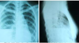
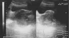
Ca bệnh phổi hiếm gặp: dị dạng nang tuyến bẩm sinh (Congenital Cystic Adenomatoid Malformation)
02/04/2020 21:41:41 | 0 binh luận
Trần Thị Kim Ng. 16 tuổi. Tiền sử gia đình bố mẹ khỏe mạnh. Con đầu lòng, đẻ thường, cân nặng 3kg. Từ khi sinh đến nay thường xuyên ho sốt. Bệnh sử bệnh nhân (BN) đều trị tại Bệnh viện Lao và bệnh phổi Nam Định. Với chẩn đoán tràn mủ màng phổi do lao thời gian 20 ngày, bệnh tiến triển chậm nên chuyển Bệnh viện phổi Trung ương. Khoa khám bệnh chẩn đoán tràn mủ màng phổi phải, chuyển khoa bệnh màng phổi. Khám: cân nặng: 41 kg, có ho, khạc đờm không có mùi hôi. Mạch: 88 lần/phút, nhiệt độ: 37 0 C. Huyết áp 90/60 mmHg, nhịp thở 22 lần/phút. Xét nghiệm máu: bạch cầu 10.000, trung tính 84%, các chỉ số khác, sinh hóa trong giới hạn bình thường. Vi khuẩn lao âm tính. Siêu âm màng phổi không có dịch, thùy dưới phổi phải có nhiều ổ dịch và giàu mạch phân bố không đều. Siêu âm các bộ phận khác không thấy bất thường X-Quang có hình mức khí dịch đáy phổi phải
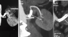
Điều trị bóc tách động mạch thận nguyên phát tại Bệnh viện Đại học Y Hà Nội
19/11/2019 16:45:52 | 0 binh luận
Bạn Đọc Quan tâm
Sự kiện sắp diễn ra

THÔNG BÁO SINH HOẠT KHOA HỌC 19/01/2024: CẬP NHẬT CHẨN ĐOÁN VÀ ĐIỀU TRỊ UNG THƯ TRỰC TRÀNG
19-01-2024 09:47:01 0 bình luận

Thông tin đào tạo
- Những cạm bẫy trong CĐHA vú và vai trò của trí tuệ nhân tạo
- Hội thảo trực tuyến "Cắt lớp vi tính đếm Photon: từ lý thuyết tới thực tiễn lâm sàng”
- CHƯƠNG TRÌNH ĐÀO TẠO LIÊN TỤC VỀ HÌNH ẢNH HỌC THẦN KINH: BÀI 3: U não trong trục
- Danh sách học viên đạt chứng chỉ CME khóa học "Cập nhật RSNA 2021: Công nghệ mới trong Kỷ nguyên mới"
- Danh sách học viên đạt chứng chỉ CME khóa học "Đánh giá chức năng thất phải trên siêu âm đánh dấu mô cơ tim"


Đơn vị hợp tác

