


Nghiên cứu giá trị chẩn đoán ung thư vú của siêu âm mode b và siêu âm đàn hồi mô - strain elastography
04/12/2019 11:59:47 | 0 binh luận
Research into the value of b-mode ultrasound and strain elastography ultrasound in the diagnosis of breast cancer SUMMARY A diagnostic test study was conducted at Bạch Mai hospital to evaluate the efficacy of Ultrasound Strain Elastography in diagnosis of breast masses. Result: 22 patients with 24 lesions were prospectively evaluated by B-mode ultrasound and strain elastography, followed by the core biopsy. The sensitivity, specificity, positive predictive value, negative predictive value, accuracyfor the B-mode Ultrasound were 94.1%, 57.1%, 84.2%, 80%, 83,3%. Elastography combined with B-mode ultrasound improved the value in diagnosis, the sensitivity, specificity, positive predictive value, negative predictive value, accuracywere 100%, 71,4%, 89,4%, 100%, 91,7%. Conclusion : Strain elastography can better diagnose BI-RADS 3 and 4a lesions, especially when combined with B-mode breast ultrasound, which may increase or decrease the BI-RADS level, improvethe accuracy from 83,3% to 91,4%. Key Words: B-mode Ultrasound, Strain Elastography, Breast cancer.
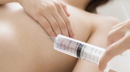
Đánh giá hiệu qua ̉bước đầu trong loại bỏ các tổn thương vú lành tính bằng sinh thiết vú có hô ̃trợ hút chân không tại Trung tâm điện quang Bệnh viện Bạch Mai
04/12/2019 14:16:10 | 0 binh luận
Aexperimentalresearch was performed in radiology center of Bach Mai hospital to evaluate the initial efficacy in the removal of benign breast lesions by vacuum-assisted biopsy SUMMARY Subjects and methods: There is a prospective intervention study in 21 female patients with 31 benign breast lesions with needle aspiration vacuumassisted biopsy under ultrasound guidance from Jan 2018 to Jun 2018. Results : The mean age is 37.5 years old.The 20-30 years old group is most common (19.3%). The average size of the lesions measuring on ultrasound is 11.7mm. The average number of samples is 10.8 with the average time of cutting is 12 minutes. The most common abnormality pathology is breast fibroadenoma (54.8%). Fibrocystic breast disease accounts for 25.8% of all lesions, which is second highest rate. The main complications after biopsy are pain and hematoma. There is a proportional correlation between the size of the lesions and other factors such as the amount of anesthetics used, the volume of the hematoma after the biopsy, the time of wound removal and the size of the biopsy needle. There is a inverse correlation between the distance from the lesion to the nipple and post-biopsy pain. The distance from the lesion to the skin surface is inversely proportional to the size of the post-biopsy hematoma. Conclusion: Vacuum-assisted breast biopsy is an effective and safe method for removal benign breast lesions. This method is also highly aesthetic. The anapathology results based on this method are reliable, especially for small lesions. Key words: Vacuum-asisted biopsy, mammotome, benign breast lesions.
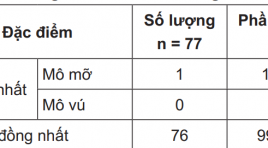
Nghiên cứu đặc điểm hình ảnh của ung thư vú tại bệnh viện Trung ương Huế
01/04/2020 12:45:52 | 0 binh luận
The ultrasonographic and mammographic appearance of breast cancer at Hue national Hospital SUMMARY Purpose: Desciber the ultrasonographic and mammographic appearance of breast cancer . Method and Material: Retrtospectively study the imaging findings based on guidline of ACR of 77 cases of breast cancer at Hue’s central hospital. Results : 1/ Ultrasonographic appearance: 99.7% patient having inhomogenous breast tissue. 90.91% tumor having irregular shape. There are only 59.7% tumor having orientation parallel to skin surface. 9.6.21% tumor having hypoechoic and heterogenous structure. Most of tumor having complet or partiasl shadowing. 2/ Mammographic appearance: Most tumor having heterogenous and dense structure. 92.3% tumor having irregular shape. 88.47% tumor having ill-defined or irregular border. Most microcalcifiction inside tumor are classified to high risk type. Conclusion: Most patient having inhomogenous breast tissue. Interpretation of mammographic film should corelate with ultrasonographic images. Most tumor were classified at level more than 3, when image findings of tumor were used according to ACR’s guidline. Keywords: Breast cancer, ultrasound, mamography.
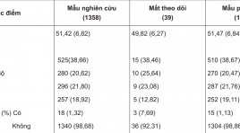
Gía trị của X quang và siêu âm trong sàng lọc ung thư vú ở phụ nữ từ 40 tuổi trở lên
30/03/2020 13:17:54 | 0 binh luận
Value of mammography, ultrasound in breast cancer screening with women ≥40 years old SUMMARY Objective: To research the value of mammography, ultrasound in breast cancer screening with women ≥ 40ys. Methods: Including of the study was 1319 women (age ≥40) in the range of six months with breast cancer screening at University Medical Center, Hochiminh city, from 1 June 2014 to 31 May 2016. Mammography and ultrasound were performed using BI-RADS (The Breast Imaging and Reporting Data System) of the American College of Radiology. Breast cancer was determined by histopathological results. Results : The proportion of breast cancer was 1.67% (22/1319). Among 22 cancers detected, 19(14.4/1000) with mammography, 18(13.65/1000) with ultrasound. The sensitivity, specificity of mammography respectively were 86.36% (IC 95%: 65.09-97.09), 99% (98.29-99.47) higher than ultrasound: 81.82% (59.71-94.81); 95.45% (94.18-95.52). When combined two tests, the sensitivity and specificity respectively were 100% (IC 97.5%:84.56-100), 95.37% (94.09-96.45); PPV was decreased (26.83%) (17.63-37.75) compared to mammography (59.38%) (40.64-76.3); NPV was increased (100%)(IC 97.5%: 99.7-100) compared to mammography (99.77%) (99.32-99.95)or ultrasound alone (99.68%)(99.18-99.91). Conclusions : The sensitivity and specificity of breast cancer screening with mammography were higher than ultrasound alone. When combined two tests, it was increased the sensitivity, decreased the specificity compared to mammography alone. Keywords: breast cancer screening, mammography, ultrasound.

Bước đầu ứng dụng hệ thống quét khối 3D vú tự động bằng siêu âm Acuson S2000-ABVS trong khảo sát bệnh lý vú tại Medic
13/04/2020 15:45:16 | 0 binh luận
Nội dung: 1.Cấu tạo máy và kỹ thuật chụp 2.Chỉ định 3.Tư thế bệnh nhân và cách đặt đầu dò 4.Workstation 5.Một số hình ảnh thực hiện tại Medic 6.Nhận xét- Kết luận

Đánh giá các yếu tố liên quan đến độ hấp thụ 8F-FDG tại khối u vú nguyên phát
12/04/2020 23:05:45 | 0 binh luận
UTV đứng đầu trong các UT ở phụ nữ Chẩn đoán UTV: Lâm sàng, cận lâm sàng PET/CT là phương pháp chẩn đoán y học hạt nhân ở mức độ tế bào PET/CT giúp chẩn đoán sớm và đánh giá chính xác giai đoạn, tiên lượng bệnh Giá trị maxSUV giúp tiên lượng bệnh Tình hình nghiên cứu trong và ngoài nước Trên thế giới: Sigato Ueda, Bong II Song… Việt Nam chưa có nghiên cứu
Bạn Đọc Quan tâm
Sự kiện sắp diễn ra

THÔNG BÁO SINH HOẠT KHOA HỌC 19/01/2024: CẬP NHẬT CHẨN ĐOÁN VÀ ĐIỀU TRỊ UNG THƯ TRỰC TRÀNG
19-01-2024 09:47:01 0 bình luận

Thông tin đào tạo
- Những cạm bẫy trong CĐHA vú và vai trò của trí tuệ nhân tạo
- Hội thảo trực tuyến "Cắt lớp vi tính đếm Photon: từ lý thuyết tới thực tiễn lâm sàng”
- CHƯƠNG TRÌNH ĐÀO TẠO LIÊN TỤC VỀ HÌNH ẢNH HỌC THẦN KINH: BÀI 3: U não trong trục
- Danh sách học viên đạt chứng chỉ CME khóa học "Cập nhật RSNA 2021: Công nghệ mới trong Kỷ nguyên mới"
- Danh sách học viên đạt chứng chỉ CME khóa học "Đánh giá chức năng thất phải trên siêu âm đánh dấu mô cơ tim"


Đơn vị hợp tác

