


Nghiên cứu giá trị của phương pháp tiêm cồn tuyệt đối dưới hướng dẫn của siêu âm trong điều trị nang tuyến vú lành tính
06/05/2021 14:18:29 | 0 binh luận
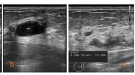
SUMMARY Objective : To investigate the effectiveness of single- session ultrasound-guided percutaneous ethanol sclerotherapy in symptom breast cysts Methods: Breast cysts symptoms patients underwent ultrasound examination and treated by ethanol sclerotherapy at Bach Mai Hospital from July 2019 to February 2020 were investigated. All patients were aspirated using 20-22G needles refilled using 99% ethanol and reaspirated completely after 10 minutes of exposure under ultrasoundguidance. Follow-ups were done by ultrasound examination at one week and 3 months to 6 months by ultrasound examination. Results : 62 breast cysts (mean volume, 5.01 ± 4.8 ml; range, 0.8 to 22 ml) of 59 patients (mean age, 44.5 years) had symptoms were treated. 7 patients (11.2%) had painful symptoms, 17 patients (27,4%) had felling burn skin, and 15 patients (24.2%) felling uncomfortable while the ethanol sclerotherapy process and all symptoms disappeared after 5 minutes. 26 breast cysts (42%) were disappeared in sonography examinations and 36 breast cysts (58%) reduced in volume (mean 96,4% volume reduced) after one week. At 3 months to 6 months, 61 cysts (98,4%) completely responded therapy as undetectable on sonography, only one cyst (1.6%) was reduced 88,6% in volume. The success of this technique was 100 %, no patient had serve complication such as hemorrhage or abscess Conclusion: Ultrasound-guided ethanol sclerotherapy is a simple, fast, and safe method in the treatment of symptom breast cysts.
Giá trị chẩn đoán của các vi vôi hóa nghi ngờ ác tính trên X quang tuyến vú
06/05/2021 12:49:48 | 0 binh luận
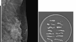
SUMMARY Objective: Diagnostic value of some microcalcifications with suspected malignancy on mammograms. Methods: The study included 60 women with microcalcifications who underwent imaging-guided biopsy between July 2019 and July 2020 at Bach Mai Hospital. Digital mammograms procured before biopsy were analyzed independently by one breast imaging subspecialists blinded to biopsy results. Results : * Micro-calcification outside of a mass – 30 cases The overall positive predictive value of biopsies was 40%. The individual morphologic descriptors predicted the risk of malignancy as follows: fine linear/branching, 7 (87.5%) of 8 cases; fine pleomorphic, 4 (25%) of 16 cases; amorphous, 1 (16.7%) of 6 cases và coarse heterogeneous, 0 cases. Fisher’s Exact testing showed statistical significance among morphology descriptors (p < 0.01) * Microcalcifications in a mass – 30 cases The overall positive predictive value of biopsies was 96.7%. The individual morphologic descriptors predicted the risk of malignancy as follows: fine linear/branching, 16 (100%) of 16 cases; fine pleomorphic, 11 (92%) of 12 cases; amorphous 2 (100%) of 2 cases và coarse heterogeneous, 0 cases. * The positive predictive value for malignancy according to BI-RADS assessment categories were as follows: category 4B, 21,1%; category 4C, 66,7%; and category 5, 94.3%. Conclusion: Morphological description and distribution of microcalcifications on mammograms helps classify BI-RADS and assess the risk of malignancy for each case for diagnosis and treatment monitoring. The positive predictive values for breast cancer increased in order of amorphous, fine pleomophic, and fine linear/ branching microcalcification.
Bước đầu đánh giá giá trị của kỹ thuật sinh thiết hút chân không trong chẩn đoán các tổn thương vi vôi hóa ở vú
06/05/2021 12:09:50 | 0 binh luận
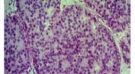
SUMMARY Objective: The purpose of this article is initial assessment the value of vaccum assited biopsy technique in diagnosis breast microcalcification lesions. Materials and Methods: T he prospective study included 17 women with 18 breast microcalcification lesions without mass that were classified BIRADS 4- 5.All lesions underwent imaging- vaccum assisted biopsy between August 2019 and July 2020 in Radiology Centre- Bach Mai Hospital. Results: The study involved 2 lesions with stereostatic vaccum assited biopsy- SVAB and 16 lesions with ultrasound guided vaccum assisted biopsy- USVAB. The mean duration of procedure was 53.22 ± 15.06 (minutes).The median number specimen was 10.9 ± 4.2. All lesions had microcalcifications in specimens. In terms of complications, the mild pain rate was 27.8% and 1/18 (5.6%) had hematoma without surgery. Biopsy results revealed 7 benign and 11 malignant lesions. The malignant rate in fine pleomorphic, amorphous and linear microcalcification were 87.5%, 33.3% and 100%, respectively.The malignant rate in regional, segmental and cluster distribution were 50%, 71.4% and 57.1%, respectively.9/11 malignant lesions had histopathologic results after surgery. The total upgrade rate at surgery was 22.2% (all for ductal carcinomas in situ- DCIS). Patients with benign lesions were followed up for a median period of 6 months, with no interval change. The sensitivity, specificity, positive predictive and negative predictive value of VAB in diagnostic breast microcalcification were 100%. Conclusion: Our first experiences: Image-guided vacuum-assisted breast biopsy is accurate, reliable and minimally invasive.It can be used as a safe approach for diagnosis in patients with breast microcalcifications. Keyword: VABB, breast microcalcification
Đánh giá giá trị chẩn đoán ung thư vú của siêu âm đàn hồi nén và sóng biến dạng
05/05/2021 17:50:03 | 0 binh luận
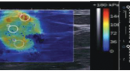
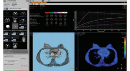
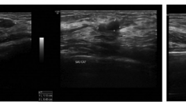
SUMMARY Objective : Evaluating the value of B-mode ultrasound and elastography ultrasound in the diagnosis of breast cancer. Methods: Breast lesion patients were classified BIRADS from 3 to 5 after underwent B-mode ultrasound and elastography ultrasound examination and done biopsy to have histopathological results at Bach Mai Hospital from July 2019 to February 2020. Results: The cut-off value of fat-to-lesion ratio is 28,4 with sensitivity (Se), specificity(Sp) and accuracy (Acc) were 76,9%; 93,3%; 85,7% respectively. The cut-off value of Elasto/B-mode ratio is 1 with Se, Sp, Acc were 100%, 73,3%; 85,7%. Se, Sp and Acc of shear-wave elastography were 100%; 97,8% and 97,5% respectively with the cut-off value is 36 kPa. Sp, Se and Acc of Tsukuba score were respectively 84,6%; 88,9%; 86,9%. B-mode ultrasound combine with shear-wave elastography has highest Se and Sp were 100%; 91,1% respectively. Conclusion: Elastography ultrasound combine with B-mode ultrasound can upgrade or downgrade the BIRADS level, so they can increase accuracy to diagnose breast cancer especially BIRADS 3 or 4a lesions.
Nghiên cứu đặc điểm hình ảnh và giá trị của cộng hưởng từ 1.5 TESLA trong chẩn đoán và định hướng điều trị ung thư vú
05/05/2021 17:32:49 | 0 binh luận
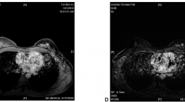
SUMMARY Objective: Study imaging characteristics and values of 1.5 Tesla Magnetic Resonance Imaging in diagnosis and treatment of breast cancer. Material and Methods: A prospective study on 52 patients with breast cancer underwent( received) breast MRI before treatment Results: The common signs is mass shape (88,5%) with irregular margins or dendrites (100%), heterogeneous contrast enhancement (96,2%) strong penetration in the first 2 minutes after contrast injection (84,6%), kinetic curve is mainly type II or type III (88,4%). Multiple lesion has 13 (25%). Invasive ductal carcinomas is 49 (94,2%) Conclusion: General features of breast cancer MRI are mainly mass with irregular margins or dendrites, and heterogeneous contrast enhancement, mainly strong penetration in the first 2 minutes after contrast injection , no case has poor absorbtion, plateau kinetics and washout kinetics are major. Key Words: Magnetic resonance imaging, breast cancer.
Đánh giá hiệu quả bước đầu trong loại bỏ các tổn thương vú lành tính bằng sinh thiết vú có hỗ trợ hút chân không tại Trung tâm Điện quang Bệnh viện BẠCH Mai
05/05/2021 17:23:21 | 0 binh luận
SUMMARY Objective: A experimental research was performed in radiology center of Bach Mai hospital to evaluate the initial efficacy in the removal of breast benign lesions by vacuum-assisted biopsy. Subjects and methods: There is a prospective intervention study in 32 female patients with 45 breast benign lesions with needle aspiration vacuum-assisted biopsy under ultrasound guidance from January 2018 to December 2018. Results: The mean age is 36.5 years old. The 20-40 years old group is most common (60.0%). The average size of the lesions measuring on ultrasound is 12.9mm. The average number of samples is 13.2 with the average time of cutting is 14.5 minutes. The most common abnormality pathology is breast fibroadenoma (62.2%). Fibrocystic breast disease accounts for 17.8% of all lesions, which is second highest rate. The main complications after biopsy are pain and hematoma in tissu. 78.8% of patients after treatment don’t have to take Paracetamol. The average size of hematoma after 1 month with 10G needle is 4.8mm; with 8G needle is 6.3mm. Conclusion: Vacuum-assisted breast biopsy is an effective and safe method for removal benign breast lesions. This method is also highly aesthetic. The anapathology results based on this method are reliable, especially for small lesions. Key words: Vacuum-assisted biopsy, mammotome, breast benign lesions.
Đặc điểm hình ảnh siêu âm của ung thư tuyến vú tại Bệnh viện Bạch Mai
05/05/2021 17:17:10 | 0 binh luận
SUMMARY Objective: Describer the ultrasoundgraphic appearance of breast cancer. Methods: Data was collected from 49 patients who underwent breast ultrasound, guided interventional and operated procedures from august 2017 to june 2019, diagnosis of breast cancer. Study the imaging findings based on guidline of ACR BI-RADS 2013. Results: Ultrasoundgraphic appearance: 100% breast cancer lesions are mass, 85,7% tumors having irregular shape. There are 75,5% tumor having not orientation parallel to skin surface. Spiculated and angular margin 63,3%, 73,5% tumors having hypoechoic, 40,8% tumor having calcification in mass. Conclusion: Most of breast cancer tumors having mass finding, irregular shape. The findings as orientation not parallel to skin surface, speculated or angular margin, hypoechoic and having calcification in mass are suspected to breast cancer. Keywords: Breast cancer, ultrasound.
Cộng hưởng từ tuyến vú ở bệnh nhân ung thư vú thể ẩn có di căn hạch nách
03/04/2020 09:02:31 | 0 binh luận
Mr imaging of the breast in patients with occult primary breast cancer presenting as an axillary metastasis SUMMARY: Purpose: To access the value of MRI in the diagnosis of occult primary cancer with axillary metastasis. Methods and Materials: 12 patients occult breast cancer with malignant axillary adenopathy and negative on mammographic, echography and physical examination findings, were underwent contrast material-enhanced MR imaging in Hanoi medical university hospital. Results: The sensitivity of MRI in the diagnosis of occult primary cancer with axillary metastases was 83%. MR imaging depicted small cancers from 3 to 12mm diameter. Of the 12 patients, three patients were underwent mastectomy, five were underwent lobectomy, and four were underwent breast-conservation therapy. Conclusion: MR imaging is very sensitive for the detection of occult breast cancer with malignant axillary adenopathy. MR imaging offers potential not only for cancer detection but also for staging the cancer within the breast, which may be useful for treatment planning. Keywords: dynamic breast MRI, occult breast cancer
Bạn Đọc Quan tâm
Sự kiện sắp diễn ra


THÔNG BÁO SINH HOẠT KHOA HỌC 19/01/2024: CẬP NHẬT CHẨN ĐOÁN VÀ ĐIỀU TRỊ UNG THƯ TRỰC TRÀNG
19-01-2024 09:47:01 0 bình luận
THÔNG BÁO SINH HOẠT KHOA HỌC 19/01/2024: CẬP NHẬT CHẨN ĐOÁN VÀ ĐIỀU TRỊ UNG THƯ TRỰC TRÀNG
19-01-2024 09:47:01 0 bình luận
prev
next
Thông tin đào tạo
- Những cạm bẫy trong CĐHA vú và vai trò của trí tuệ nhân tạo
- Hội thảo trực tuyến "Cắt lớp vi tính đếm Photon: từ lý thuyết tới thực tiễn lâm sàng”
- CHƯƠNG TRÌNH ĐÀO TẠO LIÊN TỤC VỀ HÌNH ẢNH HỌC THẦN KINH: BÀI 3: U não trong trục
- Danh sách học viên đạt chứng chỉ CME khóa học "Cập nhật RSNA 2021: Công nghệ mới trong Kỷ nguyên mới"
- Danh sách học viên đạt chứng chỉ CME khóa học "Đánh giá chức năng thất phải trên siêu âm đánh dấu mô cơ tim"


Đơn vị hợp tác

