


An toàn trong sử dụng thuốc tương phản: các tác dụng phụ thường gặp và ít gặp
25/08/2021 14:39:43 | 0 binh luận
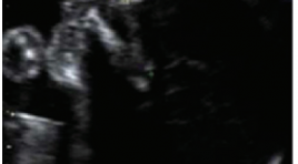
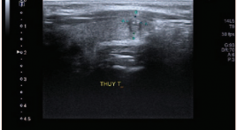
Độ dài xương mũi thai nhi ở tuổi thai từ 19-26 tuần tại Việt Nam
30/03/2020 22:28:55 | 0 binh luận
SUMMARY Aim: The purpose of this study was to establish the normal range of fetal nasal bone length (NBL) at the second trimester of gestation in Vietnamese pregnancies. Methods: A prospective cross-sectional study was carried out. A total of 2432 singleton pregnancies were evaluated for fetal NBL at 19–26 weeks 6 days between 1 January 2013 and 31 December 2014. Three measurements of NBL were taken and the largest value was chosen. Fetal anomalies and neonatal abnormalities were excluded. Results : Mean fetal NBL was 6.75 ± 0.82 mm and mean gestational age was 22.2 ± 1.4 weeks. Median NBL increased linearly with advancing gestational age: NBL (mm) = 0.26 × gestational age (weeks) + 1.03 (R2 = 0.20; P <0.000). The fifth percentile of fetal NBL ranged from 4.6 to 6.3 mm, corresponding with gestational age 19–26 weeks. Conclusion : The normal range ofNBLwas established at second trimester inVietnamese pregnancies. NBL at the fifth percentile for gestational age was different from that of other Asian ethnicities. It is necessary to define short NBL or hypoplasia of NBL with regard to ethnicity. Key words : Down syndrome, fetal nasal bone length, ultrasonography.
Nhận xét giá trị phương pháp chọc hút kim nhỏ dưới hướng dẫn siêu âm trong chẩn đoán ung thư biểu mô tuyến giáp
06/05/2021 16:49:08 | 0 binh luận
SUMMARY We conduct research on diagnostic value of ultrasound guided fine needle aspiration (FNA) in the diagnosis of thyroid carcninoma compared with post-operative pathology at Hai Phong International Hospital. Results: we had over 602 patients whose thyroid nodules had an FNA under ultrasound guidance and had cytology results graded according to Bethesda 2007, of which 99 patients underwent surgery. We found that FNA under ultrasound guidance is a simple method of high accuracy in the definitive diagnosis of thyroid cancer with a sensitivity 97.14%, specificity 93 , 1%, positive predictive value 97.14%, negative predictive value 93.1%, accuracy 95.96%. Bethesda's thyroid cytology is useful for classifying patients with thyroid nodules for prognosis, management, and treatment. The risk characteristics of ultrasound malignancy include:marked hypoechogenicity, irregular margins, taller-than-wider, and micro-calcification. Conclusion: In our study, thyroid nodule FNA under ultrasound guidance is the most important first-line diagnostic methodin diagnosis of thyroid carcinoma. Keywords : FNA - US, thyroid cancer.
Nhận xét mối tương quan giữa thể thông bào xương chũm và tình trạng thông khí của tai giữa trên cắt lớp vi tính ở tai xẹp nhĩ
06/05/2021 14:44:10 | 0 binh luận
SUMMARY Objectives : Describe characteristic imagings and comment on the correlation between types of mastoid pneumatization and the aeration status of middler ear on computed tomography in atelectatic ears. Material and methods: The study describes 74 ears of 74 atelectatic patients who had 64-128 slice temporal bone CT, at Bach Mai Hospital and National Otorhinorarynology Hospital from 12/2018 to 3/ 2020. Results: Among atelectatic ears, condensed images of the middle ear on CT scanner contain: the anterior epitympanic recess (AER) in 35.1%, the inner epitympanum in 45.9%, the lateral epitympanum in 54.1%, the mesotympanum in 20.3%, the hypotympanum in 3.5%, the antrum in 52.7%. The mastoid pneumatizations on CT scanner include sclerotic mastoid in 44.6%, diploic mastoid accounts for 41.9%, the well pneumatized mastoid accounts for 13.5%, the difference has statistical significance with p = 0.001. There is a close significantly correlation between mastoid pneumatization and condensations in middle ear spaces (anterior epitympanic recess - attic - antrum) in atelectatic ears with p <0.0001, Cramer's V = 0.957. Conclusion: There is a close statistically significant correlation between aeration status of middle ear spaces and mastoid pneumatization on CT. Sclerotic mastoid or diploic mastoid are advantageous to the appearance and development of atelectatic ear. Keywords: atelectatic ear, mastoid pneumatization, the aeration status, attic, antrum, CT of temporal bone
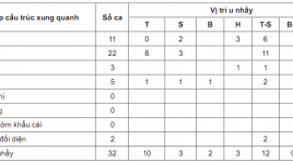
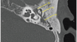
Nghiên cứu đắc điểm hình ảnh chụp cắt lớp vi tính trong chẩn đoán u nhầy mũi xoang
05/05/2021 17:27:41 | 1 binh luận
SUMMARY Objective: Our aim was to describe computed tomography image characteristic of paranasal sinus mucoccele. Method: A retrospective and prospective, axial-descriptive study in paranasal sinus mucoccele patients who were treated at National Otorhinorarynology from December 2016 to July 2019. Results: 32 patients were enrolled. Mean age was 52.9 (22-83) with M/F=1. Involved sinus distribution including 37.5% frontal-ethmoid, 31.3% frontal, 9.4% ethmoid, 6.3% ethmoid-maxillary, 6.3% sphenoid and 9.4% maxillary sinus. 96.9% tumors were hyperdense or isodense (compared to brain tissue) in pre-contrast CT Scanner. In the postcontrast image: 84.4% of tumors did not marked enhance while another 15.6% had rim enhance which could be explained due to patients clinical acute symtoms of infection. In characteristic, 87.5% tumors had erosion of sinus bone (65.6% lamina papiracea, 25% orbital roof and 25% ethmoidal roof). Regarding to the spread of mucocele: 68.75% tumors had intraorbital extension while 15.6% had intracranial extension. No record of nerve or cavernous sinus invasions. Conclusion: A sinus computed tomography scan with contrast material was highly valuable in diagnosis of paranasal sinus mucocele and contribute to the planning of surgery. Key words: paranasal sinus mucocele, computed tomography
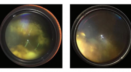
Đánh giá hiệu quả bước đầu điều trị u nguyên bào võng mạc bằng phương pháp truyền hóa chất qua đường động mạch
05/05/2021 12:48:16 | 0 binh luận
SUMMARY Retinoblastoma is the most common malignant tumor eye tumor in children which can cause blindness and even mortality if diagnosis and treatment are delayed. Intra-arterial chemotherapy (IAC) under the guidance of radiology is a new method for the treatment of intraocular retinoblastomas. Purpose: To evaluate the initial outcomes and safety of intra-arterial chemotherapy in treatment retinoblastoma. Materials and Methods: 15 patients diagnosed with intraocular retinoblastoma were clinically and subclinically examined, made imaging diagnosis (ultrasound and MRI), then define retinoblastoma stage by the 2003 international classification, whether or not it has been combine with other methods , and then is indicated for IAC, from October 2017 to June 2019. Each intervention is 3-4 weeks apart. The patients were followed immediately after treatment for the clinical symptoms and after 3-4 weeks after intervention to reassess the tumors and treatment results. Results: 15 patients (including 6 males and 9 females) corresponded to 15 study eyes with an average 35,5 ±20,8 age of months (11 months to 84 months). According to the International classification, there are 3 patients (20%) of group B retinoblastoma, 6 patients (40%) of group C retinoblastoma, 6 patients (40%) of group D retinoblastoma , no patients in group A and group E. The total number of tumors in 15 eyes is 27, which has been treated with intravenous chemotherapy combined with one or two local treatments (laser and cryotherapy) . The total number of interventions was 29, each patiens was treated from 1 to 3 times. As a result, 4 patients (26,7%) had good treatment results, 8 patients (53,3%) had average results, 3 patients (20%) had bad results, then had to enucleate; No patient were distant metastases or death during the follow-up. As a result, we saved 12/15 eyes at the risk of enucleation, 2/15 patients had complications of the treatment process, 1 patient had erythema of the eyelid and forehead skin area, 1 patient had choroidal - retinal atrophy due to occlusive vasculopathy. Conclusions : The method of intra- arterial chemotherapy in the treatment of retinoblastoma has promising, safe, and effective initial results in maximally salvaging the eye. Keyword: Retinoblastoma, Intra-arterial chemotherapy (IAC).
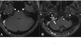
Vai trò của chuỗi xung 3D TOF MRA trong đánh giá rò động tĩnh mạch màng cứng nội sọ
05/05/2021 17:25:06 | 0 binh luận
SUMMARY Objective: We evaluate the role of 3D TOF MRA in diagnosis of the fistula location and cortical venous drainage of intracranial dural arteriovenous fistula (DAVF) in comparison with Digital Subtraction Angiography Subjects and methods: Prospective study between 1/2015 and 4/2019, 93 patients (35 male, 58 female), aged from 11 to 88 (mean 55), diagnosed of DAVF on conventional MRI with 3D TOF MRA and then underwent DSA for confirming the diagnosis. In three cases, 3D TOF MRA field of view is not enough to evaluate the fistula site but enough to evaluate the present of abnormal hyperintensity in cortical veins. Results: In our study, source images from 3D TOF MRA showed high sensitivity and positive predictive values (up to 100%, 97,6% respectively) in diagnosis of DAVF (n=90) and detected cortical venous drainage (n=93) with high specificity and high positive predictive value (100%). Cohen's Kappa coefficient showed very good agreement between 3D TOF MRA and DSA in detecting the location of DAVF. In 4 falsepositive cases, 1 case showed high-intensity area in the transverse-sigmoid venous sinous on 3D TOF MRA due to thrombosis and 3 other cases showed high-intensity area in the cavernous sinus causing misdiagnosis of DAVF. Conclusion: The use of 3D TOF MRA source images is valuable in diagnosing the location of fistulas and cortical venous drainage in intracranial DAVF. False-positive cases in this study suggested that MRA with contrast could ameliorate the limitation of 3D TOF MRA. Keywords: DAVF, dural arteriovenous fistula, cortical venous reflux, cortical venous drainage, 3D TOF MRA, DSA, false-positive.
Bạn Đọc Quan tâm
Sự kiện sắp diễn ra

THÔNG BÁO SINH HOẠT KHOA HỌC 19/01/2024: CẬP NHẬT CHẨN ĐOÁN VÀ ĐIỀU TRỊ UNG THƯ TRỰC TRÀNG
19-01-2024 09:47:01 0 bình luận

Thông tin đào tạo
- Những cạm bẫy trong CĐHA vú và vai trò của trí tuệ nhân tạo
- Hội thảo trực tuyến "Cắt lớp vi tính đếm Photon: từ lý thuyết tới thực tiễn lâm sàng”
- CHƯƠNG TRÌNH ĐÀO TẠO LIÊN TỤC VỀ HÌNH ẢNH HỌC THẦN KINH: BÀI 3: U não trong trục
- Danh sách học viên đạt chứng chỉ CME khóa học "Cập nhật RSNA 2021: Công nghệ mới trong Kỷ nguyên mới"
- Danh sách học viên đạt chứng chỉ CME khóa học "Đánh giá chức năng thất phải trên siêu âm đánh dấu mô cơ tim"


Đơn vị hợp tác

