


KẾT QUẢ ỨNG DỤNG XẠ HÌNH THẬN ĐỘNG TẠI BỆNH VIỆN NHI ĐỒNG THÀNH PHỐ
18/10/2023 11:47:33 | 0 binh luận
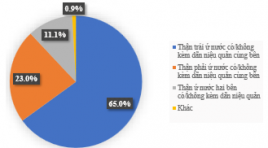
SUMMARY Background: Dynamic renal scan has become one of the most effective techniques to investigate the kidney parenchymal function, also the urine collecting and drainage system, in both adults and children. Compared with adults, it is the most commonly used test in children and accounts for more than half of the indications for nuclear medicine unit. In City Children's Hospital, dynamic renal scan has been implemented since the beginning of 2018 and initially showed its clearly role in facilitating the decision on treatment and follow-up strategy of pediatric patients. Therefore, we carry out this study to evaluate the results of dynamic renal scan application of the unit. Objective: To assess the results of application of dynamic renal scan with 99mTc-DTPA on pediatric patients in City Children's Hospital. Subject and method: We retrospectively examined data derived from 671 pediatric patients aged 01 month to 15 years in City Children’s Hospital who underwent at least 1 99Tc DTPA dynamic renal scan with diuretic challenge test from June 2018 to March 2022. The main purpose is to investigate some of the scintigraphic features related to Hydronephrosis in children and the prognostic factors of renal function loss. Results: There were a total of 740 performed scans, including 611 patients underwent once, 52 patients underwent twice, 7 patients underwent threetimes and 1 patient underwent fourtimes. Among them, there are 454 boys and 217 girls with a male/female ratio of 2/1. The age of the pediatric patients at the time of scanning varied, from 1 month to 15 years old. However, there is a clear dominant distribution for the group of patients under 5 years old, accounting for 516 cases (~69.7%%), which is more than 3 times the number for the group of patients aged 5 to 10 years and about 10 times for the group of patients over 10 years old, 175 cases (~23.7%) and 49 cases (~6.6%) respectively. Many different geographical locations in the country, where the patients come from, were noted. However the majority of cases live in Ho Chi Minh City, the number is 218 cases, accounting for ~30% of the total, followed by the west provinces and provinces from central region, as well as the Highlands area.*
KHẢO SÁT KÍCH THƯỚC KHUNG CHẬU Ở PHỤ NỮ VIỆT NAM TRƯỞNG THÀNH BẰNG CHỤP CẮT LỚP VI TÍNH
22/01/2024 16:36:43 | 0 binh luận
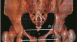
Mở đầu: Xác định các thông số khung chậu nữ bình thường hỗ trợ chẩn đoán bất xứng đầu chậu chính xác hơn, giảm tỷ lệ mổ lấy thai không cần thiết. Chụp cắt lớp vi tính (CLVT) có thể đo kích thước khung chậu. Mục tiêu : Xác định kích thước, các yếu tố ảnh hưởng ở người phụ nữ Việt Nam trưởng thành. Đối tượng và phương pháp nghiên cứu : Thực hiện cắt ngang mô tả trên 257 phụ nữ trưởng thành được chụp CLVT bụng - chậu; đo các kích thước và so sánh theo nhóm tuổi, tiền sử số lần sinh, chiều cao. Kết quả : Các kích thước trung bình của khung chậu: đường kính liên hợp sản khoa và ngang giữa là 11,8 ± 0,9 cm, 12,2 ± 0,9 cm; đường kính trước sau eo giữa và lưỡng gai là 11,2 ± 0,8 cm, 10,6 ± 0,8 cm. Các đường kính ở nhóm ≥ 40 tuổi nhỏ hơn khi so sánh với nhóm 30 – 39 tuổi và nhóm 18 -29 tuổi, p ≤ 0,05. Các đường kính eo giữa và đường kính trước sau eo dưới ở nhóm chưa sinh nhỏ hơn nhóm đã sinh, p ≤ 0,05. Các đường kính ở nhóm ≤ 145 cm nhỏ hơn nhóm > 145cm, p ≤ 0,05. Kết luận: Nghiên cứu đưa ra giá trị tham khảo cho các đường kính khung chậu phụ nữ Việt Nam trưởng thành, có thể hỗ trợ chẩn đoán bất xứng đầu chậu chính xác hơn. Từ khóa : Đường kính khung chậu nữ, CLVT

NHẬN XÉT MỐI LIÊN QUAN GIỮA ĐẶC ĐIỂM HÌNH ẢNH FDG PET/CT VỚI TÌNH TRẠNG ĐỘT BIẾN GEN EGFR Ở BỆNH NHÂN UNG THƯ PHỔI KHÔNG TẾ BÀO NHỎ GIAI ĐOẠN IV
22/01/2024 16:44:41 | 0 binh luận
TÓM TẮT Mục tiêu nghiên cứu : Nhận xét mối liên quan giữa đặc điểm hình ảnh FDG PET/CT với tình trạng đột biến gen EGFR ở bệnh nhân ung thư phổi không tế bào nhỏ giai đoạn IV. Đối tượng và phương pháp nghiên cứu : 108 bệnh nhân ung thư biểu mô không tế bào nhỏ của phổi giai đoạn IV được chụp FDG PET/CT, xét nghiệm EGFR trước điều trị từ 01/2018 đến 11/2020. Kết quả : Độ tuổi trung bình 62,1±9,2 (37 - 83), tỷ lệ nam (61,1%) nữ (38,9%), tiền sử hút thuốc lá (39,8%), không hút thuốc lá( 60,2%), giai đoạn IVa (31,5%) và giai đoạn IVb (68,5%) Tỉ lệ đột biến gen EGFR và tỷ lệ không có đột biến tương ứng 52,8% và 47,2% trong đó đột biến exon 19 và exon 21 là cao nhất với tỷ lệ là 31,5% và 18,5%. 48,1% bệnh nhân điều trị hoá trị 35,2% bệnh nhân điều trị đích. Giới, tiền sử hút thuốc, pSUVmax, kích thước u là bốn yếu tố độc lập dự báo đột biến gen EGFR với OR lần lượt là 0,19 (KTC 95%:0,08-0,45; p<0,001); 0,347 (KTC 95%: 0,156-0,770; p=0,009) 0,805 (KTC 95%:0,722-0,899; p<0,001) và 0,782 (KTC 95%:0,645–0,947; p=0,012). Kết luận : FDG PET/CT có giá trị dự đoán mức độ đột biến EGFR ở bệnh nhân ung thư biểu mô không tế bào nhỏ của phổi. Từ khoá : Ung thư phổi, biểu mô tuyến, FDG PET/CT, dự đoán, đột biến EGFR
ĐẶC ĐIỂM XÉT NGHIỆM VÀ HÌNH ẢNH CỘNG HƯỞNG TỪ TUYẾN YÊN TRÊN BỆNH NHÂN MẮC HỘI CHỨNG CUSHING PHỤ THUỘC ACTH
23/01/2024 11:48:10 | 0 binh luận
TÓM TẮT Mục tiêu : Khảo sát hình ảnh tuyến yên trên cộng hưởng từ ở những bệnh nhân mắc hội chứng Cushing phụ thuộc ACTH. Đối tượng và phương pháp nghiên cứu: Nghiên cứu mô tả cắt ngang 15 bệnh nhân (12 nữ, 3 nam) được chẩn đoán mắc hội chứng Cushing phụ thuộc ACTH và được chụp cộng hưởng từ tuyến yên từ tháng 10/2022 đến tháng 10/2023. Kết quả nghiên cứu: Tỷ lệ nam/nữ = 1/4. Tuổi trung bình 38,47 ± 10,78 (27-62). Tất cả bệnh nhân đều có nồng độ ACTH huyết tương >10pg/ml, phù hợp với chẩn đoán lâm sàng hội chứng Cushing phụ thuộc ACTH. Bệnh nhân quan sát thấy u tuyến yên trên cộng hưởng từ chiếm 80%. Kích thước u trung bình là 4,59 ± 1,77mm, 100% u là microadenoma, phân loại độ I, giai đoạn 0 theo phân loại của Hardy-Wilson. Nhóm u đồng tín hiệu trên các chuỗi xung T1W và T2W chiếm tỷ lệ cao nhất, lần lượt là 83,4% và 58,3%. Trên chuỗi xung tiêm thuốc động học, tất cả u biểu hiện là nốt ngấm thuốc kém. Kết luận: Hình ảnh u tuyến yên trên cộng hưởng từ rất đa dạng, đặc biệt là đặc điểm tín hiệu của u. Việc đánh giá kết hợp trên chuỗi xung động học sau tiêm thuốc đối quang từ là cần thiết để chẩn đoán đối với những trường hợp u kích thước nhỏ và đồng tín hiệu trên các chuỗi xung thường quy. Từ khóa: cộng hưởng từ, u tuyến yên, hội chứng Cushing phụ thuộc ACTH, microadenoma, phân loại Hardy-Wilson
ĐẶC ĐIỂM TẬP TRUNG 18FDG CỦA TỔN THƯƠNG U, HẠCH TRÊN PET/CT Ở BỆNH NHÂN UNG THƯ PHỔI KHÔNG TẾ BÀO NHỎ CÓ CHỈ ĐỊNH PHẪU THUẬT TRIỆT CĂN TẠI BỆNH VIỆN U BƯỚU HÀ NỘI
18/10/2023 11:55:43 | 0 binh luận
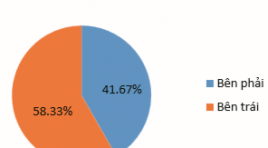
SUMMARY Purpose: To review some clinical features, characteristics of 18FDG uptake of tumors and lymph nodes on PET/CT in NSCLC patients with indications for radical surgery at Hanoi Oncology Hospital. Subjects and methods: 82 patients with primary NSCLC were taken with 18FDG PET/CT before surgery. Results: Right lung tumor 64.6%, left lung tumor 35.4%. The average size of the tumor was 2.6 ± 1.0 cm, the patient with lymph node (+) on PET/ CT had an average lung tumor size of 3.3 ± 0.9cm; larger than patients with N0 lymph nodes (p < 0.05). The mean SUVmax of lung tumors was 6.0 ± 4.5 and increased with tumor size (positive correlation, r=0.58). U ≤2cm, SUVmax = 4.1 ± 2.1. U >2-3cm, SUVmax = 5.3 ± 3.6 and U >3 -5cm, SUVmax = 8.3 ± 4.9. SUVmax increases with clinical disease stage and is higher in patients with positive lymph nodes on PET/CT. Conclusion: 18FDG PET/CT plays an important role in the diagnosis of NSCLC. SUVmax is a quantitative parameter related to tumor size, lymph node status and clinical disease stage. Keywords: NSCLC, 18FDG-PET/CT, SUVmax.
ĐÁNH GIÁ HIỆU QUẢ BAN ĐẦU ĐIỀU TRỊ THOÁT VỊ ĐĨA ĐỆM BẰNG TIÊM OZONE ĐĨA ĐỆM QUA DA VÀ PHONG BẾ RỄ BẰNG OZONE KẾT HỢP CORTICOID DƯỚI HƯỚNG DẪN CỦA CẮT LỚP VI TÍNH
18/10/2023 10:11:02 | 0 binh luận
SUMMARY Background: back pain and sciatica caused by disc herniation has a burden upon social activity. The newly minimally invasive technique, percutaneous Ozone (O2-O3) intradisal injection procedure has demonstrated safe and effective in long terms. Purpose : Evaluate the clinical effectiveness of intradiscal Ozone injection combining periradicular injection of Ozone and steroid under CT guidance for the treatment of lumbar disc herniation. Material and method: Prospective study, 100 patients symtomatic (lumbar pain, sciatica) with mild/moderate lunbar disc bulging or herniation on MRI. The patients were treated with intradiscal Ozone injection combining periradicular injection of Ozone and steroid under CT guidance. Clinical outcomes and MRI images were reviewed to evaluate at 3 months and 6 months. Results: The VAS score and the Oswestry Disability Index (ODI) before and after treatment 3months, 6 months were significant reduction. The mean improvement was 4.7 for VAS and 14 for ODI. All the procedures were technically successful. There were no adverse events associated with the treatment. Conclusion: Intradiscal injection Ozone treatment of herniated disc is an minimally invasive, easy, safe and effective procedure with low complications and side effects. Keywords: lumbar disc herniation, O3 injection
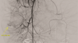
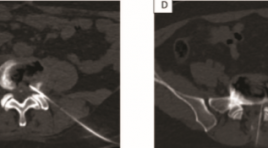
NGHIÊN CỨU BƯỚC ĐẦU KẾT QUẢ CHỤP VÀ NÚT MẠCH TRONG ĐIỀU TRỊ CHẢY MÁU TIÊU HÓA DƯỚI
17/10/2023 17:15:40 | 0 binh luận
SUMMARY Background: Acute lower gastrointestinal bleeding (ALGB) is an urgent, potentially life-threatening emergency, especially in case endoscopy is not identifiable or uncontrollable in management bleeding. Following the development of technology and applying minimal invasive modalities, endovascular treatment is more and more to be applied. Primary endpoint: Figure out the rate of technical success, the clinical success and the complication relating to bowel ischemia of embolization in ALGB. Secondary endpoint: Figure out the rate of negative bleeding finding angiograms in diagnosis of ALGB Method: Retrospective cohort study Results: From 01/2019 to 01/2022, 23 embolisms were performed in total 35 procedures of 24 patients. There were 12 angiographies with negative bleeding finding result (34.3%). In embolization procedure, first embolisms were 21/23 and two procedures were re-embolism. The rate of technical success after first embolisms was 95.2% and clinical success reaches 76.2%. There was no major complication relating to bowel ischemia. Conclusion: Transcatheter angiographic embolization is a safe and effective management for acute lower gastrointestinal bleeding with minimal invasive. Keywords : acute lower gastrointestinal bleeding, embolization, endovascular
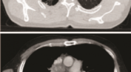
BÁO CÁO CA BỆNH: SINH THIẾT U PHỔI DƯỚI HƯỚNG DẪN SIÊU ÂM
17/10/2023 16:42:23 | 0 binh luận
SUMMARY The ultrasound-guided transthoracic biopsies are indicated for pulmonary masses or mediastinal masses located in the periphery adjacent to the pleural-thoracic surface and pleural lesions. We present a case where the lung tumor was anterior to the scapula with emphysema. Keywords: Transthoracic biopsy, Ultrasound-guided transthoracic biopsies, lung tumor
Bạn Đọc Quan tâm
Sự kiện sắp diễn ra

THÔNG BÁO SINH HOẠT KHOA HỌC 19/01/2024: CẬP NHẬT CHẨN ĐOÁN VÀ ĐIỀU TRỊ UNG THƯ TRỰC TRÀNG
19-01-2024 09:47:01 0 bình luận

Thông tin đào tạo
- Những cạm bẫy trong CĐHA vú và vai trò của trí tuệ nhân tạo
- Hội thảo trực tuyến "Cắt lớp vi tính đếm Photon: từ lý thuyết tới thực tiễn lâm sàng”
- CHƯƠNG TRÌNH ĐÀO TẠO LIÊN TỤC VỀ HÌNH ẢNH HỌC THẦN KINH: BÀI 3: U não trong trục
- Danh sách học viên đạt chứng chỉ CME khóa học "Cập nhật RSNA 2021: Công nghệ mới trong Kỷ nguyên mới"
- Danh sách học viên đạt chứng chỉ CME khóa học "Đánh giá chức năng thất phải trên siêu âm đánh dấu mô cơ tim"


Đơn vị hợp tác

