


TỰ TỐNG XUẤT DỊ VẬT (ĐINH SẮT NHỌN) TỪ PHẾ QUẢN THÙY GIỮA BÊN PHẢI BÁO CÁO TRƯỜNG HỢP HIẾM GẶP
17/10/2023 16:06:58 | 0 binh luận
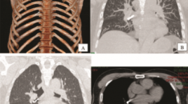
SUMMARY Tracheobronchial foreign body aspiration is an uncommon but potentially life-threatening event in adults. Symptoms typically consist of a choking event followed by cough and dyspnea. Chest radiography and computed tomography can provide information regarding the location and characteristics of foreign bodies hence aiding in diagnosis. Bronchoscopy remains the gold standard for diagnosis and management of this condition. Foreign body spontaneous expulsion rarely occurs. We present a case of 61-year-old male patient who spontaneously expulsed a sharp nail from right bronchus. Keywords : spontaneous expulsion, tracheobronchial, nail, foreign body, aspiration.
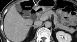
ĐÁNH GIÁ KẾT QUẢ CỦA DẪN LƯU Ổ DỊCH QUA DA TRONG ĐIỀU TRỊ VIÊM TUỴ CẤP NẶNG
13/10/2023 15:35:24 | 0 binh luận
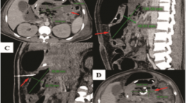
SUMMARY Objectives: Evaluating the result and complications of percutaneous drainage under the guidance of imaging in the treatment of severe acute pancreatitis. Subjects and methods: A total of 51 patients diagnosed with severe acute pancreatitis at Bach Mai hospital from 7/2021 to 7/2022 were treated by percutaneous drainage. Results: 51 patients included 41 men and 10 women, with the age from 27 to 72 years old, the success rate of drainage technique is 97,3%, the mortality rate of patients in the study is 7,8% and there were no cases of complications after drainage. Clinical and laboratory symptoms were significantly reduced compared with pre-drainage. Conclusions: Percutaneous drainage under the radiology guidance with ascite, inflammatory fluid in acute pancreatitis is a safe technique with a high success rate. Keywords : severe acute pancreatitis, percutaneous drainage
ĐÁNH GIÁ ĐẶC ĐIỂM HÌNH ẢNH CỦA CẦU CƠ ĐỘNG MẠCH VÀNH BẰNG CHỤP CLVT 256 DÃY
13/10/2023 11:00:20 | 0 binh luận
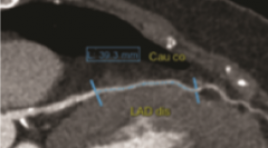
SUMMARY Purpose: to evaluate and analyze the ratio of position, depth and length of the coronary myocardial bridge, the status of pre-myocardial atherosclerosis and the degree of vascular stenosis during systole by multislice-computed tomography (MSCT) images series. Material and method : Computed tomography scan for 175 patients using a 256-row MSCT machine at Hanoi Friendship Hospital between July 2021 and August 2022. Select the best quality images in systole and diastole. Measure the length and thickness of the bridge. For each myocardial bridge segment, assess for the presence of anterior atherosclerotic plaque no more than 2 cm in length of the artery. Assess the degree of vascular stenosis during systole caused by the myocardial bridge. Result: Most of the myocardial bridges were located in the anterior interventricular artery (LAD) (98.3%). The average values of the length and thickness of the bridge are 23.1 ± 10.5mm and 1.0 ± 1.0mm. Atherosclerosis was detected in 77 cases (accounting for 43%). The average degree of tunneling artery stenosis was 23.0% systolic. Conclusion: MSCT 256 series has a high value in detecting bridges, determining the location, there by measuring the length, the thickness of the pons, the status of anterior atherosclerotic plaques as well as the degree of narrowing of the vessel lumen systole of the musculature. Keywords: myocardial bridge, coronary artery, multislice computed tomography.
ĐÁNH GIÁ HIỆU QUẢ ĐIỀU TRỊ THOÁT VỊ ĐĨA ĐỆM CỘT SỐNG THẮT LƯNG BẰNG TIÊM NGOÀI MÀNG CỨNG QUA LỖ LIÊN HỢP DƯỚI HƯỚNG DẪN CỦA CẮT LỚP VI TÍNH
12/10/2023 15:02:23 | 0 binh luận
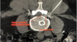
SUMMARY Objective: Evaluation of the transforaminal epidural steroid injection for the treatment of lumbar disc herniation under computed tomography-guided Subject and method : 80 patients with lumbar disc herniation were diagnosed at 108 Central Military Hospital from June 2020 to June 2021. Longitudinal follow-up and prospective study Results : Location of transforaminal L3-L4 35%, L4-L5 58 %, L5S1 7%. VAS score before intervention, 1 day, 14 days, 1 month and 3 months after intervention were 6,48 ± 1,46; 3,21 ± 1,53; 2,74 ± 1,37; 2,55 ± 1,44; 2,75 ± 1,39 and ODI score before intervention, 1 day, 14 days, 1 month and 3 months after intervention were 59,26 ± 12,18; 35,68 ± 7,97; 33,63 ± 6,14; 30,18 ± 6,01; 28,15 ± 6,46. Statistically signficant difference Conclusion: Transforaminal epidural injection is an effective method of pain management and functional improvement for patients with lumbar disc herniation Keywords: Lumbar disc herniation, epidural injection, transforaminal
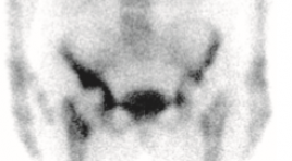
ĐÁNH GIÁ VAI TRÒ CỦA XẠ HÌNH XƯƠNG BA PHA VỚI MÁY SPECT/CT TRONG THEO DÕI BỆNH NHÂN SAU THAY KHỚP NHÂN TẠO TẠI BỆNH VIỆN ĐÀ NẴNG
12/10/2023 10:39:14 | 0 binh luận
SUMMARY Objectives: Describe the clinical and subclinical characteristics of patients after joint replacement who underwent three-phase bone scan at Danang Hospital and evaluation of the role of three-phase bone scan method with SPECT/CT in monitoring patients after joint replacement. Study subjects: 32 patients after joint replacement who underwent three-phase bone scan with SPECT/CT Hawkeye 4 produced by GE at the Department of Medicine, Danang Hospital. Methods of study : Cross-sectional descriptive, longitudinal monitoring, retrospective and prospective. Results: Mean age: 69.88 ± 7.80. Female/male ratio: 23/9. Pain is the most common symptom. The white blood cell count and CRP concentration were mostly incresed. There were no abnormal changes in the X-ray picture. The image of joint loosening accounts for the highest proportion. Most of the knee scintigraphy shows abnormalities. Sensitivity: 83.33%. Specificity: 75%. Accuracy: 81.25%. Positive predictive value: 90.91%. Negative predictive value: 60%. Conclusions: Three-phase bone scintigraphy is a simple method making it easy to perform with its high sensitivity and positive predictive value in monitoring patients after artificial joint replacement. Keywords: Three-phase bone scan, artificial joints, joint loosening.
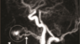
BƯỚC ĐẦU PHÂN TÍCH KẾT QUẢ ỨNG DỤNG KỸ THUẬT MRA TWIST TRONG ĐÁNH GIÁ DỊ DẠNG THÔNG ĐỘNG - TĨNH MẠCH NGOẠI BIÊN
12/10/2023 10:32:28 | 0 binh luận
SUMMARY Background: TWIST is a MRA with high time resolution, which can display the mapping of vascular structure well. The identification of the feeding and draining vasculature of peripheral arteriovenous malformation (pAVM) is important for the appropriate treatment planning. Therefore, we examined the application of MRA TWIST in evaluation pAVM in comparison with Digital Subtraction Angiography (DSA). Objective: To evaluate the feeding arteries and draining veins of pAVM with MRA TWIST in comparison with DSA. Subjects and methods: A retrospective study between January 2016 and July 2021 was conducted upon 25 patients (7 males and 18 females; mean age 22,2; age range 3-53 years) with pAVM, who obtained MRA TWIST and then underwent DSA for confirming the diagnosis. The numbers and names of the feeding arteries and early draining veins were evaluated by two independent readers. The accurate ratio and Kappa test for the interobserver agreement were calculated. Results: About the feeding arteries, MRA TWIST accurately evaluated 82,6% in head and neck, and 85,7% in limbs in comparison with DSA. About the draining veins, MRA TWIST accurately evaluated 84% in comparison with DSA. Kappa coefficient showed good agreement in detecting the feeding arteries and the draining veins between MRA TWIST and DSA. Conclusion: MRA TWIST is a reliable non-invasive imaging technique for evaluation of the feeding arteries and the draining veins of pAVM and can be useful for planning the endovascular treatment. Keywords: peripheral arteriovenous malformation (pAVM), MRA TWIST, digital subtraction angiography (DSA).
NGHIÊN CỨU ĐẶC ĐIỂM HÌNH ẢNH UNG THƯ DẠ DÀY TRÊN MÁY CHỤP CẮT LỚP VI TÍNH 256 DÃY
16/11/2021 11:27:45 | 0 binh luận
SUMMARY Purpose: This report aimed to assess the describing imaging characteristic of gastric cancer on 256 - Slice Multidetector Computed Tomography gastrography. Materials and methods: 256 - slice multidetector CT gastrography in 31 patients with histopathological findings of gastric cancer from 2020 Fer to 2021 August at Huu Nghi hospital. Results: Gastric cancer is often detected in old people. The most common position is the antrum of gastric (71%), the least common position is the cardia - fundus of gastric (3,2%).There is a correlation between diameter of tumors and T staging (p< 0,05). There are correlation between the shape, short diameters of nodes and LN metastasis (p< 0,05). Short diameters of nodes is ≥ 8 mm and round shape of nodes were moderate diagnostic ability indicator for LN metastasis in gastric cancer. Conclusion: 256 slice - MDCT gastrography is an effective method and non-invasion offers improved detected gastric cancer and evaluates lymph node metastasis. Keyword: Gastric cancer,256 slice - MDCT,short diameters of Lymph node metastasis.
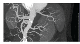
VAI TRÒ CỦA CHỤP CẮT LỚP VI TÍNH TRONG ĐÁNH GIÁ MẠCH MÁU THẬN CỦA NGƯỜI CHO THẬN SỐNG TRƯỚC PHẪU THUẬT GHÉP THẬN
16/11/2021 10:06:25 | 0 binh luận
SUMMARY Background: In laparoscopic donor nephrectomies, it is important to understand the exact anatomy of the vascular structures during minimally invasive surgery. The aim of study: to determine the accuracy of MDCT to predict vascular anatomy in living kidney donors and to reveal the prevalence of vascular variations in a VietNam population. Materials and methods: This is a retrospective cross-sectional study. One hundred and eleven living donors were included in this study, who had MDCT for the assessment of their renal vessels and laparoscopic surgery in Cho Ray hospital between February 2020 to April 2021. The initial CT results were compared with the surgical findings and repeated review sessions of CT scans were performed to determine the causes of mismatches in discordant cases. Results: The accuracy of MDCT was 98,2% to predict the number of renal vessels. One artery was missed during the initial CT interpretation due to perception error. One case is false positive. The accuracy of MDCT was 95,5% to predict the early branching of a renal artery and late confluence of a renal vein variation. The prevalence of multiple renal arteries and veins, early branching of a renal artery and late confluence of a renal vein were 20,7%, 6,8%, 13,5%, 19,8%. One case (0,9%) each of a retroaortic left renal vein and a circumaortic left renal vein were found. Conclusion: Multidetector computed tomography is a reliable technique in preoperative renal anatomy evaluation in live renal donors. Key word: living donor kidney, multidetector computed tomography (MDCT) *
Bạn Đọc Quan tâm
Sự kiện sắp diễn ra

THÔNG BÁO SINH HOẠT KHOA HỌC 19/01/2024: CẬP NHẬT CHẨN ĐOÁN VÀ ĐIỀU TRỊ UNG THƯ TRỰC TRÀNG
19-01-2024 09:47:01 0 bình luận

Thông tin đào tạo
- Những cạm bẫy trong CĐHA vú và vai trò của trí tuệ nhân tạo
- Hội thảo trực tuyến "Cắt lớp vi tính đếm Photon: từ lý thuyết tới thực tiễn lâm sàng”
- CHƯƠNG TRÌNH ĐÀO TẠO LIÊN TỤC VỀ HÌNH ẢNH HỌC THẦN KINH: BÀI 3: U não trong trục
- Danh sách học viên đạt chứng chỉ CME khóa học "Cập nhật RSNA 2021: Công nghệ mới trong Kỷ nguyên mới"
- Danh sách học viên đạt chứng chỉ CME khóa học "Đánh giá chức năng thất phải trên siêu âm đánh dấu mô cơ tim"


Đơn vị hợp tác

