


Trường hợp lâm sàng: Điều trị phình khổng lồ động mạch cảnh trong bằng STENT thay đổi dòng chảy
15/04/2020 20:50:10 | 0 binh luận
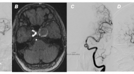
Treatment of giant intracranial aneurysm using flow - divertion stent: a case report SUMMARY We reported a difficult case of treatment of internal carotid artery giant aneurysmby using flow - diversion stent, FRED. The patient was completely recovered of clinical status and there was not the appearance of internal carotid artery aneurysm on digital subtraction angiography after 10 - month follow-up. Using flow - diversion stent is a new technique, may be effectively alternative method compared to conventional aneurysmal coiling, especially in treatment of intracranial giant aneurysmsor aneurysms at difficult accessing location. Key words: Giant intracranial aneurysm, flow - diversion stent, endovascular therapy
Ung thư biểu mô tuyến nhầy của ruột thừa - Báo cáo một trường hợp hiếm gặp và tổng kết trên y văn
15/04/2020 20:46:24 | 0 binh luận
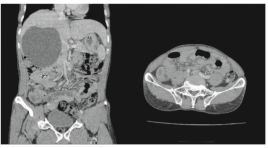
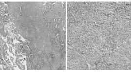
Mucinous cystadenocarcinoma appendix: a case report and review literature SUMMARY Background :Mucinous cystadenocarcinoma of the appendix is a rare disease. Detection situation is often presented with appendicitis symptoms. The appendectomy was underwent andthe result of mucinous cysadenocarcinoma appendix wasconfirmed onhistopathology. The patient is evaluated for staging to decide the next course of treatment. We report a case of mucinous cysadenocarcinoma appendix diagnosed and treated at Bach Mai Hospital and compared with the literature. Clinical case : A male patient, 65 years, admitted Bach Mai hospital because lower right quadrant. Patient was performed abdominal ultrasound and contrast enhanced computed tomography (CLVT) showed a 27mm appendix diameter, thickened around the perimeter of 7mm and lost the gastrointestinal tract, with little fluid around the appendix. Patients diagnosed before surgery: Appendicitis follow lymphoma. The patient underwent an appendectomy. Pathology results: Mucinous cystadenocarcinoma appendix. Conclusion :Mucinous cystadenocarcinoma appendix is a rare disease, clinical presentation and imaging is non-specific. Diagnosis mainly depends on pathology, then patient is evaluated to determine stage and decidednext course of treatment. Key word: Mucinous cystadenocarcinoma appendix.
Tụy lạc chỗ tại ruột non với biến chứng viêm hoại tử ruột - báo cáo một trường hợp hiếm và tổng kết trên y văn
04/12/2019 20:50:29 | 0 binh luận
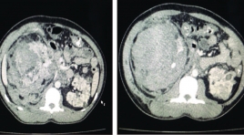
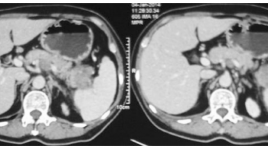
Ectopic pancreas in the wall of intestine complicated with necrotic and inflamed intestine: A case report and review literature SUMMARY Background: Ectopic pancreas is a rare congenital condition characterized by pancreatic tissues located outside normal of confines of pancreas and lacking any anatomic or vascular connection with main pancreas. It can occur anywhere in the gastrointestinal tract but rarely are found in small intestine. Its preoperative diagnosis is difficult because the clinical symptoms are often nonspecific. We introduce a case of ectopic pancreas in the intestinal wall complicated with necrotic and inflamed intestine, which received a treatment by resection. Case presentation : A 44 years old man attended to Bach Mai hospital due to acute abdominal pain in epigastrium as result of gastrointestinal perforation. Contrast enhanced computed tomography (CT) of abdomen showed a mesenteric mass surrounded by inflamed fat in the left lower quadrant abdomen. In addition, CT images also suggested necrosis of the bowel wall next to the mass caused by twisting the mesentery and mesenteric vessels (whirlpool sign). The patient underwent local surgical resection and following histology revealed ectopic pancreatic tissues in the wall of intestine and necrosis of intestine. Conclusion : Although ectopic pancreas is rare, it should be considered in the differential diagnosis of a mesenteric or intestinal mass surrounded by necrotic and inflamed intestine. Keyword: Ectopic pancreas, mesenteric mass, intestinal mass, whirlpool sign.
Nghiên cứu giá trị siêu âm trong chẩn đoán teo mật bẩm sinh ở trẻ < 4 tháng
26/03/2020 22:51:55 | 0 binh luận
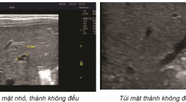
Value of Ultrasound in diagnosis of biliary atresia in infants younger than 4 months old SUMMARY Purpose: To evaluate prospectively the value of ultrasonography (US) in the diagnosis of biliary atresia (BA), with surgery as the reference standard. Material and Methods : 98 fasting infants (< 4 months old) with jaundice, acholic stools and conjugated hyperbilirubinemia underwent detailed US studies performed by an experienced pediatric radiologist with a 5MHz curvilinear transducer and a 7.5MHz linear-array transducer. The following features were prospectively recorded: gallbladder morphology, size and contraction, triangular cord sign. The radiologist was blinded to results of other investigations. Sensitivity, specificity, and positive and negative predictive values were calculated for each US variable. BA and non-BA groups were compared by means of the Fisher exact test for categorical variables and an unpaired t test for continuous variables. Result : Forty infants had surgically confirmed BA, and 58 had other documented causes of neonatal jaundice; the mean ages at US assessment were 57 and 58 days, respectively (P>0,5). Seven US features showed a significant difference between BA and non-BA groups (P< 0,01, Fisher exact test). The features with the greatest individual sensitivity and specificity, respectively, in the diagnosis of BA were triangular cord sign (87,5% and 94,8%), abnormal gallbladder wall (87,5% and 89,6%) and no contraction (90,6% and 89,6%).The gallbladder was significantly smaller in infants with BA than in those without BA (15,4mm vs 22,5 mm in length, P <0,01). Conclusion : US is valuable in diagnosis of biliary atresia if patients fast enough. Multiple US features should be used to increase the accuracy of the diagnosis Abbreviation : US: Ultrasound BA: Biliary Atresia TC: Triangular Cord Key word: Billiary atresia, Triangle Cord
Nhân một trường hợp u đặc giả nhú của tụy
01/04/2020 16:29:52 | 0 binh luận
Solid pseudopapillary tumor of the Pancreas SUMMARY Solid pseudopapillary tumors of the pancreas are rare in children. We present a 7-year-old girl with clinical presentation of intermittent abdominal pain after meal, no vomiting for 6 months. The abdominal ultrasound and computed tomography showed a mass that measuring 6,5x6,8x7 cm at the epigastric area. Total resection of the tumor were performed. The definite diagnosis was made by the post-operation pathological finding.
Nhân một trường hợp u lympho tụy được chụp và theo dõi trên PET/CT tại bệnh viện Việt Đức
01/04/2020 13:01:01 | 0 binh luận
Pancreatic lymphôma – case report SUMMARY Pancreatic Lymphôma is most commonly a B-cell sub-type of non-Hodgkin lymphôma and is classified as either primary or secondary. We describe a case secondary Pancreatic lymphôma in a 64-year-old woman who presented with abdominal pain and weight loss for one month. Her laboratory tests upon admission were as follows : white blood cell count 9.6 G/l (reference range: 4,0-10,0), red blood cell 4.5 T/l (reference range: 3.8-5.8. The tumor marker levels of CEA; CA 125, CA 15-3, CA 19-9, αFP were all within the normal range. Abdominal ultrasound usually shows only a spenlic mass and many lymphô node in the abdomen. Gastroscopy and colonoscopy were normal. Abdominal multi slice computed tomography (MSCT) revealed a mass in the pancreatic body - tail and splenn suggest infarct lesions. There were many lymphadenopathies located at the portal hilus, para-aortic region and left renal hilus. PET/CT showed all lesions on the MSCT increased metabolic activity with maximal standardized uptake values (SUVmax) ranging from 8.9 to 17.0. Laparoscopy biopsy of pancreatic mass be performed to establish the diagnosis. In reports was extranodal marginal zone B cell lymphôma. The PET/CT after treatment 2 months suggest the disease respond well to treatment.
Báo cáo trường hợp ca lâm sàng u lympho nguyên phát tại màng não
01/04/2020 12:53:29 | 0 binh luận
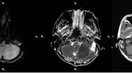
SUMMARY Diffuse large B-cell lymphoma (DLBCL) presented as a primary dural lesion is an extremely rare, which may be misdiagnosed as meningioma. We report the case of DLBCL presented as primary dural lesion. A 42 – year –old man with headache, MRI of brain showed a lesion occipital region, tracking the dural matter. The patient was treated with tumor resection and the diagnosis of DLBCL was established. Thus, intracranial dural DLBCL must be considered in differential diagnosis of meningeal lesions. Keywords: Primary dural lymphoma, Diffuse large B-cell lymphoma.
Nhân một trương hợp tràn dịch màng phổi trong bệnh cảnh xơ gan ở trẻ em
01/04/2020 09:43:23 | 0 binh luận
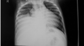
Hepatic hydrothorax in infancy - Case report
Bạn Đọc Quan tâm
Sự kiện sắp diễn ra

THÔNG BÁO SINH HOẠT KHOA HỌC 19/01/2024: CẬP NHẬT CHẨN ĐOÁN VÀ ĐIỀU TRỊ UNG THƯ TRỰC TRÀNG
19-01-2024 09:47:01 0 bình luận

Thông tin đào tạo
- Những cạm bẫy trong CĐHA vú và vai trò của trí tuệ nhân tạo
- Hội thảo trực tuyến "Cắt lớp vi tính đếm Photon: từ lý thuyết tới thực tiễn lâm sàng”
- CHƯƠNG TRÌNH ĐÀO TẠO LIÊN TỤC VỀ HÌNH ẢNH HỌC THẦN KINH: BÀI 3: U não trong trục
- Danh sách học viên đạt chứng chỉ CME khóa học "Cập nhật RSNA 2021: Công nghệ mới trong Kỷ nguyên mới"
- Danh sách học viên đạt chứng chỉ CME khóa học "Đánh giá chức năng thất phải trên siêu âm đánh dấu mô cơ tim"


Đơn vị hợp tác

