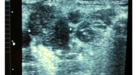
Nghiên cứu giá trị siêu âm và siêu âm hướng dẫn chọc hút tế bào bằng kim nhỏ hạch di căn trong ung thư thực quản
02/06/2020 17:26:05 | 0 binh luận
summary urpose : To survey ultrasonographic characteristics suggested benign and malignant cervical lymph node metastasized from EC methods (esophagus cancer). To find out the values of FNA in diagnosis of metastatic cervical lymph node from EC. Materials and Methods: 20 patients were diagnosed EC and ultrasound detected suspiciously malignant cervical lymph node, from 4/2012 to 6/2013. The patients were surveyed: Mode 2D, FNA, cytopathology in Hue Central Hospital. We divided the diagnosis into 2 groups: benign and malignant group. Results: In 20 patients with cytology, 8 cases are diagnosed malignant lymph node metastasized from EC. The remains, 12 patients are diagnosed with benign lymph node: lymphadenitis. In our research, the signs suggest malignant lymph node in sonography: hypoechonic, heterogeneous, loss of the fatty hyperechoic hilum, ill-defined border, size about 10mm in diameter (short axis), round-shaped, greater numbers. Se= 80%, Sp= 100%, PPV= 100%, no false negative cas. Conclusion: The combination of ultrasound and FNA is valuable in the diagnosis of suspected malignant lymph node in the neck, metastasized from EC. It is a safe and simple method for selected patients which allows diagnosis and staging in a single step.
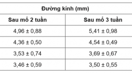
Nghiên cứu đặc điểm siêu âm đường thông động tĩnh mạch bên tận ở cẳng tay trên bệnh nhân suy thận lọc máu chu kỳ
30/03/2020 21:59:47 | 0 binh luận
Ultrasound characteristics of side-to-end arteriovenous fistula at forearm in chronic renal failureforperiodical hemodialysis SUMMARY Objective: To assess ultrasound characteristics of radiocephalic fistula at 2 and 3weeksafter operation in chronic renal failure with periodical hemodialysis. Methods: This study was conductedon 34 patients with indication of periodical hemodialysis due to chronic renal failure from April, 2016 to July, 2017 at Hemodialysis Department in Hue Central Hospital. These patients were operated to createside-to-end radiocephalic fistula at forearm and examed with ultrasound at week 2 and 3after operation. Results : The mean age was 45,79 ± 14,59 (mean ± SD); 47,10% male and 52,90% female patient. Diabetes was presented in 5,88% of the patients. The mean diameter of vein was 4,96 ± 0,88 mm at week 2 and 5,40 ± 0,99 mm at week 3 (p < 0,05);venous flow volume was 531,33 ± 162,40 ml/p and 666,56 ± 260 ml/p at week 2 and 3, respectively (p < 0,05). The rate of mature fistula at week 3 after operation was 82,35%. Most of the fistula complication was stenosis at the arterio-venous anastomosis and the draining veins. Conclusion : Ultrasound can assess the flow through arteriovenous fistula, detect several early complications which cause abnormal fistulas and help formanagement and treatmentof patients. Key word: radio-cephalic fistula, periodical hemodialysis.
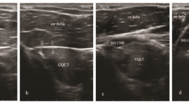
Siêu âm với tần số cao khảo sát phân nhánh thần kinh vận động của thần kinh cơ bì chi phối cơ nhị đầu cánh tay: Từ vị trí tách nhánh đến phân bố trong cơ
30/03/2020 22:19:02 | 0 binh luận
High-frequency ultrasonography for the motor branchesof the musculocutaneous nerve innervating biceps brachii: from the branching location to the distribution in the muscle SUMMARY Purpose: The aim of this study was to investigate the ability of high-frequency ultrasonography in examing the motor branches of the musculocutaneous nerveinnervating biceps brachii in the correlation with anatomical and histological knowledge. We analysed the location where they exit the main nerve trunk, penetrate the muscle epimysium and distribute inside the muscle. Methods: Sixteen healthy volunteers (eight males and eight females, ages 20-60, mean age 35) were examined on both sides of the musculocutaneous nerves and their branches innervating biceps brachii. The 5-18 MHz and 16-23 Mhz multi-frequency transducers along with the latest high-resolution ultrasound systems were used to examine the musculocutaneous nerves slowly and continuously in cross section from the coracoid process of the scapula to the elbow. By analyzing the nerve bundles inside the musculocutaneous nerve and the epimysium of biceps brachii, we observed the position where one nervebranch separated from the main trunk of the nerve, penetratedthe epimysium and distributedinside the muscle.Blood vessels were distinguished with nerves by Doppler ultrasound and compression method. Results: One right arm of a 28-year-old womanwas found with the absence of the musculocutaneous nerve and the median nerve give the motor branches to the biceps brachii. Thirty one musculocutaneous nerves and their motor branches to biceps brachii muscles were detected on ultrasound. Inside the muscle, the nerve branches were located in the hyperechoic bands while the surrounding muscular tissue was hypoechoic. In these hyperechoic bands, the nerves were identified because of hypoechoic structure and thicker than the thickness of the bands. The blood vessels were also foundin these bands. The minimum diameter of the nerve branches inside the muscles can be seen as 0.3 mm. Conclusion: High-frequency ultrasonography can examine very small nervestructure, detemine the position where the motor branches exit from the maintrunk of the nerve, penetrate the muscle epimysium and branching inside the muscle. Keywords : ultrasound, motor branch of the nerve, intramuscular nerve distribution, musculocutaneous nerve, biceps brachii muscle
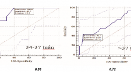
Nghiên cứu giá trị của siêu âm doppler trong tiên lượng tình trạng sức khỏe của thai ở thai phụ tiền sản giật
30/03/2020 22:11:27 | 0 binh luận
Value of doppler ultrasonography in predicting fetal well-being in pregnant women with preecclampsia SUMMARY Background: .Study on the value of some ultrasound explorations in predicting fetal well-beingin pregnant women with preeclampsia and to compare the effectiveness of different Doppler indices in predicting fetal well-being in pregnant women with preeclampsia. Methods : Study on153 patients with pre-eclampsia at Obs. & Gyn. Department - Hue Central Hospital were taken by an prospective cohort study from 12/2012 to 2/2016,. Results: Cut-off value of UTA RI for IUGR and fetal distress prediction at gestational age of 34-37 weeks was 0.6. The UTA S/D ratio cut-off value of 2.6 for fetal distress prediction at gestational age of 34-37 weeks had the sensitivity of 100% and specificity of 60%. Fetal distress prediction using UMA RI at gestational age of 34-37 weeks with cut-off value of 0.64 had the sensitivity of 90.9%, at gestational age above 37 weeks with cut-off value of 9.75 had the sensitivity of 100%. Cut-off values for UMA RI for IUGR prediction at gestational age of 34-37 weeks was 0.74 and at gestational age above 37 weeks was 0.76. Conclusion :The study found the cut-off values of PI, RI, S/D ratios of the UTA, UMA andMCA to predict fetal distress, IUGR in preeclampsia to help clinicians determine the most appropriate management to reduce perinatal morbidity and mortality rates.The study also compared the effectiveness of different Doppler indices in predicting fetal well-being. Key words : Doppler ultrosound,uterine Doppler, fetal distress, preeclampsia, IUGR
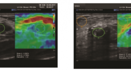
Nghiên cứu giá trị siêu âm đàn hồi bán định lượng (SEMI-QUANTITATIVE) trong chẩn đoán u vú
30/03/2020 22:04:54 | 0 binh luận
Research Value of Strain Elastography (Semi-quantitative) in Breast Tumor Diagnosis. SUMMARY Objective : Combined B-mode ultrasound and Strain Elastography Imaging (Semi-quantitative) which calculates the cutoff value of Strain Elastography in diagnosis of benign /malignant breast tumor. Method : Patients with breast tumors were combined B-mode breast ultrasound, using the WS80A equipment (Samsung), and Strain Elastography (Semi-quantitative), followed by Tsukuba-score and Ratio (B/A) (A= tumor lesion, B = fatty tissue above the lesion). From that, evaluated the accuracy, specificity, positive predictive value, accuracy and cut-off values of the Strain Elastography for diagnosis of benign/malignant breast tumors. Results : 93 women with breast tumors (67 benign tumors, 26 breast cancers), diagnosed by cytology and histopathology. The average rate of semi-quantitative in malignant and benign tumors compared to fat tissue respectively was (4.73 +/- 2.45) and (1.85 +/- 0.92). The area under the ROC curve is 0.92. The cut-off value was (2.43) has the highest sensitivity (88.5%) and the specificity (82.1%) in the diagnosis of malignant tumors. Positive predictive value (92.8%), accuracy (82.3%). Conclusion : Using Strain Elastography to measure the elasticity ratio of the breast tumor compared to fat tissue, with a cutoff values (2.43), with high sensitivity and specificity in diagnosis of benign /malignant breast tumor, which complements the breast Birads categories classification. Key words: Strain Elastography (SE), Semi-quantitative, benign /malignant breast tumor, Tsukuba-score , Ratio (B/A).
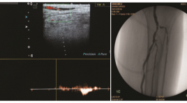
Đặc điểm hình ảnh siêu âm doppler và giá trị bổ sung của chụp mạch số hóa xóa nền trong chẩn đoán hẹp, tắc động mạch chi dưới
30/03/2020 21:49:42 | 0 binh luận
Imaging characteristics of doppler ultrasound and complementary value of digital subtraction angiography in the diagnosis of peripheral arterial occlusive disease of lower extremity SUMMARY Objectives: Describe imaging characteristics of doppler ultrasound (DUS) and evaluate the complementary value of digital subtraction angiography (DSA) in the diagnosis of peripheral arterial occlusive disease (PAOD) of lower extremity. Materials and Methods: The study is a cross sectional one and was carried out at the hospital of Hue university of medicine and pharmacy. 40 patients diagnosed with PAOD of lower limbs went through arterial assessment with DUS and DSA. The image findings of both technique were used to evaluate the diagnosis accuracy of DUS and the complementary value of DSA. Results: The sensitivity, specificity, positive predictive value and negative predictive value of DUS in PAOD is 80,65%, 92,83%, 86,21% and 89,61% respectively. DSA complemented for DUS with 6,36% additional cases of >50% stenosis or complete occlusion and 5,49% cases of low flow in occlusion suspected arteries. DSA revealed an additional of 81% collaterals in occluded arteries compared to DUS. Conclusion: DUS has high diagnostic value in PAOD of lower extremities. DSA has high complementary value for DUS in the diagnosis of PAOD of lower extremities, with the highest value at below-knee arteries. Keywords: peripheral arterial occlusive disease, doppler ultrasound, digital subtraction angiography.
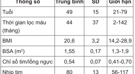
Nghiên cứu đặc điểm siêu âm tim ở bệnh nhân lọc máu chu kỳ tại bệnh viện 175
01/04/2020 16:26:53 | 0 binh luận
The evaluation of cardiac morphology and function on echocardiography of patients post dialysis at 175 Hospital SUMMARY Studies in 101 patients with these degree of kidney failure and dialysis several times, we can see the changes occurring in heart can see when echocardiography: The morphologic changes of heart, showing the dilate slight of left ventricle, left atrium and left ventricle wall thickening, increased heart muscle mass. There are signs of diastolic dysfunction on doppler ultrasound, that saw earlier by tissue doppler imaging (Em, Em / Am ...) but not affecting systolic function. There are changes in cardiac morphology and function between the dialysis groups over time towards improving. Keywords: Renal failure, dialysis, echocardiography.
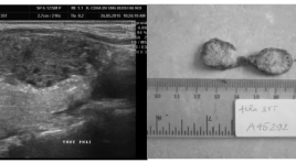
Đặc điểm hình ảnh siêu âm các tổn thương ung thư tuyến giáp
01/04/2020 16:06:30 | 0 binh luận
Ultrasound features of thyroid cancer SUMMARY urpose: To find features of thyroid malignancy nodule on the ultrasound. Material and Methods: A total 307 thyroid nodules of 272 patients (146 malignant nodules on the 146 patients, 161 benign nodules on the 126 patients), who underwent thyroid ultrasound examintation. There ultrasound features are compared with pathologic result for evaluation value. Result: The following US features showed a significant association with malignancy: solid component, hypoechogenicity (Sensitivity 80.82%; Specificity 59.01%), marked hypoechogenicity (Sensitivity 16.44%, Specificity 98.76%), irregular margins (Sensitivity 77.4%, Specificity 92.55%), microcalcifi cations (Sensitivity 69.18%, Specificity 97.52%), and taller-than-wide shape (Sensitivity 69,86%, Specificity 94.41%). Conclusion: High-resolution thyroid US is the most useful diagnostic tool for evaluating thyroid nodules. Ultrasound features of thyroid nodules malignancy include a hypoechonic, mark hypoechonic, taller than wide, irregular margins and microcalcification. Keyword: Thyroid ultrasound, thyroid, ultrasound.
Bạn Đọc Quan tâm
Sự kiện sắp diễn ra
Thông tin đào tạo
- Những cạm bẫy trong CĐHA vú và vai trò của trí tuệ nhân tạo
- Hội thảo trực tuyến "Cắt lớp vi tính đếm Photon: từ lý thuyết tới thực tiễn lâm sàng”
- CHƯƠNG TRÌNH ĐÀO TẠO LIÊN TỤC VỀ HÌNH ẢNH HỌC THẦN KINH: BÀI 3: U não trong trục
- Danh sách học viên đạt chứng chỉ CME khóa học "Cập nhật RSNA 2021: Công nghệ mới trong Kỷ nguyên mới"
- Danh sách học viên đạt chứng chỉ CME khóa học "Đánh giá chức năng thất phải trên siêu âm đánh dấu mô cơ tim"


