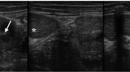
Nhân ba trường hợp xoắn lách phụ phát hiện bằng siêu âm và hồi cứu
03/04/2020 11:37:41 | 0 binh luận
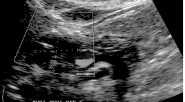
Torsion of accessory spleen in children: three case reports and literature review summa ry Objectives: To present sonographic findings of torsion of accessory spleen. Methods: Three case reports. Results: Case 1: A 2-year-old girl presented with a twoday history of left abdominal pain. Ultrasound revealed a solid, hypoechoic and avascular mass below the lower pole of the spleen. Case 2: A 14-year-old boy presented with 10 days of intermittent left upper quadrant pain. Ultrasound showed a solid, heterogeneous and hypoechoic mass below the lower pole of the spleen. This mass had poor vascularity, some small peripheral vessels, whirlpool sign at the hilum and vascular dilatation at the lower pole of the spleen. Case 3: An 8-month-old boy, ultrasound showed a hypoechoic, avascular mass in the right upper quadrant near the main spleen with hyperechogenicity of the surrounding omentum. In all three cases, we diagnosed torsion and necrosis of accessory spleen. Surgical and pathologic results confirmed the diagnosis. Conclusion s: Torsion of accessory spleen is an extremely rare entity. It may manifest as acute or subacute abdominal pain. USG is a simple and exact procedure if we consider it. Keywords: accessory spleen, torsion, ultrasound, children.
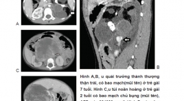
Một số đặc điểm hình ảnh khác biệt của khối sau phúc mạc ngoài thận ở trẻ em trên phim chụp cắt lớp vi tính 128 dãy đầu thu
17/03/2020 10:33:40 | 0 binh luận
The 128 MSCT imaging differentiations of retroperitoneal extra-renal masses in children SUMMARY Perpose : Find out the differrentiation of 128 MSCT characteristics of retroperitoneal extra-renal mass in children Material and methods : we had 128 MSCT images of 67 children patients in National Children’s hospital from January 2018 to April 2019 with pathologically proven of the retroperitoneal extra-renal masses. The patients was divided into 9 groups: neuroblastoma and ganglioneuroblastoma (n=42), Adrenal non-tumorigentic masses (n = 8 ), teratoma (n = 8), ganglioneuroma (n = 3), hemangioma (n = 1), neurofibroma (n = 1), rhadomyosarcoma (n = 1), undifferentiated pleomorphic sarcoma (n=1), pheochromocytoma (n = 1), york sac tumor (n = 1). We retrospectively reviewed the CT images of 67 children patients to find out the imaging differrentiation compared the enhancement level variations between groups and the enhancing of masses with muscle, liver, spleen. Results: Age of all groups is from 3 days to 11 years, the age of adrenal non-tumorigentic masses is lowest. No statitistically significant different between male and female in retro-peritoneal extra-renal masses. Diameter of adrenal non-tumorigenntic group is smallest. Neuroblastomas, teratomas, geminoma and sarcoma are usually large. Encasement of vessel, calcification, abdominal lymph nodes and far metastasis findings are common signs in neuroblastoma and ganglioneuroblastoma group. Fatty and calcified CT findings are common in teratoma group. There are statitistically significant different enhancement level variation between groups. If the enhancing of the masses was lower than the muscle, these are usually benign. If the enhancement of the masses was higher than the muscle and lower than spleen, it can be benign or malignant, neuroblastoma is very common in this level. Hemangioma enhancement is higher than spleen. Conclusions: There are differrentiations of 128 MSCT characteristics of retro-peritoneal extra-renal masses in children. Keywords: retro-peritoneal extra-renal masses, multiple slices computer tomography (MSCT).
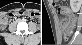
Đặc điểm hình ảnh X quang cắt lớp vi tính của viêm túi thừa đại tràng
05/12/2019 08:53:52 | 0 binh luận
The computed tomography scan characteristics of colonic diverticulitis SUMMARY Purpose: Describe the computed tomography (CT) scan characteristics of colonic diverticulitis (CD). Classification of colonic diverticulitis the World Emergency Surgery Society (WSES), and to compare computed tomography findings of right vs. left colonic diverticulitis. Methods: Retrospective studies described case series of patients diagnosed Colonic Diverticulitis at University Medical Center hospital and there was CT scan between January and December 2018. Clinical features, treatmentwere collected and assess the characteristics CT scan of Colonic diverticulitis Results : There were 104 patients, 75 right CD and 29 left CD. Mean age 46, ratio male/female 1,6. Inflamed diverticulum 89,4%; pericolic air bubbles19,2%; pericolic fluid 51,9%; abscess 1,7%; fistula 1,9%; bowel obstruction 1%. The classification of acute diverticulitis by the WSES, uncomplicated acute diverticulitis and complicated acute diverticulitisstage 1a, 1b, 2 a, 2b respectively of 48% and 39,2%; 6,9%; 4,9%; 1%. None of the complicated diverticulitis stage 3,4. Compare CT findings of right vs. left CD: Mean age (41 vs. 61), inflamed diverticulum (96% vs. 72,4%), pericolic air bubbles(8% vs. 48,3%), pericolic fluid (45,3% vs. 69%), abscess(4% vs. 31%), they differed significantly between the two groups (P < 0,05). Conclusions : Diverticulitis is often right-sided, mild in severity. Most are uncomplicate and complicated diverticulitis stage 1a by the classification of WSES. Right CD occurs in younger and lower complications compared to left CD Key words : Colonic diverticulitis,pericolic air, abscess,computed tomography findings, WSES.
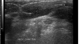
Di căn hạch của u mô đệm dạ dày ruột (GIST). Tổng quan tài liệu và nhân một trường hợp
30/03/2020 15:44:08 | 0 binh luận
Lymph node metastasis in GIST. Literature overview and case report SUMMARY GIST is considerd as rare tumor coming from Cajal cell. Due to poor clinical manifestations or vague symptoms, the disease is seen at late phase, some can be earlier by hazardous examination. The late comming with great dimension and existing metastatic lymph node generally having bad prognose. Lymph node metastatic is rare and not to be concentrated as liver and peritoneal metastasis. Absence of lymph node pre- post operation, intervention combining with adjuvant therapy is also difficult to prognose but generally can have 5 year survival. A case presenting in this paper with good prognose after treatment though lymph node metastasis after 4 years.
U mô đệm dạ dày ruột ( GIST) từ siêu âm đến cộng hưởng từ
30/03/2020 15:26:06 | 0 binh luận
Gastrointestinal Stromal Tumor: from ultrasound to MRI SUMMARY Gastrointestinal Stromal Tumor: From ultrasound to MRI. Former GIST was considered as leiomyoma, shwannoma, epithelioma. Histoimmuno logy determined with Protein KIT CD117 and the presence of interstitial cell Cajal. GIST is not rare but not be early diagnosed. Conventional X Ray, USG, CT scanner, MRI and PET can find. Though almost discovered hazardly during screening examination, we use USG for detection then CT Scanner detail study. Comment and Results : 6 cases was noted, 4 hazardly, operation and histology affirmed. Sex M/F 2/6, 4 cases under 3 cm as dimension, unique cas 30x40 cm. Metastase not yet found. All are healthy after 3 years. Conclusion: The affection is hazardly found. All imaging modalities can detected. 5 year survival depend on tumor dimension. Metastasis often to liver, mesentery but late. USG and CT is used also to follow up after interventional and target therapy. Early diagnosis rely on screening control and community care. Key words : Gastrointestinal stromal tumor, GIST, Ultrasound, CT scanner, MRI.
Chiến lược xử lý cơn đau quặn thận
23/05/2020 12:30:25 | 0 binh luận
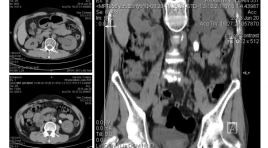
Management of renal colic SUMMARY Renal colic is a frequently situation seen in emergency. The pathophysiological mechanisms of pain in acute renal colic is due to increased intrapyelic pressure by ureteral obstruction with renal synthesis of prostaglandin E2. Intrarenal hemodynamic changes by increase the blood’s flow. The pharmacological treatment base on these pathophysiologicals mechanisms. The diagnostic and radiological mission are relatively well codified in all emergency center. For the simple renal colic, the radiograph of the abdomen without preparation can identify the calcifications of stones on urinary tract. The ultrasound can detect the presence of stones in the kidney and in the urinary tract. In somes cases, ultrasound find only indirect sign obstruction of which suspect the migration of the renal stones into urinary tract. For complex colic, considering the sensitivity, the dose of radiation, the cost and quality of the information obtained, the scanner is the test of choice of the initial diagnostic tool. Keywords: Renal colic, ureteral obstruction, simple renal colic, complex colic, radiograph, ultrasound, scanner.
Chẩn đoán thoát vị bịt trước mổ
22/03/2020 21:15:45 | 0 binh luận
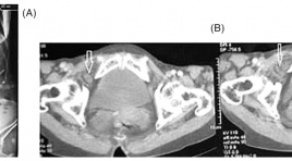
Preoperative diagnosis of obturator hernia SUMMARY The obturator hernia is a very rare type of hernia which usually presents in thin, elderly women with poor clinical signs. The preoperative diagnosis is typically difficult, with non-specific signs and symptoms which result in a delay in the diagnosis. Delayed diagnosis is a cause of increased mortality due to ruptured gangrenous bowel. We report two cases of incarcerated obturator hernia presented with a history of intestinal obstruction and the hernia was diagnosed preoperatively by computed tomography. Keywords: Obturator hernia; diagnosis, computed tomomgraphy scan.
Đặc điểm hình ảnh nang ống mật chủ ở trẻ em trên siêu âm và cộng hưởng từ 1.5T
31/03/2020 21:43:09 | 0 binh luận
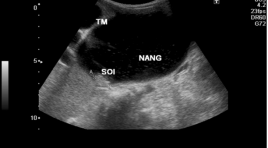
Imaging characteristics of choledochal cysts in children on ultrasound and MRI 1.5T SUMMARY: Purposes: The description of Imaging characteristics of choledochal cysts in children on Ultrasound and MRI 1.5T Material and methods: 44 patients were diagnosed choledochal cysts on Ultrasound and/or had Ultrasound and MRCP from 7/2012 to 9/2013 in National Hospital of Pediatrics. 16 patients with choledochal cysts underwent MRCP using a half –Fourier acquisition single-shot turbo spin-echo sequence. MRCP findings were compared with intraoperative cholangiography. Result: In 44 patients choledochal cysts including 34 girls and 10 boys:age range, 2 months -16 years; mean age 3.4 years. Clinical presentation: abdominal pain is the most common symptom (72.72%), vomiting (63.18%), fever (15.90%), jaundice (13.63%). The type of choledochal cysts (according to Todani): type I (93%), type IVa (7%); Type of dilatation: cystic dilatation (77.27%), fusiform dilatation (22.73%); mean measurement: 39.47mm; stone in cyst (29.5%); intrahepatic duct dilatation (43.18%); gallstone (6.8%). MRCP findings (n=16): with the most common form being typeI 87.5% (14/16), typeIVa 12.5% (2/16); cystic dialatation (93.7%), fusiform dilatation 6.3%. Cystolithiasis (75%); intrahepatic duct dilatation (56,25%). Kappa value was good agreement (k: 0.717-0.738) when compared Ultrasound and MRCP. The presence of the anomalous junction of pancreaticobiliary duct was revealed by MRCP in only 10 cases of 13 cases choledochal cysts with Kappa value was good agreement (k=0.612) when compared with intraoperative cholangiography. Conclusion: Ultrasound and MRI showed overall good accuracy in the detection and the classification of choledochal cysts and revealed associated complications. MR cholangiopancreatography provides information about anomalous pancreaticobiliary ductal union in children with choledochal cyst. Key words : Choledochal cyst, Ultrasound. Magnetic resonance cholangiopancreatography (MRCP).
Bạn Đọc Quan tâm
Sự kiện sắp diễn ra
Thông tin đào tạo
- Những cạm bẫy trong CĐHA vú và vai trò của trí tuệ nhân tạo
- Hội thảo trực tuyến "Cắt lớp vi tính đếm Photon: từ lý thuyết tới thực tiễn lâm sàng”
- CHƯƠNG TRÌNH ĐÀO TẠO LIÊN TỤC VỀ HÌNH ẢNH HỌC THẦN KINH: BÀI 3: U não trong trục
- Danh sách học viên đạt chứng chỉ CME khóa học "Cập nhật RSNA 2021: Công nghệ mới trong Kỷ nguyên mới"
- Danh sách học viên đạt chứng chỉ CME khóa học "Đánh giá chức năng thất phải trên siêu âm đánh dấu mô cơ tim"


