NGHIÊN CỨU VAI TRÒ CỦA CẮT LỚP VI TÍNH TRONG CHẨN ĐOÁN UNG THƯ LƯỠI
15/11/2021 17:18:34 | 0 binh luận
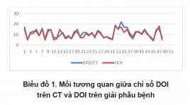
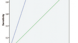
SUMMARY Objective: The objective of the study was to evaluate the value of CT in preoperative staging of tongue cancer according to AJCC 8th Methods: Cross-sectional study. We did indicate CT for 66 patients with tongue cancer at Ung Buou Hospital from 5/2019 to 5/2020. Preoperative stages on CT and histopathological stages were compared. Results: DOIs on CT were larger than the pathological DOI ( p<0.001). DOIs on CECT correlated well with pathological DOI (r=0.79, p<0.001). The correlation between CT and pathology in T staging was 0,63. In the evaluation of metastatic nodes, the sensitivity of CT was 80,9%, the specificity was 91,1%. The correlation between CT and pathology in N staging was 0,58. In the evaluation of ENE, the sensitivity of CT was 75%, the specificity was 81,8%. Conclusions: CT can determine the DOI value accurately. The correlation between CT and pathology is good in T staging and moderate in N staging. Keywords: tongue cancer, CT, AJCC 8th staging, DOI.

Đánh giá kết quả điều trị rò động tĩnh mạch màng cứng nội sọ ngoài vùng xoang hang bằng can thiệp nội mạch có sử dụng bóng chẹn bảo vệ
06/05/2021 16:21:45 | 0 binh luận
SUMMARY Purpose: To evaluate the apply the transvenous balloon protection in endovascular intervention of non - cavernous sinus dural arteriovenous fistula. Material and methods: The uncontrolled interventional study was conducted in Radiology center at Bach Mai hospital from January 2017 to August 2020. 15 patients with non - cavernous sinus dural arteriovenous fistula underwent endovascular treatment using transvenous balloon protection. Results: 15 patients were treated in 18 procedures. Among these, there were 7 males and 8 females, mean age was 48.2 ± 14.82 years. Most non - cavernous sinus dural arteriovenous fistulas were located at the transverse - sigmoid sinus (76,5%). According to the Cognard classification, Cognard IIa accounted for 47%, Cognard IIb accounted for 17,6%, Cognard IIa+b accounted for 29,4%, and Cognard IV accounted for 5,9%. Most fistulas presented with multiple feeding arteries, the most common artery was middle meningeal artery. With 18 procedures underwent tranvenous balloon protection, the sinus protection was achieved in 17 out of 18 patients. 86,7% of these patients had complete occlusion of fistula, whereas partial occlusion occurred in 13,3% of these patients. After treatment, 86,7% of these cases didn’t have complication, complete symptom remission rate was 53,3%, 26,7% showed symptom relief. Only 1 case had severe complication, accounted for 5,6%. Conclusion : Endovascular intervention using transvenous balloon protection is a safe and effective technique in the treament of non - cavernous sinus dural arteriovenous fistula. Key words: dAVF, endovascular intervention, transvenous balloon protection.

Giá trị của thang điểm EU-TIRADs 2017 và ACR-TIRADS 2017 trong đánh giá nhân tuyến giáp
06/05/2021 12:41:10 | 0 binh luận
SUMMARY Objective : Characteristic of thyroid nodules according to EU-TIRADS 2017 and ACR-TIRADS 2017 classification. Comparison among EUTIRADS and ACR-TIRADS in the diagnostic efficiency of thyroid nodules. Subject and methods : descriptive cross-sectional study was performed on 233 patients with thyroid nodule who were diagnosed with ultrasound and performed FNA at Dien Quang Center, Bach Mai Hospital from 8/2019 to 8/2020. Results: 233 patients in study with 233 thyroid nodules, 79 nodules with malignant cells (33.9%), 154 nodules had no malignant cells (66.1%). Malignant nodule are mainly solid nodule (97.5%), 100% sponges and follicles do not have malignant cells. Hypoechoic solid and irregular margins had high sensitivity and specificity (>70%) in diagnosing malignancies. Very hypoechoic solid and microcalcification has a 100% specificity in diagnosing malignancies. There is a statistically significant difference in malignancy between the nuclear group of thyroid with height> = width and height <width. ACR-TIRADS classification has sensitivity 96.2%, specificity 53.9%, PPV 51.7%, NPV 96.5%, accuracy 68.2% . EU-TIRADS classification sensitivity 87.3%, specificity 71.4%, PPV61.1%, NPV 91.7%, accuracy 76.8%. Conclusion: ACR-TIRADS and EU-TIRADS classification are well diagnostic efficiency in the diagnosis of malignant and benign thyroid nodules. The ACR-TIRADS classification has a higher sensitivity but less specificity than the EU-TIRADS classification. Key words: thyroid ultrasound, EU-TIRADS classification, ACRTIRADS classification.

Tiến bộ kỹ thuật cộng hưởng từ trong đánh giá u tế bào đệm
04/12/2019 20:45:31 | 0 binh luận
Advanced magnetic resonance imaging in evaluation of glioma SUMMARY Glioma is the most common primary cerebral tumor which has a poor prognosis, high disability and fatalityespecially in high grade gliomas. The current standard of imaging technique for evaluating glioma is conventional MRI. Basic cMRI sequences are T1W, T2W, FLAIR, T1W+Gd. Conventional MRI provides critical clinical information about gliomas. Unfortunately, conventional MRI is nonspecificity, not reflect the complicated biology, has a limited capacity to grading and differentiate gliomas from other pathologies such as: inflammation,MS… Recently, there is a development of many new MRI techniques and these application have increased such as diffusion-weighted imaging, diffusion-tensor, tractography, perfusion, spectroscopy and functional MRI. These techniques provided complementary information to cMRI for assessing tumor in cellularity, white matter invasion, hypoxia, necrosis, vascularization, permeability and relation tumor with functional areas. They give more accurate in diagnosis, planning pre-surgery and monitoring post-therapy. This lecture introduces an principle and clinical application of these advanced MRI techniques in cerebral gliomas which were performed at Choray hospital. Keywords : Glioma, conventional MRI, diffusion-weighted, diffusion-tensor, tractography, perfusion, spectroscopy, functional MRI.

Sa lồi thanh quản thể bên trong nhân một trường hợp
19/11/2019 13:58:50 | 0 binh luận
Bênh nhân nam, 58t, khàn tiếng, ngoài ra không có biểu hiện gì nên BN không quan tâm. Khám sức khoẻ định kỳ lâm sàng nghi có bất thường vùng hạ họng nên chỉ định chụp CLVT và chụp X quang phổi. Phương tiện : máy CLVT Siemens 16 dẫy, khoảng cách 3,6 mm, điểm trung tâm thường là sụn giáp, ở phía sau phải ngang mức đốt sống cổ C4. Vì chúng tôi định vị không tốt nên hình cắt ngang rõ nhưng dựng hình hướng cắt dọc (sagittal)không thấy được toàn bộ hình. Không dùng cản quang vì bờ khối đều. BN được chỉ định sử trí bằng nội soi laser.
Bạn Đọc Quan tâm
Sự kiện sắp diễn ra
Thông tin đào tạo
- Những cạm bẫy trong CĐHA vú và vai trò của trí tuệ nhân tạo
- Hội thảo trực tuyến "Cắt lớp vi tính đếm Photon: từ lý thuyết tới thực tiễn lâm sàng”
- CHƯƠNG TRÌNH ĐÀO TẠO LIÊN TỤC VỀ HÌNH ẢNH HỌC THẦN KINH: BÀI 3: U não trong trục
- Danh sách học viên đạt chứng chỉ CME khóa học "Cập nhật RSNA 2021: Công nghệ mới trong Kỷ nguyên mới"
- Danh sách học viên đạt chứng chỉ CME khóa học "Đánh giá chức năng thất phải trên siêu âm đánh dấu mô cơ tim"


