


HÌNH ẢNH BIẾN CHỨNG MẠCH MÁU TRÊN CẮT LỚP VI TÍNH ĐA DÃY Ở BỆNH NHÂN NHẬN GAN TỪ NGƯỜI CHO SỐNG
22/01/2024 16:12:28 | 0 binh luận
TÓM TẮT Mục tiêu : nhận xét đặc điểm hình ảnh cắt lớp vi tính (CLVT) trong quá trình theo dõi bệnh nhân (BN) sau ghép gan trong tháng đầu. Đối tượng và phương pháp : 67 BN ghép gan từ người cho sống tại Bệnh viện Trung ương Quân đội 108 được đánh giá bằng CLVT gan 3 pha, quá trình đánh giá được thực hiện trên máy CLVT đa dãy. Kết quả : Nghiên cứu trên 67 BN (54 nam & 13 nữ), tuổi trung bình của BN là 55 tuổi, nhỏ nhất 10 tuổi, lớn nhất 75 tuổi. Chỉ định ghép gan phần lớn là ung thư biểu mô tế bào gan (chiếm 38,8%), suy gan cấp (chiếm 35,8%) và xơ gan (chiếm 23,9%). Biến chứng động mạch xảy ra ở 06 trường hợp: 02 trường hợp huyết khối động mạch gan, 03 trường hợp hẹp động mạch gan, 01 trường hợp có đồng thời giả phình kèm huyết khối động mạch gan. Có 12 trường hợp có huyết khối tĩnh mạch gan và 01 trường hợp huyết khối tĩnh mạch chủ dưới. Kết luận : Chụp CLVT đa dãy là phương pháp chẩn đoán không xâm nhập và rất hữu ích trong việc phát hiện biến chứng mạch máu ở BN nhận gan sau ghép gan từ người cho sống, đánh giá đồng bộ về hệ mạch gan, nhu mô gan. Chụp mạch CLVT là lựa chọn tốt nhất để xác nhận các nghi ngờ về biến chứng mạch máu trên siêu âm. Việc phát hiện các biến chứng trên CLVT góp phần rất lớn trong chẩn đoán và điều trị. Từ khóa : ghép gan từ người cho sống, chụp CLVT đa dãy, biến chứng sau ghép, huyết khối động mạch gan, hẹp động mạch gan, huyết khối tĩnh mạch gan

BƯỚC ĐẦU NGHIÊN CỨU HIỆU QUẢ SỚM CỦA PHƯƠNG PHÁP ĐIỀU TRỊ UNG THƯ BIỂU MÔ TẾ BÀO GAN TIẾN TRIỂN...
22/01/2024 16:01:55 | 0 binh luận
TÓM TẮT Mục tiêu: Đánh giá kết quả sớm của phương pháp đặt cổng truyền hóa chất động mạch gan bằng phác đồ Lipiodol kết hợp với Cisplatin và 5 FU trên bệnh nhân UTBMTBG giai đoạn tiến triển tại chỗ. Đối tượng và phương pháp nghiên cứu: Chúng tôi thực hiện can thiệp lâm sàng tiến cứu, có theo dõi dọc trên đối tượng là 10 BN UTBMTBG giai đoạn tiến triển. Mỗi bệnh nhân được đặt cổng truyền hoá chất dưới da phục vụ truyền hoá chất động mạch gan. Phác đồ điều trị bao gồm Lipiodol, Cisplastin và 5FU. Sau 1 tháng điều trị, các bệnh nhân được thăm khám, chụp CLVT và phân loại đáp ứng thành đáp ứng hoàn toàn, đáp ứng một phần, bệnh ổn định và bệnh tiến triển theo tiêu chuẩn mRECIST. Kết quả : 6 BN (60%) có đáp ứng sớm sau điều trị, trong đó 1 BN (10%) có đáp ứng hoàn toàn và 5BN (50%) đáp ứng một phần. Một bệnh nhân có bệnh tiến triển sau điều trị, và 2 bệnh nhân bệnh ổn định. Kích thước phần ngấm thuốc của khối u giảm từ 102±41mm xuống còn 83±53mm, có ý nghĩa thống kê với p <0.05. Ngoài ra, HKTMC thoái triển trên 2 BN, và chức năng gan được cải thiện trên 2 BN. Có 3 BN xuất hiện biến chứng, bao gồm nhiễm trùng, tụ máu, tắc và vỡ cổng truyền. Kết luận: Truyền hoá chất động mạch gan với phác đồ Lipiodol kết hợp Cisplastin và 5FU đạt tỷ lệ đáp ứng sớm cao, có thể gây thoái triển HKTMC và cải thiện chức năng gan.
VAI TRÒ CỦA FDG PET/CT TRONG CHẨN ĐOÁN VÀ TIÊN LƯỢNG U PHYLLODES TUYẾN VÚ DI CĂN VÀ HỒI CỨU Y VĂN
18/10/2023 12:01:27 | 0 binh luận
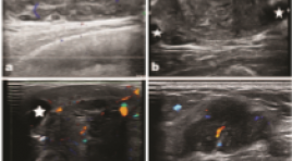
SUMMARY Metastasis of malignant phyllodes tumors is extremely rare that there are only few cases reported in literature. With the aim of providing data and diagnostic guidance, discussing treatments, we present the role of PET/CT, other radiologic modalites. We report a clinical case a 43 years-old female patient who was diagnosed with grade II phyllodes tumor of the breast and treated by surgery and radiotherapy adjuvant (2017). Following up on radiology test after 6 years (02/2023), the patient has diagnosed with bilateral lungs metastasis and left upper lobectomy was done. PET/CT scan in Choray hospital after the surgery showed multiple hypermetabolic foci in lungs, thoracic wall, sternum, pancreas, stomach, bones which suggested distant metastases from the prior tumor. Treatments for distant recurrence phyllodes tumors have not been effective and patients often have poor prognosis. However, some new target therapies may help increase life expectancy, improve quality of life.
TỰ TỐNG XUẤT DỊ VẬT (ĐINH SẮT NHỌN) TỪ PHẾ QUẢN THÙY GIỮA BÊN PHẢI BÁO CÁO TRƯỜNG HỢP HIẾM GẶP
17/10/2023 16:06:58 | 0 binh luận
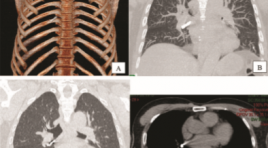
SUMMARY Tracheobronchial foreign body aspiration is an uncommon but potentially life-threatening event in adults. Symptoms typically consist of a choking event followed by cough and dyspnea. Chest radiography and computed tomography can provide information regarding the location and characteristics of foreign bodies hence aiding in diagnosis. Bronchoscopy remains the gold standard for diagnosis and management of this condition. Foreign body spontaneous expulsion rarely occurs. We present a case of 61-year-old male patient who spontaneously expulsed a sharp nail from right bronchus. Keywords : spontaneous expulsion, tracheobronchial, nail, foreign body, aspiration.
NGHIÊN CỨU GIÁ TRỊ CỦA CHUỖI XUNG TƯỚI MÁU VÀ CHUỖI XUNG PHỔ TRÊN CỘNG HƯỞNG TỪ 3 TESLA CHẨN ĐOÁN PHÂN BẬC U THẦN KINH ĐỆM
18/10/2023 10:03:50 | 0 binh luận
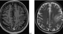
SUMMARY Purpose: To evaluate the diagnostic accuracy of magnetic resonance spectroscopy and dynamic contrast-enhanced (DCE) magnetic resonance perfusion for glioma grading. Materials and Methods: Fifteen patient confirmed pathological glioma who underwent MR spectroscopy and DCE in 3 Tesla MRI machine. The following parameters were used: Ktrans, Ve, Cho/NAA, Cho/Cre. The diagnostic accuracy for glioma grading was determined by ROC analysis. Results: There were 10 patients in the high-grade group and 5 patients in the low-grade group. Ktrans, Ve, Cho/NAA and Cho/Cre measures differed significantly between high and low-grade tumor. The AUC was 0.956 for Ktrans. Conclusion: Ktrans, Ve Cho/NAA and Cho/Cre parameters demonstrated to be useful for glioma grading. Keywords: Spectroscopy, DCE-MRI, glioma grading
ĐẶC ĐIỂM HÌNH ẢNH VÀ VAI TRÒ CỦA CẮT LỚP VI TÍNH ĐA DÃY TOÀN THÂN TRONG UNG THƯ TUYẾN TIỀN LIỆT GIAI ĐOẠN IV
18/10/2023 09:52:14 | 0 binh luận
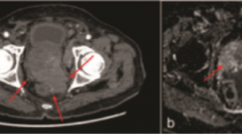
SUMMARY Purpose: The study was conducted to describe image characteristics of prostate cancer on multidetector computed tomography (MDCT) and to determine the role of MDCT in patients with stage IV prostate cancer. Objectives and subjects: A cross-sectional study was performed on 45 patients at Huu Nghi Hospital from January 2017 to May 2023. All patients had pathologically proven prostate cancer, multi-parameter magnetic resonance imaging (mp-MRI), MDCT, and bone scan and were categorized as stage IV. Results: The mean age was 78.31±5.64 years, mean Total prostatespecific antigen (tPSA) concentration was 279.78 ng/ml, mean prostate volume was 45.20 ml. 60% of patients had extra-prostatic invasion, 66.7% had regional lymph node metastases; 40% of patients had distant lymph node metastasis; 46.7% had bone metastasis; 28.9% had distant metastasis to other organs. MDCT had good to very good concordance with MRI in evaluating local invasion and good concordance with bone scintigraphy in evaluating bone metastasis, with p <0.05. Conclusion: MDCT had a high value in assessing metastatic lesions in patients with stage IV prostate cancer, especially lung lesions. Whole-body MDCT is a useful alternative to MRI and PET/CT in evaluating metastatic lesions from prostate cancer. Keywords: Prostate cancer, Multidetector computerized tomography, bone scintigraphy.
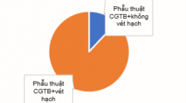
ĐÁNH GIÁ HIỆU QUẢ PHẪU THUẬT VÉT HẠCH TÁI PHÁT Ở BỆNH NHÂN UNG THƯ TUYẾN GIÁP THỂ BIỆT HOÁ SAU ĐIỀU TRỊ 131I TẠI VIỆN Y HỌC PHÓNG XẠ VÀ U BƯỚU QUÂN ĐỘI
18/10/2023 09:46:18 | 0 binh luận
SUMMARY Objectives: to describe the clinical and subclinical symptoms and evaluate the response after recurrent lymph node dissection of differentiated thyroid cancer patients after 131I treatment. Subjects: 50 differentiated thyroid cancer patients after 131I treatment, recurrence for the first time. Study methods: Retrospective and prospective description, data collection through patients’ medical records. Results: the mean age of the study patients was: 43.5 ± 12.6, the most common age is <55 years old with the rate of 80%, the female/ male ratio=3.5/1. The group of patients with Tg < 1 ng/ml accounted for the majority with 33/50 patients (66%). The median Tg before and after surgery was 2.02 ng/ml and 0.35 ng/ml, respectively. After surgery, response achieved in 98% of patients, mainly complete response and incomplete response in biochemical, accounted for 40%, 36%. Lymph node dissection reduced Tg in 78% of patients. Reccurent lymph nodes mainly observed in group IV, group III and group VI. The recurrence of lymph nodes was mainly in the central and cervical regions on the same side of the primary tumor. Conclusion: Surgery is the preferred first-line treatment for the reccurrent lymph node thyroid cancer patients. It changes response assessment, improve prognosis for reccurence patients after 131I treatment. Keywords: Differentiated thyroid cancer, response after recurrent lymph node dissection, Tg
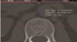
TƯƠNG QUAN GIỮA ĐƠN VỊ HOUNSFIELD Ở CỘT SỐNG THẮT LƯNG VÀ MẬT ĐỘ XƯƠNG ĐO BẰNG DEXA Ở NGƯỜI VIỆT NAM
17/10/2023 17:41:40 | 0 binh luận
SUMMARY Background: Osteoporosis is a disease that increases the risk of fractures. Evaluation of the Hounsfield Unit in the lumbar spine on computed tomography for any reason has the potential to predict bone density abnormalities that contribute to the warning of osteoporosis. Objective: The aim of this study was to evaluate the correlation between Hounsfield Units (HU) in Lumbar Spine (LS) and bone mineral density (BMD) measured by Dual-Energy X-Ray Absorptiometry (DEXA). Method: Retrospective and cross-sectional study of 150 patients comprised CT-DXA pairs within a 6-month period performed for any indication. Measure HU at lumbar vertebrae from L1 to L4. Calculate Spearman correlation coeffificient between the mean HU at lumbar vertebrae and the BMD values from DEXA scan. Using area under the ROC Curve (AUC) and finding cut-off of HU for diferentiating normal BMD and abnormal BMD, osteopenia and osteoporosis. Result: We noted correlations between the HU at LS and the BMD from DXA scan which is significant, the highest correlation at L2 (Spearman correlation coefficient = 0.68). At L2, normal BMD: ≥ 131 HU, osteopenia: 101 – 131 HU, osteoporosis: ≤ 131 HU, we also determined that threshold of 101 HU was more than 90 % sensitive, and a threshold of 171 HU was more than 90 % specific for distinguishing normal BMD. In addition, cut-off ≤ 132 HU was more than 90 % sensitive, and cut-off ≤ 62 HU was more than 90 % specific for distinguishing osteoporosis from osteopenia. Conclusion: The correlations between the HU at LS and the BMD from DXA scan is significant. Keyword : Osteoporosis, DEXA, HU at lumbar spine
Bạn Đọc Quan tâm
Sự kiện sắp diễn ra

THÔNG BÁO SINH HOẠT KHOA HỌC 19/01/2024: CẬP NHẬT CHẨN ĐOÁN VÀ ĐIỀU TRỊ UNG THƯ TRỰC TRÀNG
19-01-2024 09:47:01 0 bình luận

Thông tin đào tạo
- Những cạm bẫy trong CĐHA vú và vai trò của trí tuệ nhân tạo
- Hội thảo trực tuyến "Cắt lớp vi tính đếm Photon: từ lý thuyết tới thực tiễn lâm sàng”
- CHƯƠNG TRÌNH ĐÀO TẠO LIÊN TỤC VỀ HÌNH ẢNH HỌC THẦN KINH: BÀI 3: U não trong trục
- Danh sách học viên đạt chứng chỉ CME khóa học "Cập nhật RSNA 2021: Công nghệ mới trong Kỷ nguyên mới"
- Danh sách học viên đạt chứng chỉ CME khóa học "Đánh giá chức năng thất phải trên siêu âm đánh dấu mô cơ tim"


Đơn vị hợp tác

