


Đặc điểm hình ảnh cộng hưởng từ của u sợi và u vỏ - sợi buồng trứng
18/12/2019 09:32:38 | 0 binh luận
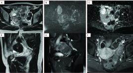
Value of magnetic resonance imaging in the diagnosis of ovarian thecomas/fibrothecomas SUMMARY Purpose: Our study aims to study the value of conventional magnetic resonance imaging (MRI) combined with DWI and Dynamic technique in the diagnosis of thecomas/fibrothecomas and differential diagnosis benign with malignant ovarian tumors. Material and method: In total, 68 thecomas/fibrothecomas, 63 malignant ovarian tumors were included in our study. All patients underwent conventional MRI, DWI in 79 cases and Dynamic enhancement (DCE) in 14 cases. The clinical features and characteristics of conventional MRI, DWI and DCE of these two groups were analyzed. Apparent diffusion coefficient (ADC) values, Tmax, MRE were measured and compared between groups. Univariate analysis, multivariate logistic regression analysis were analyzed. Sensitivity (Se), Specificity (Sp), Positive Predictive Value (PPV), Negative Predictive Value (NPV) were included. Results: All the fibromas/fibrothecomas showed hypo-isointensity on T1 weighted imaging (T1WI) and 77.9 % lesions showed hypo- to isointensity on T2 weighted imaging (T2WI). After administration of contrast medium, 82,3% tumors appeared as minor to mild enhancement, 71,4% benign tumor had type 1 curve, Tmax cutoff were 230s with Se and Sp 71,4%. MRE were not already measured because of few cases. On DWI, 68,4% fibromas/fibrothecomas manifested no signal intensity or low signal intensity. The ADC cutoff were 1.07 x 10⁻³ mm2/s to differentiate benign from malignant ovarian tumors. Multivariate logistic regression analysis showed that only T2WI and ADC were the important indicators in discriminating fibromas/fibrothecomas or benign tumors from malignant ovarian tumors. Conclusion : The combination of DWI, DCE with conventional MRI is of great value in the diagnosis of fibromas/fibrothecomas and differentiation benign ovarian tumors from malignant ovarian tumors Keywords: Fibromas/fibrothecomas, Conventional magnetic resonance imaging, Diffusion-weighted imaging, Apparent diffusion coefficient value, Dynamic contrast enhancement.
Khảo sát kết quả chụp tử cung - vòi tử cung ở các trường hợp vô sinh
31/03/2020 13:17:40 | 0 binh luận
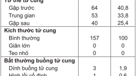
Investigation of hysterosalpingopraphy in infertile women summa ry Objectives : investigate the images of uterine cavity and fallopian tube patency by hysterosalpingopraphy in infertile women and learn the involved factors. Subjects and methods : Cross-sectional descriptive study on 157 infertile women examined at Hue University Hospital with clinical examination, history and hysterosalpingopraphy. Results : in 157 infertile women with mean-age 30.4 ± 5.4, there was 5.7% cases with abnormal uterine cavity and 13.4% abnormal tubal patency. In which, proximal blockage was 4.5%, distal blockage and hydrosalping were 8.9%. Some factors included age over 35 years (p = 0.02; OR = 2.49), secondary infertility (p=0.029; OR = 2.27), past history with tubal operation (p = 0.016) or high pressure of pushing fluid (p=0.004; OR = 4.005) associated with abnormal findings on hysterosalpingopraphy. Conclusion: hysterosalpingopraphy helps detect many abnormalities in the uterine cavity and the fallopian tubes, especially in people over 35 year old, with a history of tubal surgery, secondary infertility. It is recommended to indicated hysterosalpingopraphy routinely for these cases for early detection of abnormalities.
Đánh giá sự thay đổi tinh dịch đồ, hormon sinh dục sau can thiệp điều trị suy giãn tĩnh mạch thừng tinh
18/12/2019 14:36:31 | 0 binh luận
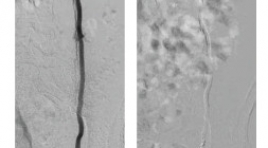
Evaluation of changes in semen, sex hormones after intervention in varicocele SUMMARY Purpose: Evaluating the technical feasibility an semen quality after intervention of varicocele. Subjects and research methods: Among 101 patients treated with varicocele emboization during 2 years. With 50 patients were followed up before and after embolization 6 months by the size of the vein, testes, and semen index including concentration, morphology, mobility, hormonal (including Testosterol, LH, FSH). Technical details, reasons for the failure of the procedure are noted. Using pairing comparison algorithm before and after treatment. Results: 100% technical success. The indicators of spermatozoa including sperm concentration, morphology and mobility were improved with the reliability of 95%, especially sperm motility index, PR - sperm rised signigicantly with Z = 4,1 ± 1,51 (before and after treatment). The index of testosterol hormones increased significantly after treatment with Z = 1.81 ± 1.32, LH and TSH hormonal indexes did not change significantly. Conclusion : Intervention of varicococele has been shown to be effective in improving semen and testosterone index. Key word : Intervention of varicocele, Sperm, testosterone index.
Mối liên quan giữa đặc điểm lâm sàng, xét nghiệm tinh dịch đồ với siêu âm doppler trên bệnh nhân giãn tĩnh mạch tinh ở người trưởng thành dưới 40 tuổi
18/12/2019 09:45:58 | 0 binh luận
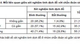
The relationship between clinical characteristics, semen test with color Doppler ultrasound in adult varicocele patient under 40 years old SUMMARY Purpose: Determine the relationship between clinical characteristics and semen test with color Doppler ultrasound in adult varicocele patientsunder 40 years old. Methods: A total of 56 patients with clinical diagnosis of varicocele were clinically graded according to Dubin and Amelar [1], calculated body mass index (BMI), did semen test and color Doppler ultrasound scrotum. Then they were calculated according to Chiou's scoring system [2] to determine varicocele. To assess the relationship we use Chi-Square test, two qualitative variables were identified as relevant when the p-value<0.05. Result s: There was arelationship between diagnosis of Doppler ultrasound according to Chiou with clinical varicocele (p<0.05). There was no relationship between BMI and diagnosis of varicose veins Doppler (p> 0.05). There was a relationship between the semen test and the diagnosis of varicocele on color Doppler ultrasound (p<0.05). Conclusion : Our study has shown a relationship between color Doppler ultrasound diagnosis based on Chiou's scoring system with clinical grade and semen test in adult varicose veins under 40 years old. Keywords : Varicocele, Color Doppler ultrasound, male infertility.
Đặc điểm hình ảnh cộng hưởng từ của rò hậu môn
05/12/2019 09:17:02 | 0 binh luận
M agnetic resonance imaging characterization of perianal fistula SUMMARY Background: Perianal fistula is a common disease in the anorectal region, just after hemorrhoids. The disease often recurs because of missed lesions at surgery, especially internal openings and secondary tracts, which can be identified by MRI. Surgery guided by Magnetic Resonance imaging (MRI) significantly reduces the recurrence of fistula. Aims : To describe the MRI features of perianal fistula and study the role of T2W TSE, post- contrast FS T1W TSE Gadolinium sequences in detecting the characterizations of the fistula tract. Subjects and methods : 367 patients with perianal fistulae, diagnosed and treated at University medical center hospital, between 1/1/2016 and 1/31/2018, were included in this study. This study is a retrospective analysis. We compared the imaging features with the surgical findings. Results : The mean age was 39.3 years (range, 12 – 84 years), male/ female = 9/1. A total of 411 fistulas were found at surgery. Agreement between the surgical findings and MR imaging for classification of the primary track, detecting secondary track was very good with kappa = 0.89 (0.84,0.93), 0.94 (0.90,0.97), respectively. The sensitivity and specificity of MRI in correctly detecting internal openings was found to be 99% and 85.2% respectively; for abscess, it was 100%. Both T2W TSE, post-contrast FS T1W TSE Gadolinium sequences have high sensitivity and specificity for the depiction of primary tracts, internal openings, and secondary tracts. Post- contrast FS T1W Gadolinium sequence was very good in detecting abscess; differentiating between abscess and inflammation, active inflammatory components with scars. Conclusions : Magnetic Resonance Imaging (MRI) was highly accurate in assessment of surgically important parameters of perianal fistulae. With the very good agreement between the surgical findings and MR imaging for classification of the primary track, detecting secondary tract, high accuracy in detecting internal openings and abscess as well, MRI was considered the reliable technique of choice for establishing preoperative fistula map. If no contraindications exist, administration of contrast material is necessary for detecting abscess and secondary tracts, differentiating active inflammatory components with scars, especially in case of complex or recurrent fistula or having surgery around the anus before. Keywords: fistula, anal, MR imaging.
Nghiên cứu đặc điểm hình ảnh và giá trị CHT chẩn đoán phân độ giai đoạn T của ung thư bàng quang
04/12/2019 21:29:46 | 0 binh luận
Researching imaging characteristics and assessing values of MRI in the diagnosis of bladder cancer at T-stage SUMMARY Purpose: Describing imaging characteristics and assessing values of MRI in the diagnosis of bladder cancer at T-stage. Subjects and methods : 43 patients with bladder tumors were selected to be in adescriptive study (38 of whom had tumors from tumor histopathology and 5 patients had histology from other organs or benign paraganglioma bladder tumor), in which they got diagnosed, operated (transurethral resection or radical cystectomy) and had pathology results from May 2017 to June 2018 at K hospital in Tan Trieu. All MRI films were evaluated preoperatively and compared with histopathology postoperativelydistinguishing superficial tumors (T1 or lower) and invasive tumors (T2 or higher). Results : Among 38 patients being studied, the mean age is 56 ± 13.24 andthe gender ratio is M/F ≈ 7/1. Out of 61 tumors, the most common tumor location is bilateral bladder 30.7%, mostly one tumor (26/38). Featured images: mean size 23.47 ± 14.09 mm, max size 68mm, min size 7 mm; most frequently found polype-shaped tumor 25/38 patients (65.8%). According to the assessment of MRI results with T2W, DCE and DWI distinguishing T staging, tumors in T1 or lower 30/38 patients (78.9%) and T2 or higher 8/38 patients (21,1%, in which 4 patients in T2, one in T3 and 3 in T4). There is no correlation between the number of tumors and T staging (p>0.05). There is a strong correlation between the shape of tumors and T staging (p<0.001). When combining T2W with DCE, the sensitivity, specificity and overall accuracy of two observers for differentiating Tis to T1 tumor from T2 to T4 tumors were 79.3 %, 100% and 84.2% respectively. When using all three image types together (T2W, DCE and DWI) to assess T staging, the sensitivity rose up to 96.5%, specificity 66.7%, overall accuracy 89.5% and PPV 90.3%. The Cohen’s Kappa score of 0.685 had a good correlation between MRI results and histopathology in distinguishing T-staging of bladder cancer (p<0.001). In addition, a total 36 patients had urothelial carcinoma, ADC values of 29/36 patients at T1 or lower were 1.138 ×10⁻³mm2/s and 7/36 patients at T2 or higher were 0.79 × 10⁻³mm2/s, and this difference of ADC was significant between Grade 1 and Grade 3. The total of 29/38 patients (76.3%) underwent TUR and deep muscle biopsy was performed at the base tumor, 9/38 patients underwent radical cysectomy or chemotherapy before surgery. Key words : Bladder Cancer, MRI of Bladder cancer.
Nghiên cứu đặc điểm hình ảnh và giá trị của cắt lớp vi tính 64 dãy trong chẩn đoán u tuyến thượng thận
31/03/2020 21:59:44 | 0 binh luận
Study of imaging characteristic and value of 64-slice CT in diagnosis of Adrenal tumor SUMMARY: Objective: This study aims to describe the characteristic and value of diagnostic adrenal tumor on 64-slice CT scanner Materials and Methods: Prospective and cross-sectional descriptive study of 39 patients were diagnosed adrenal tumor on 64-slice CT scanner. The receiver operating characteristic (ROC) curves for the value of size, the attenuation values at the unenhanced CT and the relative percentage washout (RPW) and absolute percentage washout (APW) values for the differential diagnosis of adenoma from nonadenoma. Results: Adrenal tumor was common as adenoma (16/38) with the feature of often homogeneous, clearly limited and small size (2.5 cm), and about 11.5 HU on precontrast attenuation. Pheocromocytoma was often heterogeneous, large size of 4.76cm and in conjunction with high levels of dynamic enhancement (108.3 Hu and 111.7 Hu). Tumor adrenal malignant was large size of 8.83cm, often unknown limits, infiltrates surrounding and invading IVC (2/3 cases). The area under the ROC curve and cut off with size were 0.896 and 3.29 cm, the attenuation values at the unenhanced CT were 0.984 and 24 Hu, APW were 0.937 and 57.6% and RPW were 0.994 and 43.7% (p <0.001). The sensitivity, specificity and accuracy at the threshold size of 3.29 cm were 88.2%, 90.9% and 89.7%, respectively; at threshold 24 Hu were 94.1%, 95% and 94.6%; at threshold APW = 57.6% and RPW = 43.7% were 94.1%, 85%, 89.2% and 94.1%, 100%, 97.3%, respectively. Conclusion: 64-slice CT scanner is effective methods to characterize and diagnose adrenal tumor. The relative percentage washout (RPW), absolute percentage washout (APW), the attenuation values at the unenhanced CT and the size criteria appear optimal for discriminating an adrenal adenoma from a nonadenoma. Keywords: Adrenal Gland Neoplasms, Adrenal Glands, radiography.
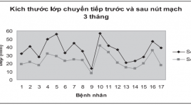
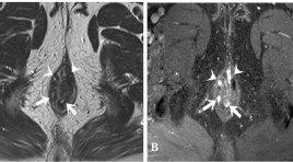
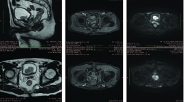
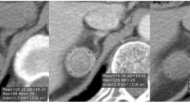
Bước đầu đánh giá kết quả điều trị lạc nội mạc tử cung trong cơ tử cung bằng phương pháp nút động mạch tử cung
02/04/2020 22:42:04 | 0 binh luận
Preliminary evaluation of uterine artery embolization for treatment of adenomyosis SUMMARY: The uterine arterial embolization was carried out for 17 patients who suffered from adenomyosis, in which of them, 10 cases were followed-up by MRI at 1 and 3 months after embolization. The results showed that: free from abdominal pain were about 87.5 and 81.25% respectively and free from menorrhagia were 66.7% and 50% respectively. The thickness of transitional zone was reduced from 33 to 22.1mm. Average reducing of uterine volume was from 274.4 to 215.9cm3, corresponding to 19.5% of decreased volume. No contrast enhancement on MRI were found at 7 patients (7/10). 100% patients felt abdominal pain at the end of the procedure, especially 94,1% strongly pain. Hospitalization time was only 1-2 days in 94,1% of patients.
Bạn Đọc Quan tâm
Sự kiện sắp diễn ra

THÔNG BÁO SINH HOẠT KHOA HỌC 19/01/2024: CẬP NHẬT CHẨN ĐOÁN VÀ ĐIỀU TRỊ UNG THƯ TRỰC TRÀNG
19-01-2024 09:47:01 0 bình luận

Thông tin đào tạo
- Những cạm bẫy trong CĐHA vú và vai trò của trí tuệ nhân tạo
- Hội thảo trực tuyến "Cắt lớp vi tính đếm Photon: từ lý thuyết tới thực tiễn lâm sàng”
- CHƯƠNG TRÌNH ĐÀO TẠO LIÊN TỤC VỀ HÌNH ẢNH HỌC THẦN KINH: BÀI 3: U não trong trục
- Danh sách học viên đạt chứng chỉ CME khóa học "Cập nhật RSNA 2021: Công nghệ mới trong Kỷ nguyên mới"
- Danh sách học viên đạt chứng chỉ CME khóa học "Đánh giá chức năng thất phải trên siêu âm đánh dấu mô cơ tim"


Đơn vị hợp tác

