


Độ dài xương mũi thai nhi ở tuổi thai từ 19-26 tuần tại Việt Nam
30/03/2020 22:28:55 | 0 binh luận
SUMMARY Aim: The purpose of this study was to establish the normal range of fetal nasal bone length (NBL) at the second trimester of gestation in Vietnamese pregnancies. Methods: A prospective cross-sectional study was carried out. A total of 2432 singleton pregnancies were evaluated for fetal NBL at 19–26 weeks 6 days between 1 January 2013 and 31 December 2014. Three measurements of NBL were taken and the largest value was chosen. Fetal anomalies and neonatal abnormalities were excluded. Results : Mean fetal NBL was 6.75 ± 0.82 mm and mean gestational age was 22.2 ± 1.4 weeks. Median NBL increased linearly with advancing gestational age: NBL (mm) = 0.26 × gestational age (weeks) + 1.03 (R2 = 0.20; P <0.000). The fifth percentile of fetal NBL ranged from 4.6 to 6.3 mm, corresponding with gestational age 19–26 weeks. Conclusion : The normal range ofNBLwas established at second trimester inVietnamese pregnancies. NBL at the fifth percentile for gestational age was different from that of other Asian ethnicities. It is necessary to define short NBL or hypoplasia of NBL with regard to ethnicity. Key words : Down syndrome, fetal nasal bone length, ultrasonography.
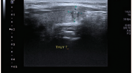
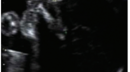
Nhận xét giá trị phương pháp chọc hút kim nhỏ dưới hướng dẫn siêu âm trong chẩn đoán ung thư biểu mô tuyến giáp
06/05/2021 16:49:08 | 0 binh luận
SUMMARY We conduct research on diagnostic value of ultrasound guided fine needle aspiration (FNA) in the diagnosis of thyroid carcninoma compared with post-operative pathology at Hai Phong International Hospital. Results: we had over 602 patients whose thyroid nodules had an FNA under ultrasound guidance and had cytology results graded according to Bethesda 2007, of which 99 patients underwent surgery. We found that FNA under ultrasound guidance is a simple method of high accuracy in the definitive diagnosis of thyroid cancer with a sensitivity 97.14%, specificity 93 , 1%, positive predictive value 97.14%, negative predictive value 93.1%, accuracy 95.96%. Bethesda's thyroid cytology is useful for classifying patients with thyroid nodules for prognosis, management, and treatment. The risk characteristics of ultrasound malignancy include:marked hypoechogenicity, irregular margins, taller-than-wider, and micro-calcification. Conclusion: In our study, thyroid nodule FNA under ultrasound guidance is the most important first-line diagnostic methodin diagnosis of thyroid carcinoma. Keywords : FNA - US, thyroid cancer.
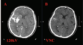
Giá trị của CLVT hai mức năng lượng trong tiên lượng nguy cơ chảy máu não sau lấy huyết khối cơ học
06/05/2021 17:04:08 | 0 binh luận
SUMMARY Purpose: Evaluate the characteristics of Dual-energy CT of the brain performed after mechanical thrombectomy. Acess the capability of iodine extravasation quantification on DECT to predict hemorrhagic complications. Material and methods: Retrospective descriptive study. Thirty consecutive patients who underwent brain dual-energy CT right after mechanical thrombectomy for acute ischemic stroke between July 2019 and September 2020 in Radiology Center, Bach Mai hospital, were included. Maximum iodine concentration was measured. Follow-up CT or MRI examinations performed at 24hrs after intervention were reviewed for intracerebral hemorrhage development. The correlation between dualenergy CT parameters and intracerebral hemorrhage development was analyzed by the Mann-Whitney U test and Fisher exact test. Receiver operating characteristic curves were generated for continuous variables. Result: Nineteen of 30 patients (63.3%) developed hemorrhage in different grades. On postoperative dual-energy CT, parenchymal hyperdensities and iodine extravasation were present in 19 (100%) of the 19 patients who developed intracerebral hemorrhage and in 7 (63.6%) of the 11 patients who did not (P = 0.0012). Signs of bleeding were present in 5 (45.4%) of the 19 patients who developed intracerebral hemorrhage and in none of the patients who did not. Median density of contrast extravasation in hemorrhage and non-hemorrhage is 108.8HU and 33.6HU (P=0.001). Median maximum iodine concentration was 2.9 mg/mL in the patients who developed intracerebral hemorrhage and 0.59 mg/mL in the patients who did not (P = 0.003). Maximum iodine concentration showed an area under the curve of 0.9 for identifying patients developing intracerebral hemorrhage. Conclusion : DECT helps differentiating haemorrhage from contrast extravasation. The presence of parenchymal hyperdensity with a maximum iodine concentration of >1.1 mg/mL may identify patients developing intracerebral hemorrhage with 94.7% sensitivity and 81.8% specificity. Key words: Dual – energy CT, thrombectomy, iodine extravasation.
Giá trị của CLVT hai mức năng lượng trong tiên lượng nguy cơ chảy máu não sau lấy huyết khối cơ học
06/05/2021 15:29:32 | 0 binh luận
SUMMARY Purpose: Evaluate the characteristics of Dual-energy CT of the brain performed after mechanical thrombectomy. Acess the capability of iodine extravasation quantification on DECT to predict hemorrhagic complications. Material and methods : Retrospective descriptive study. Thirty consecutive patients who underwent brain dual-energy CT right after mechanical thrombectomy for acute ischemic stroke between July 2019 and September 2020 in Radiology Center, Bach Mai hospital, were included. Maximum iodine concentration was measured. Follow-up CT or MRI examinations performed at 24hrs after intervention were reviewed for intracerebral hemorrhage development. The correlation between dualenergy CT parameters and intracerebral hemorrhage development was analyzed by the Mann-Whitney U test and Fisher exact test. Receiver operating characteristic curves were generated for continuous variables. Result: Nineteen of 30 patients (63.3%) developed hemorrhage in different grades. On postoperative dual-energy CT, parenchymal hyperdensities and iodine extravasation were present in 19 (100%) of the 19 patients who developed intracerebral hemorrhage and in 7 (63.6%) of the 11 patients who did not (P = 0.0012). Signs of bleeding were present in 5 (45.4%) of the 19 patients who developed intracerebral hemorrhage and in none of the patients who did not. Median density of contrast extravasation in hemorrhage and non-hemorrhage is 108.8HU and 33.6HU (P=0.001). Median maximum iodine concentration was 2.9 mg/mL in the patients who developed intracerebral hemorrhage and 0.59 mg/mL in the patients who did not (P = 0.003). Maximum iodine concentration showed an area under the curve of 0.9 for identifying patients developing intracerebral hemorrhage. Conclusion: DECT helps differentiating haemorrhage from contrast extravasation. The presence of parenchymal hyperdensity with a maximum iodine concentration of >1.1 mg/mL may identify patients developing intracerebral hemorrhage with 94.7% sensitivity and 81.8% specificity. Key words : Dual – energy CT, thrombectomy, iodine extravasation
Đánh giá kết quả đặt stent điều trị hẹp mạch nội sọ tại Trung tâm Điện quang Bệnh viện Bạch Mai
06/05/2021 16:47:06 | 0 binh luận
SUMMARY Background & Aims : Evaluation the results of the stenting on treatment of Intracranial Atherosclerosis. Methods: A prospective, non-controlled intervention study in intracranial artery stenosis patients with or without symptoms. The patients were indicated for treatment with stent placement from June 2017 to June 2020 at Radiology Center of Bach Mai Hospital. Results : The study was performed on 18 patients, including 14 patients have acute celebral ischemic stroke with intracranial stenosis and 4 patients have simple intracranial stenosis. : The study was performed including 10 men (55.6%) and 8 women (44.4%). The mean age of patients was 66.28 ± 10.87 years. The rate of successful interventions for intracranial artery stenosis was 94.4%. There are 2 patients (11.11%) had acute or immediately post intervention. Symptoms and complications, especially related to intracranial artery stenosis, were observed in 4 patients (22.22%). After an average of 3 months of follow-up, 1 patient died from perforation causing cerebral hemorrhage (5.56%) and 03 patients from stent-obstructive cerebral infarction after intervention (16.67%). Results of clinical recovery after stenting based on mRs scores with mortality, good recovery and slow recovery were 22.22%, 44.45% and 33.33%, respectively. Conclusion: The results of stent treatment for intracranial artery stenosis in our research have a high success rate. The safety of the intervention and post treatment clinical recovery rate are high. Key words : ICAD, PTAS
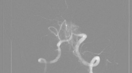
Đánh giá hiệu quả giảm đau trong tiêm phong bế rễ thần kinh thắt lưng dưới hướng dẫn của DSA
06/05/2021 15:13:57 | 0 binh luận
SUMMARY Objection. Evaluation of clinical efficacy of lumbar selective nerve root block under DSA guidance Subject and methods: Prospective study on 160 patients at Duc Giang hospital from May 2019 to May 2020 with signs of lumbar radiculopathy. Subjects were undergone lumbar selective nerve root block under DSA guidance and were assessed with visual analog scale (VAS) and Owestry disability questionaire (ODI) prior to procedure and at 3 days, 1 week, 2 weeks, 1 month and 2 months follow-up. Results: In 160 patients who undergone lumbar selective nerve root block under DSA guidance, average of age was 63.1±12.8. Female account for 60.6% of participants. VAS and ODI score were significant improve (p<0.05). Conclusion: Lumbar selective nerve root block under DSA guidance was an effective method in the treatment of sciatica. Key words: Lumbar selective nerve root block, DSA, lumbar radiculopathy.
Nghiên cứu đặc điểm hình ảnh và giá trị của cộng hưởng từ 1.5 tesla trong chấn thương dây chằng, sụn chêm khớp gối
06/05/2021 17:39:25 | 0 binh luận
SUMMARY Objective: Image Characteristic of ligament and meniscus injuries of the kneee on 1.5Tesla magnetic resonance imaging at Duc Giang hospital. Methods: The cross-sectional retrospective study of 98 patients with diagnosed of knee injuries by magnetic resonance imaging (MRI) at Duc Giang Hospital from January 2018 to January 2020, aimed to comments on MRI appearance features in knee injury. The cross sectional study on the statistical basis of data to make comments on the MRI features in the diagnosis of knee injury, using Siemen Essenza 1.5 Tesla MRI with knee joint coil. Result: The most common age is from 20 to 40 years old. Male prominent. MRI can detect osseous edema with 35.7% in tibia and Femur is 23,5%, 94.9% anterior cruciate ligament (ACL) and 5.1% posterior cruciate ligament (PCL) injury. On the other hand, there were 45,9% medial meniscal and 25,5% lateral meniscal injury. We saw 2% tibial collateral ligament and 1% fibular collateral ligament. Conclusion: MRI imaging features play an important role in diagnosing and accessing the severity of knee injury. Keywords: ligament, meniscus, knee injury, MRI, ACL, PCL
Bạn Đọc Quan tâm
Sự kiện sắp diễn ra

THÔNG BÁO SINH HOẠT KHOA HỌC 19/01/2024: CẬP NHẬT CHẨN ĐOÁN VÀ ĐIỀU TRỊ UNG THƯ TRỰC TRÀNG
19-01-2024 09:47:01 0 bình luận

Thông tin đào tạo
- Những cạm bẫy trong CĐHA vú và vai trò của trí tuệ nhân tạo
- Hội thảo trực tuyến "Cắt lớp vi tính đếm Photon: từ lý thuyết tới thực tiễn lâm sàng”
- CHƯƠNG TRÌNH ĐÀO TẠO LIÊN TỤC VỀ HÌNH ẢNH HỌC THẦN KINH: BÀI 3: U não trong trục
- Danh sách học viên đạt chứng chỉ CME khóa học "Cập nhật RSNA 2021: Công nghệ mới trong Kỷ nguyên mới"
- Danh sách học viên đạt chứng chỉ CME khóa học "Đánh giá chức năng thất phải trên siêu âm đánh dấu mô cơ tim"


Đơn vị hợp tác

