


HỘI NGHỊ KHOA HỌC "CẬP NHẬT CHUYỂN ĐỔI SỐ VÀ CÔNG NGHỆ TRONG CHẨN ĐOÁN HÌNH ẢNH - 2024"
13/12/2023 11:20:58 | 0 binh luận
ĐẶC ĐIỂM TẬP TRUNG 18FDG CỦA TỔN THƯƠNG U, HẠCH TRÊN PET/CT Ở BỆNH NHÂN UNG THƯ PHỔI KHÔNG TẾ BÀO NHỎ CÓ CHỈ ĐỊNH PHẪU THUẬT TRIỆT CĂN TẠI BỆNH VIỆN U BƯỚU HÀ NỘI
18/10/2023 11:55:43 | 0 binh luận
SUMMARY Purpose: To review some clinical features, characteristics of 18FDG uptake of tumors and lymph nodes on PET/CT in NSCLC patients with indications for radical surgery at Hanoi Oncology Hospital. Subjects and methods: 82 patients with primary NSCLC were taken with 18FDG PET/CT before surgery. Results: Right lung tumor 64.6%, left lung tumor 35.4%. The average size of the tumor was 2.6 ± 1.0 cm, the patient with lymph node (+) on PET/ CT had an average lung tumor size of 3.3 ± 0.9cm; larger than patients with N0 lymph nodes (p < 0.05). The mean SUVmax of lung tumors was 6.0 ± 4.5 and increased with tumor size (positive correlation, r=0.58). U ≤2cm, SUVmax = 4.1 ± 2.1. U >2-3cm, SUVmax = 5.3 ± 3.6 and U >3 -5cm, SUVmax = 8.3 ± 4.9. SUVmax increases with clinical disease stage and is higher in patients with positive lymph nodes on PET/CT. Conclusion: 18FDG PET/CT plays an important role in the diagnosis of NSCLC. SUVmax is a quantitative parameter related to tumor size, lymph node status and clinical disease stage. Keywords: NSCLC, 18FDG-PET/CT, SUVmax.
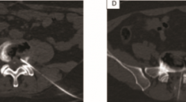
ĐÁNH GIÁ HIỆU QUẢ BAN ĐẦU ĐIỀU TRỊ THOÁT VỊ ĐĨA ĐỆM BẰNG TIÊM OZONE ĐĨA ĐỆM QUA DA VÀ PHONG BẾ RỄ BẰNG OZONE KẾT HỢP CORTICOID DƯỚI HƯỚNG DẪN CỦA CẮT LỚP VI TÍNH
18/10/2023 10:11:02 | 0 binh luận
SUMMARY Background: back pain and sciatica caused by disc herniation has a burden upon social activity. The newly minimally invasive technique, percutaneous Ozone (O2-O3) intradisal injection procedure has demonstrated safe and effective in long terms. Purpose : Evaluate the clinical effectiveness of intradiscal Ozone injection combining periradicular injection of Ozone and steroid under CT guidance for the treatment of lumbar disc herniation. Material and method: Prospective study, 100 patients symtomatic (lumbar pain, sciatica) with mild/moderate lunbar disc bulging or herniation on MRI. The patients were treated with intradiscal Ozone injection combining periradicular injection of Ozone and steroid under CT guidance. Clinical outcomes and MRI images were reviewed to evaluate at 3 months and 6 months. Results: The VAS score and the Oswestry Disability Index (ODI) before and after treatment 3months, 6 months were significant reduction. The mean improvement was 4.7 for VAS and 14 for ODI. All the procedures were technically successful. There were no adverse events associated with the treatment. Conclusion: Intradiscal injection Ozone treatment of herniated disc is an minimally invasive, easy, safe and effective procedure with low complications and side effects. Keywords: lumbar disc herniation, O3 injection
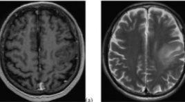
NGHIÊN CỨU GIÁ TRỊ CỦA CHUỖI XUNG TƯỚI MÁU VÀ CHUỖI XUNG PHỔ TRÊN CỘNG HƯỞNG TỪ 3 TESLA CHẨN ĐOÁN PHÂN BẬC U THẦN KINH ĐỆM
18/10/2023 10:03:50 | 0 binh luận
SUMMARY Purpose: To evaluate the diagnostic accuracy of magnetic resonance spectroscopy and dynamic contrast-enhanced (DCE) magnetic resonance perfusion for glioma grading. Materials and Methods: Fifteen patient confirmed pathological glioma who underwent MR spectroscopy and DCE in 3 Tesla MRI machine. The following parameters were used: Ktrans, Ve, Cho/NAA, Cho/Cre. The diagnostic accuracy for glioma grading was determined by ROC analysis. Results: There were 10 patients in the high-grade group and 5 patients in the low-grade group. Ktrans, Ve, Cho/NAA and Cho/Cre measures differed significantly between high and low-grade tumor. The AUC was 0.956 for Ktrans. Conclusion: Ktrans, Ve Cho/NAA and Cho/Cre parameters demonstrated to be useful for glioma grading. Keywords: Spectroscopy, DCE-MRI, glioma grading
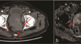
ĐẶC ĐIỂM HÌNH ẢNH VÀ VAI TRÒ CỦA CẮT LỚP VI TÍNH ĐA DÃY TOÀN THÂN TRONG UNG THƯ TUYẾN TIỀN LIỆT GIAI ĐOẠN IV
18/10/2023 09:52:14 | 0 binh luận
SUMMARY Purpose: The study was conducted to describe image characteristics of prostate cancer on multidetector computed tomography (MDCT) and to determine the role of MDCT in patients with stage IV prostate cancer. Objectives and subjects: A cross-sectional study was performed on 45 patients at Huu Nghi Hospital from January 2017 to May 2023. All patients had pathologically proven prostate cancer, multi-parameter magnetic resonance imaging (mp-MRI), MDCT, and bone scan and were categorized as stage IV. Results: The mean age was 78.31±5.64 years, mean Total prostatespecific antigen (tPSA) concentration was 279.78 ng/ml, mean prostate volume was 45.20 ml. 60% of patients had extra-prostatic invasion, 66.7% had regional lymph node metastases; 40% of patients had distant lymph node metastasis; 46.7% had bone metastasis; 28.9% had distant metastasis to other organs. MDCT had good to very good concordance with MRI in evaluating local invasion and good concordance with bone scintigraphy in evaluating bone metastasis, with p <0.05. Conclusion: MDCT had a high value in assessing metastatic lesions in patients with stage IV prostate cancer, especially lung lesions. Whole-body MDCT is a useful alternative to MRI and PET/CT in evaluating metastatic lesions from prostate cancer. Keywords: Prostate cancer, Multidetector computerized tomography, bone scintigraphy.
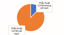
ĐÁNH GIÁ HIỆU QUẢ PHẪU THUẬT VÉT HẠCH TÁI PHÁT Ở BỆNH NHÂN UNG THƯ TUYẾN GIÁP THỂ BIỆT HOÁ SAU ĐIỀU TRỊ 131I TẠI VIỆN Y HỌC PHÓNG XẠ VÀ U BƯỚU QUÂN ĐỘI
18/10/2023 09:46:18 | 0 binh luận
SUMMARY Objectives: to describe the clinical and subclinical symptoms and evaluate the response after recurrent lymph node dissection of differentiated thyroid cancer patients after 131I treatment. Subjects: 50 differentiated thyroid cancer patients after 131I treatment, recurrence for the first time. Study methods: Retrospective and prospective description, data collection through patients’ medical records. Results: the mean age of the study patients was: 43.5 ± 12.6, the most common age is <55 years old with the rate of 80%, the female/ male ratio=3.5/1. The group of patients with Tg < 1 ng/ml accounted for the majority with 33/50 patients (66%). The median Tg before and after surgery was 2.02 ng/ml and 0.35 ng/ml, respectively. After surgery, response achieved in 98% of patients, mainly complete response and incomplete response in biochemical, accounted for 40%, 36%. Lymph node dissection reduced Tg in 78% of patients. Reccurent lymph nodes mainly observed in group IV, group III and group VI. The recurrence of lymph nodes was mainly in the central and cervical regions on the same side of the primary tumor. Conclusion: Surgery is the preferred first-line treatment for the reccurrent lymph node thyroid cancer patients. It changes response assessment, improve prognosis for reccurence patients after 131I treatment. Keywords: Differentiated thyroid cancer, response after recurrent lymph node dissection, Tg
TƯƠNG QUAN GIỮA ĐƠN VỊ HOUNSFIELD Ở CỘT SỐNG THẮT LƯNG VÀ MẬT ĐỘ XƯƠNG ĐO BẰNG DEXA Ở NGƯỜI VIỆT NAM
17/10/2023 17:41:40 | 0 binh luận
SUMMARY Background: Osteoporosis is a disease that increases the risk of fractures. Evaluation of the Hounsfield Unit in the lumbar spine on computed tomography for any reason has the potential to predict bone density abnormalities that contribute to the warning of osteoporosis. Objective: The aim of this study was to evaluate the correlation between Hounsfield Units (HU) in Lumbar Spine (LS) and bone mineral density (BMD) measured by Dual-Energy X-Ray Absorptiometry (DEXA). Method: Retrospective and cross-sectional study of 150 patients comprised CT-DXA pairs within a 6-month period performed for any indication. Measure HU at lumbar vertebrae from L1 to L4. Calculate Spearman correlation coeffificient between the mean HU at lumbar vertebrae and the BMD values from DEXA scan. Using area under the ROC Curve (AUC) and finding cut-off of HU for diferentiating normal BMD and abnormal BMD, osteopenia and osteoporosis. Result: We noted correlations between the HU at LS and the BMD from DXA scan which is significant, the highest correlation at L2 (Spearman correlation coefficient = 0.68). At L2, normal BMD: ≥ 131 HU, osteopenia: 101 – 131 HU, osteoporosis: ≤ 131 HU, we also determined that threshold of 101 HU was more than 90 % sensitive, and a threshold of 171 HU was more than 90 % specific for distinguishing normal BMD. In addition, cut-off ≤ 132 HU was more than 90 % sensitive, and cut-off ≤ 62 HU was more than 90 % specific for distinguishing osteoporosis from osteopenia. Conclusion: The correlations between the HU at LS and the BMD from DXA scan is significant. Keyword : Osteoporosis, DEXA, HU at lumbar spine
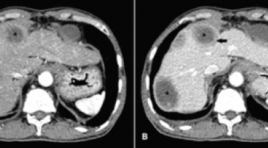
MÔ TẢ ĐẶC ĐIỂM HÌNH ẢNH DI CĂN GAN Ở BỆNH NHÂN UNG THƯ ĐẠI TRỰC TRÀNG TRÊN CLVTV
17/10/2023 17:47:51 | 0 binh luận
SUMMARY Background: Colorectal cancer is a common cancer that has a high morbidity and mortality rate, and the liver is the most frequent metastatic organ. Dynamic computed tomography (CT) can be used to diagnose colorectal liver metastases based on their imaging characteristics and enhancing patterns which are different from other liver lesions. Objective: To describe the imaging characteristics of colorectal liver metastases on dynamic CT. Methods: A cross–sectional study was conducted on 39 colorectal cancer patients with pathologically proven liver metastases. All patients underwent triphasic CT. Other lesions sharing similar imaging characteristics were considered metastases. Patient demographic and CT findings were documented Results : 39 patients and 92 lesions were included in the analysis. Liver metastases can be solitary (48.7%) or multiple (51.3%). Most of them are located in the right lobe (62%) more than the left lobe. Lesions less than 5 cm have the highest rate (80.5%). Liver metastases are often well–defined (54.4%) with non-lobulated borders (58.7%). Calcification is rare (5.4%). Compared to the surrounding liver parenchyma, colorectal liver metastases have a lower density in the pre-contrast phase (89.3%), and are also less enhanced in arterial and venous phases (93.5% and 98.9% gradually). Contrast-enhanced lesions are usually heterogeneously (91.3%). Rim enhancement at the peripheral area is often observed (78.2%). Of 51 lesions, it appears on both arterial and venous phases (55.4%). Central necrosis is about 41.3% of the total lesions and dominantly appears in lesions larger than 3 cm. Conclusions: Imaging characteristics of the colorectal liver metastases mostly observed are the thin rim enhancement at the peripheral area, low density in the pre-contrast phase, less enhancement than the normal surrounding liver parenchyma on both arterial and venous phases, central necrosis in the large lesions, and heterogeneously enhanced pattern. Keywords : colorectal liver metastases, dynamic CT, imaging characteristics.
Bạn Đọc Quan tâm
Sự kiện sắp diễn ra


THÔNG BÁO SINH HOẠT KHOA HỌC 19/01/2024: CẬP NHẬT CHẨN ĐOÁN VÀ ĐIỀU TRỊ UNG THƯ TRỰC TRÀNG
19-01-2024 09:47:01 0 bình luận
THÔNG BÁO SINH HOẠT KHOA HỌC 19/01/2024: CẬP NHẬT CHẨN ĐOÁN VÀ ĐIỀU TRỊ UNG THƯ TRỰC TRÀNG
19-01-2024 09:47:01 0 bình luận
prev
next
Thông tin đào tạo
- Những cạm bẫy trong CĐHA vú và vai trò của trí tuệ nhân tạo
- Hội thảo trực tuyến "Cắt lớp vi tính đếm Photon: từ lý thuyết tới thực tiễn lâm sàng”
- CHƯƠNG TRÌNH ĐÀO TẠO LIÊN TỤC VỀ HÌNH ẢNH HỌC THẦN KINH: BÀI 3: U não trong trục
- Danh sách học viên đạt chứng chỉ CME khóa học "Cập nhật RSNA 2021: Công nghệ mới trong Kỷ nguyên mới"
- Danh sách học viên đạt chứng chỉ CME khóa học "Đánh giá chức năng thất phải trên siêu âm đánh dấu mô cơ tim"


Đơn vị hợp tác

