Case lâm sàng: Tràn dịch màng ngoài tim do dò dưỡng chấp, nhân một trường hợp được điều trị tại bệnh viện Nhi Trung Ương
20/04/2021 13:28:35 | 0 binh luận
Đang cập nhật
Can thiệp điều trị phình động mạch não cổ rộng vị trí đỉnh thân nền bằng web: báo cáo ca lâm sàng
06/05/2021 17:44:59 | 0 binh luận
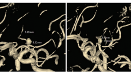
SUMMARY A basilar tip artery bifurcation aneurysm was diagnosed incidentally in a 69-year-old female patient. She was a normal her medical history. The MRI revealed a non-ruptured aneurysm with a maximum fundus diameter 4.9mm. Selective cerebral angiography confirmed the presence of a wide-necked aneurysm with a mean transverse diameter of 5mm and a mean height of 3mm; the neck width was 4.9mm. With this complex anatomy, the multidisciplinary team recommended endovascular treatment of the aneurysm with the WEB device. The procedure was performed with dual antiplatelet therapy (75 mg aspirin PO daily 3 days and 180 mg ticagrelor PO daily 2 days prior to the intervention). Immediately after placement of the WEB device, DSA showed complete occlusion of the aneurysm but has thromboembolic event in P3 posterior cerebral artery and no deficit. Postoperatively the patient received dual antiplatelet therapy for 5 days and continued 75 mg aspirin PO for 2 weeks, ticagrelor was discontinued. The patient was discharged 2 days after the intervention without any neurological symptoms. Follow-up by MRI at 3, 6, by MRI and DSA 12 months after treatment confirmed the stable obliteration of the aneurysm without recurrence or reperfusion. The value of the WEB device in the endovascular treatment of wide-necked basilar tip artery bifurcation aneurysms is the main topic of this chapter.

Nhân một trường hợp chảy máu ổ bụng tự phát do tổn thương động mạch vị phải được chẩn đoán bằng chụp cắt lớp vi tính
31/03/2020 16:24:54 | 0 binh luận
Acute abdominal hemorrage by gastric atery aneurysm rupture- Case report SUMMARY We show the case of acute abdominal hemorrhage by gastric atery aneurysm rupture that was diagnosed and treated in Viet Tiep Hospital, Hai Phong. The female patient who was attended to hospital in acute lost blood volume. The patient was examined by US and abdominal CT scanner. The diagnosis was gastric atery aneurysm rupture. The patient was treated by surgery 4/5 gastroectomy because of gastric necrosis. In our experience, the large blood clot in lesser sac, increase arterial diameter and leak of contrast agent were suggested of diagnosis.
Không có thân chung động mạch vành trái trên CLVT 256 dãy: nhân một trường hợp và tổng hợp y văn
04/12/2019 20:56:05 | 0 binh luận
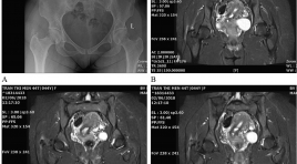
Absent left main coronary artery on MDCT 256 Slices: a case report and literature review SUMMARY Absent left main coronary artery is rare congenital cardiacvascular anomalous. Absent LMCA has been detected by digital substration angiography (DSA) or operating. Nowadays,multi detector computer tomography (MDCT) can be easily findingand non - invasive procedure. We report a 80 - year – old male was admitted to our hospital with atypical chest pain. An MDCT 256 slices showedseparate origin of left anterior descending (LAD) and left circumflex artery (LCx) from left sinus of Valsalva, absent LMCA. Keywords : Absent left main coronary artery, anomalus congenital cardiacvascular, multi detector computer tomography (MDCT).
sinh thiết, chọc tế nào khối tụy và quanh tụy
17/03/2020 11:18:58 | 0 binh luận
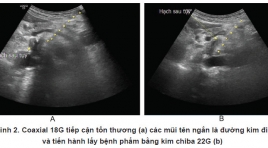
Ultrasound-guided fine needle aspiration and core needle biopsy for pancreatic neoplasms and surrounding-pancreatic neoplasms SUMMARY Solid pancreatic or peripancreatic lesions are common diseases . All most of them need to pathology diagnosis. provides an alternative pathway for adequate specimen acquisition. Because of the deep retroperitoneal location, we usually use endoscopic ultrasound (EUS)-guided fine needle aspiration (FNA) or inderect percutaneous core needle biopsy (CNB) . Both of them have disadvantages on their own. the sensitivity and specificity of EUS-guided FNA (EUS-FNA) for pancreatic neoplasms has were 85% and 98%, respectively and The complication rate of EUS-FNA is approximately 1%–2% [1]. However, one limitation related to this technique is that it often only provides a cytologic specimen with scant cellularity and lack of histologic architecture, which restrains us from making a complete tissue analysis for diagnosis and grade differentiation. the sensitivity and specificity of inderect percutaneous CNB for pancreatic neoplasms has were 90,4% and 92% respectively [2], overcome the disadvantages of EUS-FNA [3]. But it is relatively risky and difficult especially for those who do not have much experience. Reaching through the liver, spleen and kidneys increases the risk of bleeding. Approaching through the stomach and intestine, the incidence of complications can reach 15.3% [4], [5] including: infection, peritonitis and gastrointestinal perforation. Access through the gallbladder has a high risk of cholestatic and cholecystitis [4]. Derect percutaneous CNB/ FNA is a new technique which can make good the risks of two technique before. In this review, we will present some cases of FNA and biopsy of solid pancreatic or peripancreatic lesions by direct access approach, thereby showing about techniques, diagnostic effectiveness as well as complications of percutaneous CNB/ FNA. Key word: Fine needle aspiration (FNA), Core needle biopsy (CNB), Pancreas.
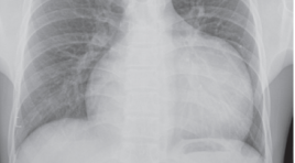
Phình khổng lồ tiểu nhĩ trái: báo cáo một trường hợp hiếm gặp tại bệnh viện Bạch Mai
30/03/2020 15:35:19 | 0 binh luận
Giant left atrial appendage aneurysm with radiology: case report and literature SUMMARY We report a extremely rare case of 16-year-old boy with a giant left atrial appendage aneurysm. Patient‘s symptoms are dyspnea and atrial tachyarrhythmia. The exact diagnosis is make by chest X-ray, echocardiography (transthoracic and transoesophageal), magnetic resonance imaging, and multislice computer tomographic angiography. The patient was successfully treated with surgical resection of the aneurysm. For its rarity, this case is reported and review about literature of aneurysm of left atrial appendage. Because of dangerous of atrial aneurysm (dangerous fifth chamber cardiac) with supraventricular arrhythmias and systemic thromboembolism, surgical resection is the best method to avoid further complication and recurrence. Key word : Giant left atrial aneurysm.
Sling động mạch phổi trái: nhân 5 trường hợp
13/04/2020 15:33:22 | 0 binh luận
Mô tả lần đầu 1958 (Contro và cs). Nguyên nhân hiếm gặp của hẹp khí-phế quản bẩm sinh (4% các bất thường mạch máu). ĐM phổi trái xuất phát từ mặt sau ĐM phổi phải,chạy giữa khí quản-thực quản để đến phổi trái. 50% gây hẹp khí quản. Phôi thai học: Tuần thứ 2 đến tuần thứ 7. 2 ĐM chủ lên và 2 ĐM chủ xuống nối với nhau bởi 6 cung mạch. Cung mạch số VI phát triển thành ĐM phổi và ống ĐM. Các chồi mạch của cung mạch số VI và mầm phổi nối với nhau tạo thành 2 ĐM phổi. Chồi mạch bên trái kết nối thất bại với mầm phổi -> Sling ĐM phổi.
Nghiên cứu biến thể giải phẩu và kích thước các nhánh chính động mạch vành trên hình ảnh MSCT 64
13/04/2020 12:38:14 | 0 binh luận
Nội dung: Đặt vấn đề Tổng quan Mục tiêu nghiên cứu Phương pháp nghiên cứu Kết quả và bàn luận Kết luận
Bạn Đọc Quan tâm
Sự kiện sắp diễn ra
Thông tin đào tạo
- Những cạm bẫy trong CĐHA vú và vai trò của trí tuệ nhân tạo
- Hội thảo trực tuyến "Cắt lớp vi tính đếm Photon: từ lý thuyết tới thực tiễn lâm sàng”
- CHƯƠNG TRÌNH ĐÀO TẠO LIÊN TỤC VỀ HÌNH ẢNH HỌC THẦN KINH: BÀI 3: U não trong trục
- Danh sách học viên đạt chứng chỉ CME khóa học "Cập nhật RSNA 2021: Công nghệ mới trong Kỷ nguyên mới"
- Danh sách học viên đạt chứng chỉ CME khóa học "Đánh giá chức năng thất phải trên siêu âm đánh dấu mô cơ tim"


