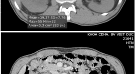
Nghiên cứu đặc điểm hình ảnh và giá trị của cắt lớp vi tính 64 dãy trong chẩn đoán u đường bài xuất tiết niệu cao
01/04/2020 13:44:43 | 0 binh luận
Imanging characteristics and the value of 64-slice computed tomography in diagnosis of upper urinary tract tumor SUMMARY Objective: To describe the imanging characterise and analyze the value of 64-slice computed tomography (CT) in the evaluation of upper urinary tract tumors. Patients and Methods: Retrospective review of the patients who underwent 64-slice CT examination, was operated in VietDuc Hospital with upper urinary tract tumor in pathologic result from 8/2013 to 8/2015. This was observation study comparing the imaging finding with the result from surgery and pathology. Result: 56 patients with upper urinary tract tumor (32men, 24women), age 63.9±11.1. The sensitivity of 64-slice CT for upper urinary tract tumor was 92.86%. The accuracy of 64- slice CT in diagnosing T stage was 82.14%. Conclusion: 64-slice CT had high value in diagnosing upper urinary tract tumors. Keywords: 64-slice computed tomography, upper urinary tract tumor.
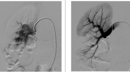
Điều trị dị dạng động tĩnh mạch thận bẩm sinh bằng can thiệp nội mạch
01/04/2020 11:38:23 | 0 binh luận
Transarterial embolization in management of congenital renal arteriovenous malformation SUMMARY Objective: To evaluate the safety and efficacy of transarterial embolization (TAE) in management of congential renal arteriovenous malformation (AVM). Patients and Methods: Between December 2007 and June 2015, 11 patients with congential renal AVM treated with TAE was investigated for clinical presentation, imagine features, treatment methods and complications in Viet Duc hospital. Results: 11 patients (9 women/2 men) with 10/11 gross hematuria, 5/11 flank pain and 1/11 hypertension underwent 11 sessions of treatment, TAE was performed with histoacryl + lipiodol in 7 patients, micro-coils in 3 patients, absolute alcohol and histoacryl in 1 patient. Technical and clinical success were obtained in all patients. There was only 1 patient with fever, renal function was normal in all patient pre - embolization and post - embolization. Conclusion: TAE treatment was safe and effective, it should be recommended as the first choice to treat congential renal AVM. Key word : renal arteriovenous malformation, embolization.

Kết quả điều trị phình động mạch não tuần hoàn sau bằng can thiệp nội mạch
01/04/2020 13:24:26 | 0 binh luận
Results of endovascular treatment of posterior fossa intracranial aneuryms SUMMARY Purpose : To evaluate the results of endovascular treatment of posterior fossa intracanial aneuryms. Method and materials: Fourty one patients harboring fourty five posterior fossa intracranial aneuryms were treated by endovascular therapy from 2012 to 2015 at Bach Mai Hospital. Clinical outcomes and follow-up of aneurysm occlusion’s result were classified by modified Rankin Scale and on MRI imaging. Results: Twenty eight patients presented subaranoidien hemorrhage and thirteen patients without SAH. Different technics were used such as coiling embolization (44.4%), coiling with balloon remodeling technics (22.2%), coiling with stenting (2.3%), flow-diverter stenting (6.7%) and parent artery occlusion (24.4%). The rate of complete aneurysm occlusion, neck residue and partial occlusion were 80%; 11.1% and 8.9% respectively. Result of good outcomes (mRS 0-2) was 85.4% and mortality was 12.2%. Almost of aneurysms post treatment were stability (84.6%), only 15,4% aneurysm were slight recanalization. The hospitalization duration of unruptured and ruptured aneurysm were 9.3 and 17.8 days, respectively. Conclusion: Endovascular treatment of posterior circulation aneurysms showed efficace of clinical outcome and stability with low morbility and mortality. Keywords: Aneurysm, posterior circulation aneurysm, embolization.
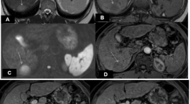
Đặc điểm hình ảnh và giá trị của cộng hưởng từ trong chẩn đoán ung thư gan nguyên phát ≤ 3 cm ở bệnh nhân xơ gan
01/04/2020 13:18:51 | 0 binh luận
Imaging characteristics and values of MRI for diagnosis of hepatocellular carcinoma ≤ 3cm in cirrhotic patients SUMMARY Purpose: To describe the MR imaging features and to evaluate values of MRI for diangosis of hepatocellular carcinomas ≤ 3cm in cirrhotic patients. Materials and methods : 35 patients with final diagnosis of hepatocellular carcinoma (HCC)≤ 3cm undergone MRI 1.5 Tesla at Radiology Departement (Bach MaiHospital) from August 2014 to July 2015. Results : The average diameter was 21.14mm, all were solitary, most of tumor locate in right liver. Among 35 tumors detected: 100% was hyperintense on Diffusion; 97.1% hyperintense on T2W; on inphase T1W 11.4% hyperintense, 57.2% hypointense, 31.4% isointense; on outphase T1W 2.9% hyperintense, 82.8% hypointense, 14.3% isointense; 40% tumors have fatty content inside. After contrast gadolium injection, at the arterial phase 71.4% of tumors showed enhancement; at the portal phase 48.57% of tumors showed contrast wash out; at the late phase 68.6% of tumors had contrast wash out and 68.6% of tumors had a rim enhancement. MRI dectected 100% of tumors and diagnosed 54.3% of tumors based on the characteristic arterial contrast enhancement and portal or late phase contrast washout. The combination of ≥3 or ≥4 morphological and hemodynamic signs could increase the diagnostic sensitivity to 91.4% and 60% respectively. Conclusion : Among 35 HCCs ≤ 3cm in cirrhotic livers: 97.1% and 100% appeared hyperintense on T2W and Diffusion respectively, most tumors were hyposignal on outphase T1W, 40% had fatty content. After gadolium contrast injection, 71.4% tumors had arterial contrast enhancement, 68.6% tumors showed portal or late contrast washout, 68.6% had rim enhancement. MRI detected 100% tumors and diagnosed 54.3% of all tumors. Keywords: Hepatocellular carcinoma, HCC, cirrhosis, MRI.
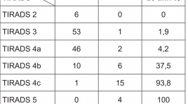
Cập nhật chẩn đoán hình ảnh phân loại bướu nhân tuyến giáp lành tính và ác tính
01/04/2020 12:49:56 | 0 binh luận
Nghiên cứu đặc điểm hình ảnh của ung thư vú tại bệnh viện Trung ương Huế
01/04/2020 12:45:52 | 0 binh luận
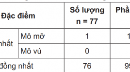
The ultrasonographic and mammographic appearance of breast cancer at Hue national Hospital SUMMARY Purpose: Desciber the ultrasonographic and mammographic appearance of breast cancer . Method and Material: Retrtospectively study the imaging findings based on guidline of ACR of 77 cases of breast cancer at Hue’s central hospital. Results : 1/ Ultrasonographic appearance: 99.7% patient having inhomogenous breast tissue. 90.91% tumor having irregular shape. There are only 59.7% tumor having orientation parallel to skin surface. 9.6.21% tumor having hypoechoic and heterogenous structure. Most of tumor having complet or partiasl shadowing. 2/ Mammographic appearance: Most tumor having heterogenous and dense structure. 92.3% tumor having irregular shape. 88.47% tumor having ill-defined or irregular border. Most microcalcifiction inside tumor are classified to high risk type. Conclusion: Most patient having inhomogenous breast tissue. Interpretation of mammographic film should corelate with ultrasonographic images. Most tumor were classified at level more than 3, when image findings of tumor were used according to ACR’s guidline. Keywords: Breast cancer, ultrasound, mamography.
Nghiên cứu đặc điểm hình ảnh trên MRI 3.0 Tesla trong bệnh lý u vùng khoang miệng và hầu họng trên xương móng tại bệnh viện ung thư Đà Nẵng
01/04/2020 12:40:19 | 0 binh luận
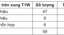
MRI imaging in oral and pharyngeal cancer in Danang cancer hospital SUMMARY Background: Oral and pharyngeal tumor are more common today and it has complex structure limiting for the paraclinic examination. CT scan was a first choiced to examine the stage of tumor, especially invasion of tumor to the skull base but nowaday CT scan has been displaced by MRI, which has high value to detect soft tissue tumor with high sensitive and acurracy. MRI also gives the best informstion about anatomy in 2D and 3D. Object and Method: Cross study, clinical examination find out tumor in the oral or pharyngeal then takes the MRI picture, we except the patient without hystopathology and treated for cancer before. Coletting the MRI images data in T1W, T2W, STIR, T1W Gd. Object: MRI machine Siemens 3.0Tesla Model Verio A Tim System T-class, Coil 3T neck A Timy System, Dotarem 10ml. Method: We decribe every characteristics of MRI images in TIRM Cor, Ax và Sag T1W; Ax và Sag T2W; Ax, Cor và Sag T1 FS+Gd pulse then comparing this characteristics with grade histopathology. Result: Age: 59.6; male/female=2.5/1; tumor in oral cavity: 35.6%; in hypopharyngeal 23.8%; nasopharyngeal 22% and oropharyngeal 18%. Diameter max: 3.17cm ±1.6. Characteristics in MRI: 80% hypointensity in T1W, 76% hypersignal in T2W, 81% hyperintensity in STIR, 79% medium - strong enhance in T1W Gd with this feature the sensitives and acuracy to diagnostic degree malignant of tumor: sensitives and acuracy in T1W: 86% and 71%; in T2W is 84% and 85%, STIR: 90% and 85%; T1W Gd: 86% and 71%. Conclusion: MRI has high value to diagnostic in oral and pharyngeal cancer. Especially, MRI play an important role to determine the grade of cancer with high sensitive and acuracy. Keywords: Oral and pharyngeal tumor, MRI
Sinh thiết ngực dưới hướng dẫn của cắt lớp vi tính liều thấp
01/04/2020 12:03:47 | 0 binh luận
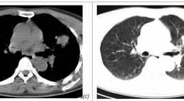
The application of low dose CT guided thoracic biopsy SUMMARY Purpose: Study the application of low dose CT guided thoracic biopsy. Materials and Methods : 192 patients with CT guided thoracic biopsy including 100 patients with rutine protocol (group A) and 92 patients with low dose protocol (group B) from 4/2014 to 4/2015. Results: the average total dose length product is 274.3+/- 172.26 mGy in group A and 93.8+/-32.87 mGy in group B (p<0.001). Effective dose is 3.8+/-2.41 mSv in group A and 1.3 +/-0.46 mSv in group B (p <0.01). Pneumothorax are 17 (17%) in group A and 11 (11.96%) in group B. Three cases in group A and two cases in group B have instantly pneumothorax drainage. Hemothorax is 1 (1%) in group A and 1 (1.1%) in group B. All the complication are stable without intervention. Conclusion: low dose CT guided thoracic biosy reduces remarkable radiation dose, neither increases the complication nor degrades image quality or diagnostic accuracy. Key words: CT guided thoracic, low dose CT guided thoracic biosy.
Bạn Đọc Quan tâm
Sự kiện sắp diễn ra
Thông tin đào tạo
- Những cạm bẫy trong CĐHA vú và vai trò của trí tuệ nhân tạo
- Hội thảo trực tuyến "Cắt lớp vi tính đếm Photon: từ lý thuyết tới thực tiễn lâm sàng”
- CHƯƠNG TRÌNH ĐÀO TẠO LIÊN TỤC VỀ HÌNH ẢNH HỌC THẦN KINH: BÀI 3: U não trong trục
- Danh sách học viên đạt chứng chỉ CME khóa học "Cập nhật RSNA 2021: Công nghệ mới trong Kỷ nguyên mới"
- Danh sách học viên đạt chứng chỉ CME khóa học "Đánh giá chức năng thất phải trên siêu âm đánh dấu mô cơ tim"


