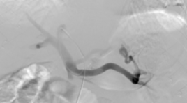
CAN THIỆP XUYÊN GAN QUA DA NÚT BÚI GIÃN TĨNH MẠCH DẠ DÀY VỠ: BÁO CÁO CA LÂM SÀNG
12/10/2023 14:14:24 | 0 binh luận
SUMMARY Background: Upper gastrointestinal bleeding from gastroesophageal varices is a frequent complication in patients with liver cirrhosis and portal hypertension. Gastric varices are difficult to control with endoscopic while endovascular intervention is an effective method. We report a case of ruptured gastric varices that was successfully treated by percutaneous transhepatic obliteration. Case presentation : A 50-year-old male patient, with a history of cirrhosis, was admitted to the hospital with a large amount of blood vomiting and black stools. Gastroscopy showed ruptured gastric varices and blood sprayed. The patient was occluded with gastric varices by percutaneous transhepatic obliteration. After 2 days, the patient's condition improved, yellow stools, and was discharged. Conclusion: Obliteration of gastric varices in patients with cirrhosis by percutaneous transhepatic obliteration (PTO) is a safe and effective technique, a good choice in the absence of gastrorenal shunt. The advantage is intervention in emergencies, no need to examine the vessels first with computed tomography. Keywords: Gstric varices, percutaneous transhepatic obliteration.
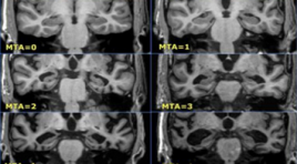
NGHIÊN CỨU MỐI LIÊN QUAN GIỮA SUY GIẢM NHẬN THỨC VÀ SA SÚT TRÍ TUỆ VỚI TỔN THƯƠNG NÃO TRÊN CỘNG HƯỞNG TỪ
12/10/2023 13:01:23 | 0 binh luận
SUMMARY Purpose: To study the association between cognitive impairment, dementia, and brain features on magnetic resonance imaging. Retrospective and Prospective study: A cross-sectional descriptive study with 100 patients above 60 years, made MMSE and tested a brain MRI at Quang Tri Provincial General Hospital from 1/2021 to 10/2021. Results: In 100 studied patients, tested by MMSE, 38% had normal cognitive status, 51% had mild cognitive impairment, and 11% had dementia. Cognitive decline is associated with age but not sex. Cognitive impairment and dementia are associated with global brain atrophy, medial temporal cephalic atrophy, frontal lobe atrophy, parietal lobe atrophy, and occipital lobe atrophy but not white matter hyperintensities (small vessel disease) and lacunar infarction. The higher the GCA (global cortical atrophy scale), the higher the MTA (mesial temporal atrophy scale), and the more severe the cognitive impairment. Conclusion: Magnetic resonance imaging is a strongly important role in the diagnosis of cognitive decline and dementia, with features such as total cerebral atrophy, medial temporal brain atrophy, parietal lobe atrophy, frontal lobe atrophy, and occipital lobes atrophy are all involved in the patient's cognitive status, and the assessment of these features on magnetic resonance imaging help to the early diagnosis and prediction of future dementia. Keyword: Dementia, mild cognitive impairment, magnetic resonance imaging
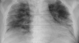
ĐẶC ĐIỂM HÌNH ẢNH X QUANG NGỰC Ở BỆNH NHÂN COVID-19 TỬ VONG TẠI PHÒNG CẤP CỨU BỆNH VIỆN CHỢ RẪY
12/10/2023 12:49:46 | 0 binh luận
SUMMARY Background: COVID-19 is a global pandemic with very high mortality. Patients are hospitalized with rapid disease progression and early death in the Emergency Department. Chest X-rays play an important role in the diagnosis and prognosis of mortality. Purpose: (1) describe the chest X-ray findings of deceased covid-19 patients; (2) evaluate the relationship between Brixia score to mortality of COVID-19 patients in the Emergency Department. Methods: Retrospective study of COVID-19 patients who died at the Emergency Department, Cho Ray Hospital from August 1, 2021 to August 31, 2021, diagnosed with COVID-19 by RT-PCR technique. Clinical features and radiographic features were collected. Results: There are 226 cases in our study. We found that almost lesions were distributed in the bilateral lung (99%), in all 6 lung regions (78.3%), and diffuse distribution (94,2%). Interstitial, alveolar and consolidation lesions accounted for 69.5%, 61.9% and 94.7%, respectively. Pleural effusion occurs in 10.9% of cases. Pneumothorax and pneumomediastinum accounted for only 0.9% and 1.3% of cases. The mean Brixia score was 15. The mild, moderate, and severe subgroups of the Brixia score were 6 (2.7%), 31 (13.8%), and 188 (83.5%) cases, respectively. There is a relationship between Brixia score and gender. Conclusions: Characteristics of chest X-rays in deceased COVID-19 patients mostly had consolidation, multifocal, many lung fields involved, and diffuse distribution in both lungs. Other features such as pleural effusion, pneumothorax, subcutaneous emphysema, and pleural thickening can be observed. Most of the patients who died had high Brixia scores. Keywords: Chest X-ray, COVID-19, deceased, Emergency Department, Brixia score.
NGHIÊN CỨU GIÁ TRỊ CỦA CỘNG HƯỞNG TỪ TƯỚI MÁU TRONG CHẨN ĐOÁN VÀ PHÂN LOẠI MÔ BỆNH HỌC UNG THƯ VÚ
12/10/2023 12:44:10 | 0 binh luận
SUMMARY Objective: To investigate wherethe the perfusion parameters of dynamic contrast enhanced magnetic resonance imaging (DCE-MRI) had correlation with molecular subtypes of breast invase ductal carcinoma (IDC). Methods : Forty patients underwent 3T MRI examination following biopsied pathological immunohistochemistry at Bach Mai Hospital from March 2021 to July 2022 were retrospectively investigated. The volume transfer constant (Ktrans), the rate constant (Kep) and the plasma volume ratio (Ve) were calculated from DCE-MRI base on the extended Tofts model. The mean and standard deviation of each perfusion parameters and the pathological immunohistochemistry, subtypes correlations were assessed by Mann-Whitney U test and One-way ANOVA. Results : 40 IDC patients (mean age, 55,5 ± 11.6; range, 38 to 82 years) were investigated. The mean of Kep was higher in tumors with estrogen receptor (ER) negative (p = 0.03), progesterone receptor (PR) negative (p = 0.03), human epidermal growth factor receptor 2 (HER-2) positive (p = 0.02) than that in tumors with ER-positive, PR-positive, and HER-2-negative. Also, Kep was significantly higher in HER-2 enriched tumors than that in luminal B tumors (p = 0.01) and triple-negative tumors (p < 0.05). Conclusion: DCE-MRI may be used as the non-invase tool to assess the molecular biological expression and the molecular subtypes of invase ductal breast cancer. Keywords: Dynamic contrast enhancement, magnetic resonance imaging, subtypes breast cancer, quantitative value.

ĐÁNH GIÁ KẾT QUẢ SỚM ĐIỀU TRỊ NANG TUYẾN VÚ BẰNG VI SÓNG TẠI ĐƠN VỊ TUYẾN VÚ BỆNH VIỆN CHỢ RẪY
12/10/2023 12:34:22 | 0 binh luận
SUMMARY Background- Objective : Breast cyst is common in premenopausal women (61.5 %) in comparison with the menopause population (39.4%) and the most common age is 35 to 50. The treatment of breast cysts using microwave has a lot of advantages and should be a considered method in clinical practice. Our objective is to evaluate the early result of breast cyst treatment with microwave under ultrasound guidance in the breast unit of Cho Ray hospital. Method: We prospective reviewed patients who underwent breast cyst treatment using microwave ablation in Breast unit of Cho Ray hospital Ho Chi Minh city from 08/2018 to 03/2020. Result: 58 patients are reviewed. The average age is 45.3. Simple breast cyst, complicated breast cyst, and complex breast cyst are 80%, 16.93 %, and 3.07 % prospectively. The average diameter of ablated cysts is 23.86 mm. All of the patients have a complete response after treatment, and the decrease in size ratio has reached more than 90 % after 6 months. No complication is recorded in the treatment process. There is one case, with calcification of the cyst wall, that does not respond to microwave ablation. Conclusion: The microwave ablation procedure is a promising method for symptom breast cyst with a high rate of success and low rate of complication. Breast cyst with calcified wall should have a careful assessment before the procedure. Key Word: Breast cyst, Microwave ablation
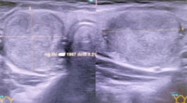
KẾT QUẢ ĐIỀU TRỊ ĐỐT U TUYẾN GIÁP LÀNH TÍNH BẰNG VI SÓNG TẠI VIỆN Y HỌC PHÓNG XẠ VÀ U BƯỚU QUÂN ĐỘI
12/10/2023 12:29:19 | 0 binh luận
SUMMARY Objective: To evaluate the early results of Microwave ablation in the treatment of benign thyroid tumors at the Vietnamese Military Institute Of Medical Radiology And Oncology from 10/2020 to 3/2022. Subjects and Methods : Prospective, descriptive studies of 32 patients with benign thyroid tumors treat by microwave ablation at the Vietnamese Military Institute Of Medical Radiology And Oncology from 10/2020 to 3/2022. Results: Mean of age: 40.91 ± 17.00, female (90,63%), male (9,37%). The solitary tumor occults 62.5%, the thyroid tumors in both glands: are 37.49%, and the average tumor size: is 24.27 ± 7.65 mm. The tumors classified into TIRADS 3 by ultrasound was 96,87%. The mean operating time was 27 ± 5 minutes, all patients were coming home after treatment. Post-operative infection: 3,13%, minor skin burn: 3.13%. The disease symptoms were lost after 3 months of therapy. Conclusions: Using microwave ablation in the treatment of benign thyroid tumors was quite safe, and achieved high cosmetic outcomes. Keyword: Thyroid nodule, Microwave ablation
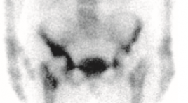
ĐÁNH GIÁ VAI TRÒ CỦA XẠ HÌNH XƯƠNG BA PHA VỚI MÁY SPECT/CT TRONG THEO DÕI BỆNH NHÂN SAU THAY KHỚP NHÂN TẠO TẠI BỆNH VIỆN ĐÀ NẴNG
12/10/2023 10:39:14 | 0 binh luận
SUMMARY Objectives: Describe the clinical and subclinical characteristics of patients after joint replacement who underwent three-phase bone scan at Danang Hospital and evaluation of the role of three-phase bone scan method with SPECT/CT in monitoring patients after joint replacement. Study subjects: 32 patients after joint replacement who underwent three-phase bone scan with SPECT/CT Hawkeye 4 produced by GE at the Department of Medicine, Danang Hospital. Methods of study : Cross-sectional descriptive, longitudinal monitoring, retrospective and prospective. Results: Mean age: 69.88 ± 7.80. Female/male ratio: 23/9. Pain is the most common symptom. The white blood cell count and CRP concentration were mostly incresed. There were no abnormal changes in the X-ray picture. The image of joint loosening accounts for the highest proportion. Most of the knee scintigraphy shows abnormalities. Sensitivity: 83.33%. Specificity: 75%. Accuracy: 81.25%. Positive predictive value: 90.91%. Negative predictive value: 60%. Conclusions: Three-phase bone scintigraphy is a simple method making it easy to perform with its high sensitivity and positive predictive value in monitoring patients after artificial joint replacement. Keywords: Three-phase bone scan, artificial joints, joint loosening.
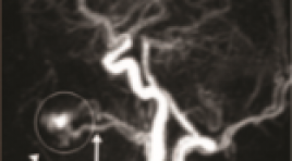
BƯỚC ĐẦU PHÂN TÍCH KẾT QUẢ ỨNG DỤNG KỸ THUẬT MRA TWIST TRONG ĐÁNH GIÁ DỊ DẠNG THÔNG ĐỘNG - TĨNH MẠCH NGOẠI BIÊN
12/10/2023 10:32:28 | 0 binh luận
SUMMARY Background: TWIST is a MRA with high time resolution, which can display the mapping of vascular structure well. The identification of the feeding and draining vasculature of peripheral arteriovenous malformation (pAVM) is important for the appropriate treatment planning. Therefore, we examined the application of MRA TWIST in evaluation pAVM in comparison with Digital Subtraction Angiography (DSA). Objective: To evaluate the feeding arteries and draining veins of pAVM with MRA TWIST in comparison with DSA. Subjects and methods: A retrospective study between January 2016 and July 2021 was conducted upon 25 patients (7 males and 18 females; mean age 22,2; age range 3-53 years) with pAVM, who obtained MRA TWIST and then underwent DSA for confirming the diagnosis. The numbers and names of the feeding arteries and early draining veins were evaluated by two independent readers. The accurate ratio and Kappa test for the interobserver agreement were calculated. Results: About the feeding arteries, MRA TWIST accurately evaluated 82,6% in head and neck, and 85,7% in limbs in comparison with DSA. About the draining veins, MRA TWIST accurately evaluated 84% in comparison with DSA. Kappa coefficient showed good agreement in detecting the feeding arteries and the draining veins between MRA TWIST and DSA. Conclusion: MRA TWIST is a reliable non-invasive imaging technique for evaluation of the feeding arteries and the draining veins of pAVM and can be useful for planning the endovascular treatment. Keywords: peripheral arteriovenous malformation (pAVM), MRA TWIST, digital subtraction angiography (DSA).
Bạn Đọc Quan tâm
Sự kiện sắp diễn ra
Thông tin đào tạo
- Những cạm bẫy trong CĐHA vú và vai trò của trí tuệ nhân tạo
- Hội thảo trực tuyến "Cắt lớp vi tính đếm Photon: từ lý thuyết tới thực tiễn lâm sàng”
- CHƯƠNG TRÌNH ĐÀO TẠO LIÊN TỤC VỀ HÌNH ẢNH HỌC THẦN KINH: BÀI 3: U não trong trục
- Danh sách học viên đạt chứng chỉ CME khóa học "Cập nhật RSNA 2021: Công nghệ mới trong Kỷ nguyên mới"
- Danh sách học viên đạt chứng chỉ CME khóa học "Đánh giá chức năng thất phải trên siêu âm đánh dấu mô cơ tim"


