


Giá trị cắt lớp vi tính trong chẩn đoán phân biệt các u nguyên phát thường gặp ở ruột non
04/12/2019 20:32:12 | 0 binh luận
SUMMARY
Objectives:
The purpose of the this study was to analyze imaging roles to assessthe diagnostic capacity for differentiating the common primary small bowel tumors.
Methods: We performed a retrospective study from the medical database from January 2015 to May 2018 at University medical center and Cho Ray hospital. The inclusion criteria were as follows: pathologically proven primary small bowel neoplasms andpatients were performed MDCT with intravenous contrast media. Radiologist were blinded to the pathological information, reviewed the image findings according to the data collection paper. Radiologist collects the characteristics of neoplasm such as anatomical distribution, growth, enhancement, wall thickening patterns, size, hyperplasia vascular on tumor surfaces and lymph node characteristics. Then, comparing each findings to pathology report to access specificity, sensitivity and positive predictive value (PPV) of them.
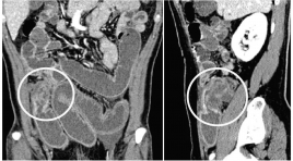
Results: A total of 98 patients met the criteria for analysis in thepresent retrospective study, 31 adenocarcinomas, 22 lymphomas, 30 GISTs anf 15 others. The extramural growth pattern isreliable prediction of GIST, with PPV of 82.3%. All of GISTs show moderate to avid enhancement. Tumor density of greater than or equal to 110 HU is likely to be GIST, with PPV of84.9%. Proliferation of blood vessels on tumor surfaces can help discriminate GIST from the others, with PPV of92%. Bowel wall thickening is the common patternof adenocarcinoma and lymphoma. Apple-core-like, shoulder defect and focal involvement are probably findings of adenocarcinoma, with PPV of 81.8%, 71.4% and 76.9%, respectively. Aneurysmal dilatation of the lumen and marked thickening wall bowel equal or greater than 25mm can strongly suggest lymphoma, with PPV of 87.5% and 72.7%, respectively. Enlarged lymph node with shorter axis greater than 20mmor multiple lymph nodes fused together forming a bulky massare likely to be lymphoma, with specificity of 100%.
Conclusion: MDCT findings could potentially be useful to differentiate the common primary small bowel neoplasms based on analyzing specific imaging characteristics of each tumor after classifying by growth pattern lesion.
Keywords: Small bowel neoplasm, differentiate
Bạn Đọc Quan tâm
Sự kiện sắp diễn ra

THÔNG BÁO SINH HOẠT KHOA HỌC 19/01/2024: CẬP NHẬT CHẨN ĐOÁN VÀ ĐIỀU TRỊ UNG THƯ TRỰC TRÀNG
19-01-2024 09:47:01 0 bình luận

Thông tin đào tạo
- Những cạm bẫy trong CĐHA vú và vai trò của trí tuệ nhân tạo
- Hội thảo trực tuyến "Cắt lớp vi tính đếm Photon: từ lý thuyết tới thực tiễn lâm sàng”
- CHƯƠNG TRÌNH ĐÀO TẠO LIÊN TỤC VỀ HÌNH ẢNH HỌC THẦN KINH: BÀI 3: U não trong trục
- Danh sách học viên đạt chứng chỉ CME khóa học "Cập nhật RSNA 2021: Công nghệ mới trong Kỷ nguyên mới"
- Danh sách học viên đạt chứng chỉ CME khóa học "Đánh giá chức năng thất phải trên siêu âm đánh dấu mô cơ tim"


Đơn vị hợp tác

