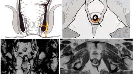
Vai trò của hình ảnh học trong nhiễm trùng quanh hậu môn
03/12/2019 14:22:51 | 0 binh luận

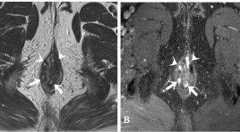
Đặc điểm hình ảnh cộng hưởng từ của rò hậu môn
05/12/2019 09:17:02 | 0 binh luận
Magnetic resonance imaging characterization of perianal fistula
SUMMARY
Background: Perianal fistula is a common disease in the anorectal region, just after hemorrhoids. The disease often recurs because of missed lesions at surgery, especially internal openings and secondary tracts, which can be identified by MRI. Surgery guided by Magnetic Resonance imaging (MRI) significantly reduces the recurrence of fistula.
Aims: To describe the MRI features of perianal fistula and study the role of T2W TSE, post- contrast FS T1W TSE Gadolinium sequences in detecting the characterizations of the fistula tract.
Subjects and methods: 367 patients with perianal fistulae, diagnosed and treated at University medical center hospital, between 1/1/2016 and 1/31/2018, were included in this study. This study is a retrospective analysis. We compared the imaging features with the surgical findings.
Results: The mean age was 39.3 years (range, 12 – 84 years), male/ female = 9/1. A total of 411 fistulas were found at surgery. Agreement between the surgical findings and MR imaging for classification of the primary track, detecting secondary track was very good with kappa = 0.89 (0.84,0.93), 0.94 (0.90,0.97), respectively. The sensitivity and specificity of MRI in correctly detecting internal openings was found to be 99% and 85.2% respectively; for abscess, it was 100%. Both T2W TSE, post-contrast FS T1W TSE Gadolinium sequences have high sensitivity and specificity for the depiction of primary tracts, internal openings, and secondary tracts. Post- contrast FS T1W Gadolinium sequence was very good in detecting abscess; differentiating between abscess and inflammation, active inflammatory components with scars.
Conclusions: Magnetic Resonance Imaging (MRI) was highly accurate in assessment of surgically important parameters of perianal fistulae. With the very good agreement between the surgical findings and MR imaging for classification of the primary track, detecting secondary tract, high accuracy in detecting internal openings and abscess as well, MRI was considered the reliable technique of choice for establishing preoperative fistula map. If no contraindications exist, administration of contrast material is necessary for detecting abscess and secondary tracts, differentiating active inflammatory components with scars, especially in case of complex or recurrent fistula or having surgery around the anus before.
Keywords: fistula, anal, MR imaging.
Bạn Đọc Quan tâm
Sự kiện sắp diễn ra
Thông tin đào tạo
- Những cạm bẫy trong CĐHA vú và vai trò của trí tuệ nhân tạo
- Hội thảo trực tuyến "Cắt lớp vi tính đếm Photon: từ lý thuyết tới thực tiễn lâm sàng”
- CHƯƠNG TRÌNH ĐÀO TẠO LIÊN TỤC VỀ HÌNH ẢNH HỌC THẦN KINH: BÀI 3: U não trong trục
- Danh sách học viên đạt chứng chỉ CME khóa học "Cập nhật RSNA 2021: Công nghệ mới trong Kỷ nguyên mới"
- Danh sách học viên đạt chứng chỉ CME khóa học "Đánh giá chức năng thất phải trên siêu âm đánh dấu mô cơ tim"


