


KẾT QUẢ BAN ĐẦU SINH THIẾT VÚ HÚT CHÂN KHÔNG DƯỚI HƯỚNG DẪN SIÊU ÂM CÓ KẾT HỢP ĐỊNH VỊ KIM CHO TỔN THƯƠNG VI VÔI HÓA
11/10/2023 14:38:52 | 0 binh luận
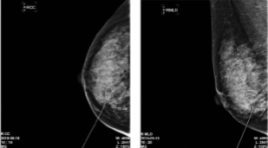
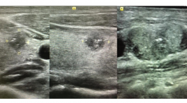
SUMMARY Purpose: This study examined the usefulness of ultrasound-guided vacuum-assisted breast biopsy for mammographic microcalcification. Methods : case series from June- 2019 to June-2020 at Breast department Cho Ray hospital. The patients with BI-RADS Category 4 Mammographic microcalcification were included. Most microcalcifications were not observed on ultrasound. Sono-guided J-wire localization was first performed for the suspicious microcalcification area, and the location of the J-wire and calcification was determined with mammography in most cases. Sono-guided VABB was performed after removing the J-wire without a stereotactic device. On the other hand, Sono-guided VABB was performed directly without J-wire localization when microcalcification lesions were identified by mass on ultrasonography. In all cases, calcification was confirmed by specimen mammography and the pathology was performed. A follow-up examination was performed to confirm the presence of complications. Results: A total of 20 lesions of 18 patients with BI-RADS Category 4 Mammographic microcalcification were included. Mean age: 49,44 ± 9,49 (35-66). Mean size of lesions 10,83 ± 3,60 mm, (4-15mm). In 20 lesions, 6 lesions (30%) were diagnosed as a malignancy (2 cases of ductal carcinoma in situ, 3 cases of ductal carcinoma invasive, 1 case of atypical ductal hyperplasia). The remaining 14 lesions (70%) were diagnosed as benign (fibroadenoma: 4; fibrocystic exchange 7, fibrocystic desease: 1, typical hyperplasia : 2). There were no significant complications during follow up after VABB. Conclusion: Sono-guided VABB can be used effectively if combined with wire localization for microcalcification lesions. Keywords: Mamography, Calcification, VABB, Image-guides biopsy, J Wire localization
CHỤP CẮT LỚP VI TÍNH HAI MỨC NĂNG LƯỢNG PHÁT HIỆN TẮC ĐM PHỔI: LỢI ÍCH THÊM VÀO CỦA BẢN ĐỒ IODINE
11/10/2023 12:57:49 | 0 binh luận
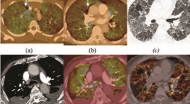
Summary Purpose: To determine if there is an added benefit of using iodine maps from dual-energy (DECT) in addition to conventional CT angiography images to diagnose pulmonary embolism (PE). Materials and Methods : In this retrospective analysis, 49 consecutive dual-energy CT angiography examinations performed from August through July 2020 at Bach Mai Hospital to evaluate for PE were reviewed. The 49 examinations included 49 patients (mean age, 59.73 years; range, 22–99 years). First, the location, level, and type (occlusive vs nonocclusive) of PEs on conventional CT angiograms were recorded. Iodine maps were then reviewed for defects suggestive of PE. Last, CT angiograms were rereviewed to detect additional PEs suggested by the iodine map. Results: 19/49 (38.8%) patients were diagnosed with PE, a total of 247 PEs were detected at initial review. After review of the DECT iodine map, 16 additional PEs were found on 8 of 49 (16.3%) patients in which 2 of 49 (4 %) patients had a new diagnosis of PE after review of the DECT iodine maps, 4/49 (8%) patients were diagnosed PE before. Of the 16 additional PEs, 8 (50%) were segmental, 8 (50%) were subsegmental, 3 (18.8 %) were occlusive, and 13 (81.2%) were nonocclusive Conclusion: Dual-energy CT iodine maps show a small incremental benefit for the detection of occlusive segmental and subsegmental pulmonary emboli. Keywords: CTPA= computed tomography pulmonary angiography, DECT= dual-energy computed tomography, PE= pulmonary embolism, iodine map.
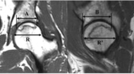
GIÁ TRỊ CỦA CỘNG HƯỞNG TỪ TỐI THIỂU TRONG CHẨN ĐOÁN HOẠI TỬ VÔ KHUẨN CHỎM XƯƠNG ĐÙI GIAI ĐOẠN SỚM Ở NHỮNG BỆNH NHÂN CÓ YẾU TỐ NGUY CƠ
11/10/2023 12:20:14 | 0 binh luận
Summary Purpose: Evaluate the agreement between limited MRI, which using T1W sequence or STIR sequence in coronal direction, with standard MRI in diagnosis early femoral head necrosis(FHN) occurring in high risk patients. Subjects and methods : descriptive cross-sectional study was performed on 58 patients, who warediagnosed of femoral head osteonecrosis at stage 2 or higher according to the Arlet Ficat classification. The patients were performed hip joints MRI at the Radiology Center, Bach Mai Hospital from June 2020 to August 2021. Results: The agreement in FHN staggingbetween limited MRI using T1Wsequence, or limited MRI using STIR sequence with the standard MRI was 0.98 and 0.86, respectively. The agreement in measurement extent of osteonecrosis areabetween limited MRI using T1Wsequence, or limited MRI using STIR sequence with the standard MRI was 0.98 and 0.85. Conclusion : There was excellent agreement between the full and limited MR examinations both forstagging and determining the extent of osteonecrosis area. The time and potential cost reduction achieved when taking limited MRImay lead to more widespread using in patient care. Key words: femoral head osteonecrosis, hip MRI, limited hip MRI.
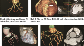
NHẬN XÉT MỘT SỐ BIẾN THỂ VÀ BẤT THƯỜNG ĐỘNG MẠCH VÀNH TRÊN CẮT LỚP VI TÍNH 128 DÃY
11/10/2023 12:17:03 | 0 binh luận
Summary Purpose: To describe the variants and the anormalies of coronary artery on 128-row computed tomography (CT). Material and method : 90 patients were treated at 103 hospital from Oct. 2020 to Jul. 2021. The characteristic figures of coronary artery assessed on 128-row CT included: the site of ostium, course, abnormal enlargement of the branch… Results : 84.4% of the patients had a right dominant system and Ramus intermedius branch was seen in 57.8%. The anomalous origins of LM and RCA were found in 3.3% for each branch. The atrophies of LCx and RCA were 7.8% and 2.2%, respectively. Conclusion : 128-row detector CT images is helpful for detecting the variants and the anormalies of coronary artery. Key words: computed tomography, coronary artery, variant, anormaly.
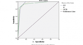
SO SÁNH GIÁ TRỊ CÁC PHÂN LOẠI EU-TIRADS, K-TIRADS VÀ ACR-TIRADS TRONG CHẨN ĐOÁN TỔN THƯƠNG DẠNG NỐT TUYẾN GIÁP
15/11/2021 17:22:56 | 0 binh luận
SUMMARY Purpose: To compare the ultrasound results of thyroid nodules in the application of EU-TIRADS, K-TIRADS, ACR-TIRADS systems with post-operative histological results. To compare diagnostic values of the three TIRADS systems. Material and methods: This is a cross-sectional study with convenient sampling. We recruited thyroid nodules that were performed pre-operative ultrasound (applied EU-TIRADS, K-TIRADS, ACRTIRADS systems) and removal surgery in Hue University of Medicine and Pharmacy Hospital between September 2019 and July 2020. Results: 138 thyroid nodules of 122 patients were enrolled. The malignancy rate was 22.5%. The majority of the lesions were classified as EU-TIRADS 3 (47.8%), K-TIRADS 3 (47.8%) và ACR-TIRADS 2 (38.4%.). The AUC of the EU-TIRADS, K-TIRADS và ACR-TIRADS systems were 0.957, 0.951 and 0.956, respectively. Among the three systems, EU-TIRADS had the highest sensitivity (Se) and negative predictive value (NPV) (100%) while its specificity (Sp), positive predictive value (PPV) and accuracy (Acc) for malignancy were lowest. K-TIRADS showed the best Sp, PPV and Ac (97.2%; 89.7% and 94.2%, respectively) and the lowest other values. Conclusion: The ability to distinguish between the malignant thyroid nodules and the benign ones of the three TIRADS systems was at a very good level. EU-TIRADS showed the most effective diagnostic performance in Se and NPV, while K-TIRADS yielded the best Sp, PPV and Acc. Key words: thyroid nodules, diagnostic values, EU-TIRADS, K-TIRADS, ACR-TIRADS.
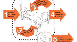
Bác sĩ siêu âm cần làm gì để hạn chế sự lây lan của COVID-19
18/05/2020 15:53:44 | 1 binh luận
Covid-19 (coronavirus disease 19) đang là một trong những thách thức lớn nhất cho thế giới trong thời điểm hiện tại. Những tác động của coronavirus (SARS-CoV-2) không chỉ đơn thuần lên sức khỏe, mà còn ảnh hưởng nghiêm trọng đến kinh tế và cuộc sống của người dân trên toàn cầu. Một vài nước đã thông báo rằng họ đang trải phải qua một trong những tình huống tồi tệ nhất trong hơn 100 năm vừa qua.
GIÁ TRỊ SIÊU ÂM TRONG CHẨN ĐOÁN UNG THƯ TUYẾN GIÁP
15/11/2021 18:00:40 | 0 binh luận
SUMMARY Objective: To determine the value of ultrasound in the diagnosis of thyroid cancer. Subjects and methods: A cross-sectional descriptive study on 98 patients with thyroid nodules who came for treatment at the Military Institute of Medical Radiology and Oncology, from April 2020 to March 2021. Results: The size of malignant nodules was mainly less than 2cm. Thyroid cancer lesions were mainly characterized by highly hypoechoic on ultrasound (61.19%). Thyroid cancer nodules had taller-than-wide feature (Sensitivity: 76.12%; specificity: 86.96%; positive predictive value: 89.47%; negative predictive value: 71.43%; accuracy: 80.53%.); irregular border feature (sensitivity: 98.51%; specificity: 86.96%; positive predictive value: 91.67%; negative predictive value: 97.56%; accuracy: 93.81%); microcalcification characteristics (sensitivity: 73.13%; specificity: 91.30%; positive predictive value: 92.45%; negative predictive value: 70.00%; accuracy: 80,53%). TIRADS value in thyroid cancer diagnosis with sensitivity: 94.03%; specificity: 86.96%; positive predictive value: 91.30%; negative predictive value: 90.91%; Accuracy: 91.15%. Conclusion: The features of hypoechoic nodules, taller-than-wide shape, irregular border, microcalcification characteristics, and TIRADS 4, TIRADS 5 scores had high prognostic value for thyroid cancer. Keywords: Ultrasound, thyroid cancer.
ĐẶC ĐIỂM HÌNH ẢNH VÀ KẾT QUẢ ĐIỀU TRỊ NÚT MẠCH HÓA CHẤT UNG THƯ BIỂU MÔ TẾ BÀO GAN Ở BỆNH NHÂN DƯỚI 40 TUỔI
16/11/2021 11:24:31 | 0 binh luận
SUMMARY Objectives: Describe the characteristics of ultrasound images, computed tomography, angiography and results of chemoembolization in patients with HCC under 40 years old at the Radiology Center of Bach Mai Hospital. Methods: A cross-sectional study was carried out on 25 patients with a confirmed HCC who treated chemoembolization at the Radiology Center - Bach Mai Hospital from August 2018 to August 2021. Results: On 2-dimensional ultrasound, the majority of HCC tumors had hypoechoic and mixed sound images (72%) and increased angiogenesis on color Doppler ultrasound, reaching 68.0%. On computed tomography images, tumor density decreased 64% and increased angiogenesis 68.0%. Tumors of HCC have increased angiogenesis on angiography, reaching 92%. After treatment AFP tends to decrease in the first 6 months, then increase in the 9th month. Bilirubin tends to decrease in the first 3 months, then increase at 6 and 9 months. Tumor size tends to decrease at 1, 3 and increase at 6, 9 months. Median survival was 13 months with interquartile range [5.5 ÷ 22], the longest survival time with continued follow-up was 57 months. Conclusion: Chemoembolization therapy is effective in reducing tumor size, bilirubin, AFP and prolonging survival time for patients under 40 years old with HCC. Keywords: chemoembolization therapy, hepatocellular carcinoma
Bạn Đọc Quan tâm
Sự kiện sắp diễn ra

THÔNG BÁO SINH HOẠT KHOA HỌC 19/01/2024: CẬP NHẬT CHẨN ĐOÁN VÀ ĐIỀU TRỊ UNG THƯ TRỰC TRÀNG
19-01-2024 09:47:01 0 bình luận

Thông tin đào tạo
- Những cạm bẫy trong CĐHA vú và vai trò của trí tuệ nhân tạo
- Hội thảo trực tuyến "Cắt lớp vi tính đếm Photon: từ lý thuyết tới thực tiễn lâm sàng”
- CHƯƠNG TRÌNH ĐÀO TẠO LIÊN TỤC VỀ HÌNH ẢNH HỌC THẦN KINH: BÀI 3: U não trong trục
- Danh sách học viên đạt chứng chỉ CME khóa học "Cập nhật RSNA 2021: Công nghệ mới trong Kỷ nguyên mới"
- Danh sách học viên đạt chứng chỉ CME khóa học "Đánh giá chức năng thất phải trên siêu âm đánh dấu mô cơ tim"


Đơn vị hợp tác

