


BƯỚC ĐẦU NGHIÊN CỨU ĐẶC ĐIỂM HÌNH ẢNH VÀ VAI TRÒ CỦA 18FDG-PET/CT TRONG CHẨN ĐOÁN GIAI ĐOẠN UNG THƯ ĐẠI TRỰC TRÀNG TRƯỚC ĐIỀU TRỊ
11/10/2023 16:44:42 | 0 binh luận
SUMMARY Objectives: To evaluate the imaging characteristics and the role of 18FDG-PET/CT in pre-treatment colorectal cancer staging. Subjects and methods : Study on 39 colorectal cancer patients without specific treatment who had taken 18FDG-PET/CT for staging diagnosis, at the Department of Nuclear Medicine, Imaging Center of Military Hospital 103, from February 2017 to November 2020. Results: The average age was 62.77 ± 14.07. The ability to detect primary tumor lesions in the colorectal of 18FDG-PET/CT was 97.44%. The majority of patients had rectal tumors, the tumor size was mainly in the range of 5-10 cm and the T3 stage accounted for the most proportion with 60.5%. There was 46.2% of patients had regional lymph node metastasis, in which the group of lymph nodes over 10 mm and the N2 group accounted for a lower proportion, but the mean SUVmax was statistically significantly higher than others group. Distant metastasis was occured mainly in liver, lung-pleural and bone. There were 59.0% patients in stage III and IV. Diagnosis by 18FDG-PET/CT was accurately at 80.77% for T stage and 66.67% for N stage. The sensitivity and specificity, positive predictive value and negative predictive value of 18FDG-PET/CT in the diagnosis of regional lymph node metastasis were 100% and 60%; 55.55% and 100%. Keywords : Colorectal cancer, 18FDG-PET/CT, SUVmax.
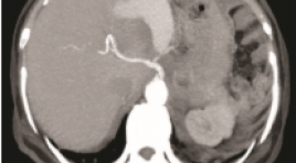
ĐÁNH GIÁ KẾT QUẢ NÚT TẮC ỐNG NGỰC ĐIỀU TRỊ BIẾN CHỨNG RÒ DƯỠNG CHẤP SAU MỔ UNG THƯ TUYẾN GIÁP
11/10/2023 16:24:55 | 0 binh luận
SUMMARY Background: chylous leakage after operation of head and neck is rare but well-known complication. Almost patients with this complication can be treated conservatively but in patients with high flow leakage, the following treatments will be very complicated. Purpose : to report the results of percutaneous thoracic duct embolization (TDE) treatment for chylous leakage of the neck in patients post thyroidectomy and cervical lymph node dissection due to thyroid cancer. Methods : 15 consecutive patients with high flow chylous leak post thyroidectomy were sent to our hospital after failed conservative treatment. All patient were undergone intra nodal lymphagiography then thoracic duct embolization. Results: Fifteen patients with cervical chylorrhea through drainage more than 300 ml/day during average 2 weeks (1 to 5 weeks) were included in this study. TDE was archived in 15/15 patients in which 14/15 TD were embolized ategrade and 1/15 TD was embolized retrograde. One patient had recurrent chylous leakage after 1 week due to recanalization of TD. She was then undergone TD sclerosis injection under CT scanner guidance. All patients had clinical successful with no chylorrhea after intervention. No major complication was noted. All patients were discharged in following weeks after intervention. Conclusion: TDE is minimal invasive and effective treatment for cervical chylous leakage post thyroidectomy. Keywords: chylous leak, chylorrhea, thoracic duct embolization
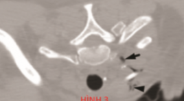
BÁO CÁO CA LÂM SÀNG: ĐIỀU TRỊ TÚI GIẢ PHÌNH ĐỘNG MẠCH VỊ TÁ TRÀNG DỌA VỠ BẰNG PHƯƠNG PHÁP CAN THIỆP NỘI MẠCH
11/10/2023 15:28:24 | 0 binh luận
SUMMARY Gastroduodenal artery (GDA) pseudoaneurysm is a rare surgical entity that causes various symptoms. In the case of rupture, it usually presents an ominous prognosis and mortality rate of up to 40%. Although open surgical procedure is a mainstay, endovascular intervention is emerging a promising treatment in recent years, due to its advantages and safety.We present a case of upper gastrointestinal bleeding caused by GDA pseudoaneurysm threatened rupture in a 71-year-old woman, with medical episodes of acute pancreatitis, a pancreatic body tumor removal surgery was performed, and now the tumor is relapsing and metastasizing. The treatment approach is blocking off the pseudoaneurysm by a covered stent. The procedure was successful and the patient is asymptomatic. Two months later, the pseudoaneurysm reduces its size and completely excluded from the preservation of the blood flow in the artery. Endovascular interventional treatment in the case of GDA aneurysms is considered a promising alternative not only to open surgery but also to an effective emerging technique even in the acute setting. Keywords : Gastroduodenal artery, Covered stent, Pseudoaneurysm
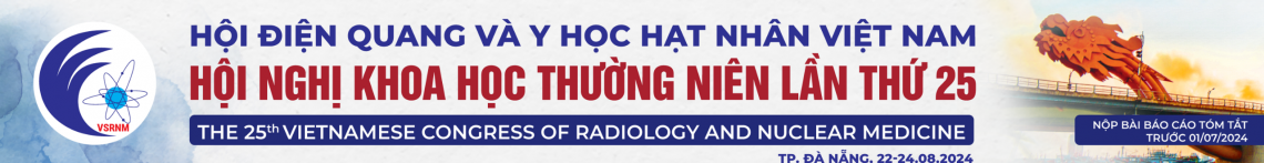
GIẢ PHÌNH ĐỘNG MẠCH PHỔI: NHÂN 3 TRƯỜNG HỢP KHÁM VÀ ĐIỀU TRỊ TẠI BỆNH VIỆN PHỔI TRUNG ƯƠNG
11/10/2023 15:21:47 | 0 binh luận
SUMMARY Pulmonary artery pseudoaneurysms (PAPs) is a rare abnormality of the pulmonary artery system. Beside, PAPs have no specific clinical presentation or may be asymptomatic, which may be present in congenital anomalies or in a variety of conditions such as pneumonia, pulmonary neoplasm, pulmonary tuberculosis, or lung fungus... [1]. We present 3 cases of PAPs examined and treated at the National Lung Hospital with PAPs appearing on the background of 3 different diseases. Both of these 3 cases have nonspecific symtoms as cough, chestpain and dyspnea with 2 cases of hemoptysis. They are all detected by computed tomography (CT) angiography. There are 2 cases treated by surgery that have good results and 1 case who continued the TB regimen received medical treatment. Keywords: pulmonary artery pseudoaneurysms, pulmonary artery, hemoptysis.
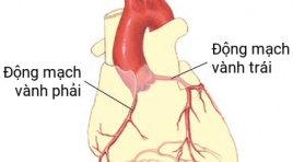
GIÁ TRỊ CỦA CẮT LỚP VI TÍNH 256 DÃY TRONG CHẨN ĐOÁN BỆNH ĐỘNG MẠCH VÀNH Ở BỆNH NHÂN RUNG NHĨ
11/10/2023 15:05:26 | 0 binh luận
SUMMARY Objectives: To determine the diagnostic value of 256 MDCT in the diagnosis of coronary artery disease in patients with atrial fibrillation. Materials and methods: We prospectively enrolled 48 patients with atrial fibrillation who underwent 256 MDCT coronary CT angiography scan at Huu Nghi Hospital from 07/2020 to 07/2021 and evaluated diagnosis value of this technique using invasive coronary angiography as golden standard. Results: At patient-based analysis, sensitivity, specificity, positive predictive value and accuracy are 100%, 83.3% and 83.3%, respectively. At vessel-based analysis, sensitivity, specificity, positive predictive value, negative predictive value and accuracy are 98.4%, 81.7%, 80.3%, 98.5% and 88.9%, respectively. At segment-based analysis sensitivity, specificity, positive predictive value, negative predictive value and accuracy are 96.7%, 93.8%, 78.4%, 99.2 % and 94.4%, respectively. Conclusion: 256 MDCT coronary angiography scan is a diagnostic method with high accuracy in diagnosing coronary artery disease in patients with atrial fibrillation. Keywords: atrial fibrillation, coronary CTA, diagnostic value
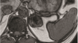
VAI TRÒ CỦA CỘNG HƯỞNG TỪ TRONG CHẨN ĐOÁN U TUYẾN THƯỢNG THẬN
11/10/2023 15:01:16 | 0 binh luận
SAMMARY Purpose: to evaluate the role of MRI in diagnosis adrenal tumors. Subject and methods: our cross–sectional descriptive study included 25 patients who were diagnosed as adenoma hyperplasia in pathology. All patients underwent MRI examination in Bach Mai hospital from June, 2020 to July, 2021. Results: mean age of all patient was 49,2±11,8. Male/female ratio was 1.5/1. 25 adrenal tumor on MRI and identified on the diagnosed as adenoma hyperplasia in pathology. These adenomas consisted 12 adrenal cortical adenoma (ACA) and 13 non – adrenal cortical adenoma (NACA): 09 pheochromocytomas, 01 adrenocortical carcinoma, 01 ganglioneuroma and 02 myelolipoma. Mean size of all adrenal tumors was 38,8 ± 23,5mm, therein, mean size of ACA group and NACA group was 23,2 ± 7,6 mm and 52,2 ± 25,7 mm respectively. Chemical Shift Imaging (CSI) allowed differential diagnose between ACA and NACA. Qualitative analysis based on signal reduction on out – phase compared to in – phase allows 11/12 ACA, but 2/13 NACA have been misdiagnosed as ACA. Quantitative analysis based on SII index (SII threshold > 16,1%) allowed correctly diagnose 11/12 ACA and 01 NACA. ASR index (ASR threshold <0,71) allowed correctly diagnose 11/12 ACA and 01 NACA. There are 02 adrenal tumors that invaded to the surrounding area in imaging and was demonstrated by surgery and pathology reports. Conclusion : MRI has an important role in adrenal mass diagnosis with sensitivity and specificity were 100% and 96,1% respectively. CSI enable to distinguish ACA and NACA with sensitivity and specificity were 91,7% and 92,3% respectively by using SII and ASR index. MRI also help to assess invasive and metastatic properties with high accuracy. Key words : adrenal tumors, adrenal MRI, chemical shift imaging (CSI).
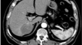
ĐÁNH GIÁ HIỆU QUẢ ĐIỀU TRỊ UNG THƯ BIỂU MÔ TẾ BÀO GAN BẰNG NÚT MẠCH VI CẦU PHÓNG XẠ YTTRIUM-90
11/10/2023 14:52:30 | 0 binh luận
SUMMARY Background: Hepatocellular carcinoma (HCC) is the second most common cause of cancer-related deaths worldwide. Radioembolization with Yttrium-90 is a safe, effective locoregional therapy for unresectable HCC. Purpose: This study aimed to evaluate the safety and efficacy of radioembolization with Yttrium-90 for the treatment of HCC. Materials and methods: A total of 93 patients were diagnosed with HCC and treated by radioembolization with Yttrium-90 from 2013 to 2021 at Bach Mai Hospital. Radiographic findings on computed tomography (CT) or magnetic resonance imaging (MRI) were assessed by using modified Response Evaluation Criteria in Solid Tumors (mRECIST). Patient survival was assessed using the Kaplan-Meier method, and prognostic factors affecting survival were assessed using Cox proportional hazards regression. Results: The mean age of 93 patients was 59.0±9.8 years. Mean tumors diameter was 6.5 ± 2.7 cm, the mean of tumor marker AFP is 1812 ± 6577 ng/ml. Complete response and partial response to treatment were observed in 21 (40.4%) and 15 patients (28.8%) of 52 follow-up patients. The median overall survival (OS) was 12.0 [95% CI: 5.0-25.0] months. Factors associated with longer OS included BCLC stage A-B, ECOG 0 and no portal vein thrombosis. Conclusion: Radioembolization with Y-90 is safe and effective in patients with unresectable HCC. BCLC stage A-B, ECOG 0 and no portal vein thrombosis were positive prognostic factors. Keywords: Hepatocellular carcinoma, radioembolization, Selective internal radiation therapy, Yttrium-90, Tumor response
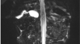
ĐẶC ĐIỂM HÌNH ẢNH CHT 4 CA TEO ĐƯỜNG MẬT BẨM SINH THỂ CÓ NANG GIỐNG U NANG ỐNG MẬT CHỦ
11/10/2023 14:44:26 | 0 binh luận
SUMMARY Purpose: to describle in detail the significant signs in hepatobiliary MRI, that help to give different diagnosis between biliary atresia (BA) with big cyst at hepatic hilar and choledochal cyst in small childrent and infant. Method: presenting 4 cases with clinical diagnosed of biliary atresia that based on clinical signs and US, MRI results, all 4 cases were operated, taken cholangiogram in surgery to confirm the diagnosis of cystic biliary atresia and had pathological results. Results: All 4 patients were under 3 months old, all are female with enlarged liver; gallbladder were in the normal size limit but all of them had abnormal shape: deformation, irregular wall, communicated directly with common bile duct. All 4 cases have cystic diameter over 10mm. No case of all has further biliary signal that above or under the cyst on MRI but one case has contrast material filling intraliver bile duct draining directly to cystic lesion on cholangiogram intraoperation . These are 3 cases of cystic BA type III and 1 cases of cystic BA type I. 2 cases has positive triangular cord sign (TC sign). Conclusion : Hepatobiliary MRI with its basic sequences, specially with MRCP is helpful to give difirential diagnosis of cystic BA and choledochal cyst in the young childrent. Keyword: Biliary atresia, cystic biliary atresia
Bạn Đọc Quan tâm
Sự kiện sắp diễn ra

THÔNG BÁO SINH HOẠT KHOA HỌC 19/01/2024: CẬP NHẬT CHẨN ĐOÁN VÀ ĐIỀU TRỊ UNG THƯ TRỰC TRÀNG
19-01-2024 09:47:01 0 bình luận

Thông tin đào tạo
- Những cạm bẫy trong CĐHA vú và vai trò của trí tuệ nhân tạo
- Hội thảo trực tuyến "Cắt lớp vi tính đếm Photon: từ lý thuyết tới thực tiễn lâm sàng”
- CHƯƠNG TRÌNH ĐÀO TẠO LIÊN TỤC VỀ HÌNH ẢNH HỌC THẦN KINH: BÀI 3: U não trong trục
- Danh sách học viên đạt chứng chỉ CME khóa học "Cập nhật RSNA 2021: Công nghệ mới trong Kỷ nguyên mới"
- Danh sách học viên đạt chứng chỉ CME khóa học "Đánh giá chức năng thất phải trên siêu âm đánh dấu mô cơ tim"


Đơn vị hợp tác

