


BÁO CÁO 2 TRƯỜNG HỢP SPECT/CT HẠCH GÁC TRONG UNG THƯ VÚ
16/11/2021 11:30:55 | 0 binh luận
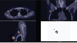
SUMMARY Sentinel lymph node is defined as the first lymph node or group of first nodes to which cancer cells are likely to spread from the primary tumor. Cancer cells are often found in sentinel lymph node(s) before metastasizing to other nodes or organs. Therefore, sentinel lymph node biopsy plays an important role in identification and management of metastasis lesions, especially in breast cancer, testicular cancer and melanoma. Sentinel lymph node scintigraphy with SPECT/CT using 99mTechnetiumnanocolloid is a useful imaging technique for nodal determination and localization. With negative histopathological results of sentinel lymph node dissection, axillary lymph node dissection can be avoided for patients thereby reducing the risk of lymphedema and other complications. In this article, two patients in Vinmec Health Care System, Times City with breast cancer who underwent SPECT/CT sentinel lymph node scintigraphy were reported. Keywords: breast cancer, sentinel lymph node, SPECT/CT, 99mTc-nanocolloid
NGHIÊN CỨU ĐẶC ĐIỂM HÌNH ẢNH UNG THƯ DẠ DÀY TRÊN MÁY CHỤP CẮT LỚP VI TÍNH 256 DÃY
16/11/2021 11:27:45 | 0 binh luận
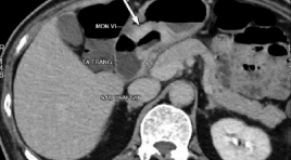
SUMMARY Purpose: This report aimed to assess the describing imaging characteristic of gastric cancer on 256 - Slice Multidetector Computed Tomography gastrography. Materials and methods: 256 - slice multidetector CT gastrography in 31 patients with histopathological findings of gastric cancer from 2020 Fer to 2021 August at Huu Nghi hospital. Results: Gastric cancer is often detected in old people. The most common position is the antrum of gastric (71%), the least common position is the cardia - fundus of gastric (3,2%).There is a correlation between diameter of tumors and T staging (p< 0,05). There are correlation between the shape, short diameters of nodes and LN metastasis (p< 0,05). Short diameters of nodes is ≥ 8 mm and round shape of nodes were moderate diagnostic ability indicator for LN metastasis in gastric cancer. Conclusion: 256 slice - MDCT gastrography is an effective method and non-invasion offers improved detected gastric cancer and evaluates lymph node metastasis. Keyword: Gastric cancer,256 slice - MDCT,short diameters of Lymph node metastasis.
ÁP DỤNG BẢNG PHÂN LOẠI CAD-RADS TRONG ĐÁNH GIÁ BỆNH ĐỘNG MẠCH VÀNH MẠN TÍNH TRÊN CẮT LỚP VI TÍNH ĐA DÃY
16/11/2021 11:19:01 | 0 binh luận
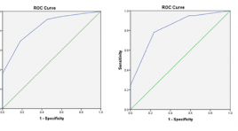
SUMMARY Purpose: To apply the CAD-RADS classification in the assessment of chronic coronary artery disease on multislice computed tomography. Material and methods: Cross-sectional description of 179 patients undergoing coronary computed tomography angiography, diagnosed according to CAD-RADS classification by two physicians independently, the intra-class correlation was used to test the inter-reviewer agreement (IRA), and comparing with results invasive coronary angiography. Results: There was an excellent IRA between the two for CADRADS (κ=0.904), by each CAD-RADS classification (κ=0.827-1.00), by coronary artery stenosis (κ=0.878-0.931). There is an excellent IRA for modifiers G (κ=1), and S (κ=1), moderate IRA for V (κ=0.574). The best cutoff value for predicting significant CAD was ≥ CAD-RADS 3. The diagnostic value of CAD-RADS classification according to the common segment: sensitivity 77.54%, specificity 87.23%, positive predictive value 89.92%, negative predictive value 72.56%. Conclusion: There is an excellent inter-reviewer agreement when applying the CAD-RADS classification to clinical practice, with high sensitivity, specificity, and accuracy when compared with invasive coronary angiography. Keywords: coronary artery, CAD-RADS, CT angiography, graft, stent
VAI TRÒ CỦA CHỤP CẮT LỚP VI TÍNH TRONG ĐÁNH GIÁ MẠCH MÁU THẬN CỦA NGƯỜI CHO THẬN SỐNG TRƯỚC PHẪU THUẬT GHÉP THẬN
16/11/2021 10:06:25 | 0 binh luận
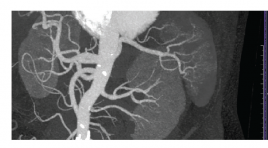
SUMMARY Background: In laparoscopic donor nephrectomies, it is important to understand the exact anatomy of the vascular structures during minimally invasive surgery. The aim of study: to determine the accuracy of MDCT to predict vascular anatomy in living kidney donors and to reveal the prevalence of vascular variations in a VietNam population. Materials and methods: This is a retrospective cross-sectional study. One hundred and eleven living donors were included in this study, who had MDCT for the assessment of their renal vessels and laparoscopic surgery in Cho Ray hospital between February 2020 to April 2021. The initial CT results were compared with the surgical findings and repeated review sessions of CT scans were performed to determine the causes of mismatches in discordant cases. Results: The accuracy of MDCT was 98,2% to predict the number of renal vessels. One artery was missed during the initial CT interpretation due to perception error. One case is false positive. The accuracy of MDCT was 95,5% to predict the early branching of a renal artery and late confluence of a renal vein variation. The prevalence of multiple renal arteries and veins, early branching of a renal artery and late confluence of a renal vein were 20,7%, 6,8%, 13,5%, 19,8%. One case (0,9%) each of a retroaortic left renal vein and a circumaortic left renal vein were found. Conclusion: Multidetector computed tomography is a reliable technique in preoperative renal anatomy evaluation in live renal donors. Key word: living donor kidney, multidetector computed tomography (MDCT) *
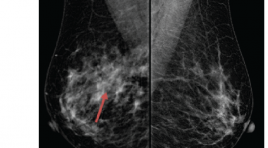
NGHIÊN CỨU GIÁ TRỊ CỦA X-QUANG CẮT LỚP TRONG CHẨN ĐOÁN UNG THƯ VÚ Ở BỆNH NHÂN CÓ VÚ ĐẶC HOẶC BẤT XỨNG KHU TRÚ
16/11/2021 09:56:31 | 0 binh luận
VAI TRÒ CỦA CẮT LỚP VI TÍNH VÀ CỘNG HƯỞNG TỪ XƯƠNG THÁI DƯƠNG TRONG CHỈ ĐỊNH CẤY ỐC TAI ĐIỆN TỬ
16/11/2021 09:50:00 | 0 binh luận
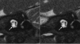
SUMMARY Objective: To describe CT scanner and MRI imaging characteristics of temporal bone of sensorineural hearing loss patients to select patients for cochlear implantation. Material and Methods: Description of inner ear and cochlear nerve imaging combined with hearing assessment to give cochlear implant indication in 132 sensorineural hearing loss patients. Cochlear nerve was evaluated on high resolution T2 3D gradient-echo MRI. Inner ear image was evaluated on high resolution MRI and CT scanner. Results: The study included 132 patients with 264 ears in which 161 ears (61%) with no inner ear malformations, 34 ears (12,9%) with normal cochlear and cochlear nerve deficiency, 65 ears (24,6%) with inner ear malformation and 4 ears (1,5%) with labyrinthine ossification. The patients with cochlear nerve aplasia and no V ABR wave on hearing assessment, the patients with severe cochlear malformation and severe cochlear ossification are not indicated for cochlear implantation. Conclusion: Indications for cochlear implantation depend on the condition of the inner ear and the presence of cochlear nerve on imaging or auditory response on hearing assessment. Key words: Inner ear malformation, cochlear nerve deficiency, cochlear implant indication.
ĐẶC ĐIỂM HÌNH ẢNH FDG PET/CT CỦA U LYMPHO KHÔNG HODGKIN TẾ BÀO T
16/11/2021 09:46:25 | 0 binh luận
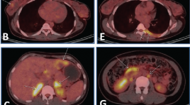
SUMMARY Objective: T-cell non-Hodgkin lymphomas (NHL) is a group of uncommon lymphomas, accounting for about 12% of all cases of non-Hodgkin lymphoma. The purpose of this study is to investigate the sites and metabolic activity of lesions in T-cell NHL on FDG PET/CT images, and to determine the correlation between metabolic activity and Ki67. Patients and methods: A retrospective study of patients with T-cell NHL who underwent FDG PET/CT examination for initial disease staging from 2009 to 2019. Results: A total of 62 patients (37 men and 25 women) with mean age of 44.8 were included in this study. There were 30/62 patients with histopathological subtypes, in which the highest frequency was peripheral T-cell lymphoma, not otherwise specified, and extranodal NK/T-cell lymphoma, nasal type with 8 cases (26.7%) each. PET/CT images showed the frequency of lesions in the lymph node regions in descending order of neck (67.7%), axillary (41.9%), mediastinum (40.3%), abdomen (40.3), inguinal area (37.1%). There were 32 extranodal sites/organs involved in T-cell NHL, most commonly in the spleen (27.4%), nasal cavity (24.2%), bone marrow (22.6%), nasopharynx (21%) with a frequency greater than 20%. The mean SUVmax of lymph nodes in descending order was 9.9 (in the abdominal lymph nodes), 7.7 (in the cervical lymph nodes), 7.7 (in the axillary lymph nodes), 6.2 (in the mediastinal lymph nodes), 5.9 (in the inguinal lymph nodes). The majority (81%) of lesions in extranodal sites/organs had a mean SUVmax ≥5. Sites/organs with high frequency of lesions having the highest mean SUVmax were skeletal muscle (17.3), nasal cavity (12.3), skin/subcutaneous tissue (11.7), nasopharynx (10.2), lung (6.9), pleura (6.8), bone marrow (5.3), liver (5.0) and spleen (5.0). Analysis of the linear correlation between SUVmax of the lesion at the biopsy site and the percentage of Ki67 was performed in 24 patients, and the results showed no correlation (r=0.03). Conclusion: In patients with T-cell NHL, FDG PET/CT is useful for detecting lesions in many sites/organs in the body. The metabolic activity of the lesions was high, but there was no correlation between SUVmax and Ki67. Keywords: T-cell non-Hodgkin lymphoma, FDG PET/CT, Ki67.
NGHIÊN CỨU ỨNG DỤNG QUY TRÌNH CHỤP 18FDG PET/CT CHO BỆNH NHÂN LAO PHỔI
16/11/2021 09:40:38 | 0 binh luận
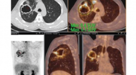
SUMMARY The study was performed on a PET/CT system provided by Philips Health Care System, radioactive drug 18FDG of the 108 Military Hospital. The procedure was based on recommendations of EANM (European Association of Nuclear Medicine) and protocol. PET/CT scan of the Ministry of Health. The technique was performed on 15 patients with a confirmed diagnosis of pulmonary TB by microbiological results being treated with anti-tuberculosis drugs. Results: There are two patterns of 18FDG uptake in TB patients: the lung parenchyma and the lymphatic system. Before treatment: Metabolic change in the mean SUVmax index found in lung parenchymal lesions was 6.75±3.01. Mediastinal lymph nodes were found in 10/15 patients (66.7%), in which 8/10 lymph nodes increased FDG uptake with mean SUVmax of 3.14±1.5. Keywords: 18FDG PET/CT, pulmonary tuberculosis.
Bạn Đọc Quan tâm
Sự kiện sắp diễn ra

THÔNG BÁO SINH HOẠT KHOA HỌC 19/01/2024: CẬP NHẬT CHẨN ĐOÁN VÀ ĐIỀU TRỊ UNG THƯ TRỰC TRÀNG
19-01-2024 09:47:01 0 bình luận

Thông tin đào tạo
- Những cạm bẫy trong CĐHA vú và vai trò của trí tuệ nhân tạo
- Hội thảo trực tuyến "Cắt lớp vi tính đếm Photon: từ lý thuyết tới thực tiễn lâm sàng”
- CHƯƠNG TRÌNH ĐÀO TẠO LIÊN TỤC VỀ HÌNH ẢNH HỌC THẦN KINH: BÀI 3: U não trong trục
- Danh sách học viên đạt chứng chỉ CME khóa học "Cập nhật RSNA 2021: Công nghệ mới trong Kỷ nguyên mới"
- Danh sách học viên đạt chứng chỉ CME khóa học "Đánh giá chức năng thất phải trên siêu âm đánh dấu mô cơ tim"


Đơn vị hợp tác

