


GIÁ TRỊ CÁC THÔNG SỐ BÁN ĐỊNH LƯỢNG CỦA CỘNG HƯỞNG TỪ ĐỘNG HỌC TRONG CHẨN ĐOÁN PHÂN BIỆT TỔN THƯƠNG VÚ LÀNH TÍNH VÀ ÁC TÍNH
15/11/2021 17:08:47 | 0 binh luận
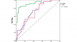
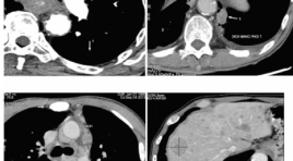
SUMMARY Objective: The aims of this study were to determine the value of the semi-quantitative parameters obtained by dynamic contrast enhancement magnetic resonance imaging (DCE-MRI) in differentiation between benign and malignant breast lesions. Methods: A retrospective study was performed on 63 females (with 72 breast lesions) underwent DCE-MRI before treatment at Cho Ray hospital from Jan 2019 to Feb 2020. The value of semi-quantitative parameters (signal intensity slope (SIslope), maximum slope of increase (MSI), percentage of peak enhancement (Epeak)) were evaluated. The diagnostic value of the time intensity curve according to 5th edition ACR (2013) was compared with the value based on semi-quantitative methods. The results of each DCE-MRI parameter were correlated with histopathology. Results: There were 63 patients with 72 breast lesions including 40 benign lesions and 32 malignant lesions. The area under the ROC curve of SIslope, MSI and Epeak were 0,908; 0,702 and 0,734, respectively. The sensitivity, specificity and accuracy of the time intensity curve according to ACR and semi-quantitative methods were 68,8% and 87,5%; 87,5% and 85%; 79,2% and 86,1%, respectively. Conclusion: Our study reinforces the importance of the semi-quantitative parameters of DCE-MRI in distinguishing between benign and malignant breast lesions. Semi-quantitative analysis of the time intensity curve helps to increase the diagnostic accuracy compared with the methods of ACR. Key words: breast lesions; semi-quantitative parameters; time intensity curve.
NGHIÊN CỨU ĐẶC ĐIỂM HÌNH ẢNH VÀ KẾT QUẢ ĐIỀU TRỊ PHÌNH ĐỘNG MẠCH THÔNG TRƯỚC ĐÃ VỠ BẰNG CAN THIỆP NỘI MẠCH
15/11/2021 17:05:19 | 0 binh luận
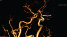
SUMMARY Background: Anterior communicating artery aneurysms (Acom) accounts for 23 - 40% of ruptured intracranial aneurysms. A ruptured cerebral aneurysm is a medical and neurological emergency that requires early diagnosis and prompt management to reduce mortality and sequelae. Material and method: Retrospective description of 40 patients who were diagnosed of ruptured anterior communicating aneurysms based on the clinical characteristics, imaging and results of endovascular treatment. Clinical outcomes were evaluated on a modified Rankin scale. Results: Patients suffering from ruptured Acom aneurysms presented headache (100%), thunderclap headache (45.0%), vomiting with or without nausea (60%), nuchal rigidity (67.5%). Aneurysms’s size was under 5mm, 5-15mm and over 15mm accounting for 52.4%, 45.0% and 2.5% respectively; None of the patients had giant aneurysms. Dome and neck ratio of <1.2 ; 1.2 - 1.5 and ≥1.5 account for 37.5%; 32.5% and 30.0% respectively. Diameter of Acom ruptured aneurysm’s neck under and above 4mm accounted for 80.0% and 20.0%, respectively. Successful coiling embolization of Acom ruptured aneurysms without complications achieved in 80.0%. The patient had a good clinical recovery of 92,5% after 3 to 6 months follow-up. Conclusion: Coiling embolization of Acom ruptured aneurysms were effective and safe. Keywords: Anterior Communicating Artery Aneurysms, coiling, intervention. *
NÚT BÚI GIÃN TĨNH MẠCH DẠ DÀY XUÔI DÒNG QUA DA Ở BỆNH NHÂN TĂNG ÁP LỰC TĨNH MẠCH CỬA DO XƠ GAN
15/11/2021 16:56:32 | 0 binh luận
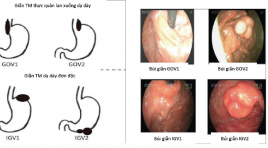
SUMMARY Abstract: Gastrointestinal bleeding due to rupture of esophageal varices and gastric varices in cirrhosis is quite common and has a high mortality rate if untreated. Gastric varices are difficult to control under endoscopy, intravascular intervention is a highly effective method. In patients who don't have or an inappropriate gastrorenal shunt, antegrade transvenous obliteration method is the preferred method of treatment. Purpose: To evaluate the initial results in antegrade transvenous obliteration method in cirrhotic patients with gastric varices. Material and methods: 13 patients diagnosed with cirrhosis of the liver had gastric varices from June 2020 to June 2021 received an antegrade transvenous obliteration intervention. The varices were assessed by endoscopy and MSCT before treatment, immediate effect after intervention on DSA imaging and clinical improvement. Results: 13 cirrhosis patients with gastric varices performed antegrade transvenous obliteration, of which 3 patients were treated with a combination of both PARTO and ATO. Results 12/13 patients were occluded from all branches, there was no case of acute gastrointestinal bleeding within 3 days after intervention account for 92,31%. 1/12 patients with complete occlusion of the feeding branches had recurrent gastrointestinal bleeding during the follow-up period > 3 months account for 8,33%. There were 3 patients who went to the examination again after 3 months, endoscopy or MSCT scan showed reduction of phlegmon dilated, no gastrointestinal bleeding. Conclusion: antegrade transvenous obliteration intervention is an effective method in patients with gastric varices rupture but without gastrorenal shunt or modified gastrorenal shunt cannot perform simple PARTO method. Keyword: Gastric varices, antegrade transvenous obliteration, retrograde transvenous obliteration, gastrorenal shunt

Mô tả bất thường giải phẫu nửa sau vòng động mạch não trên chụp cắt lớp vi tính
06/05/2021 16:04:23 | 0 binh luận
SUMMARY Objective: To describe the anatomical variations in posterior half of the circle of Willis and to determine the rate of the anatomical variability of these arteries. Materials and Methods: We conducted a cross-sectional study in 480 people who were performed CT angiography of the cerebral arteries at the University Medical Center Hospital between January 2019 and December 2020. The blood vessels to be investigated included: posterior communicating arteries, posterior cerebral arteries, basal artery, anterior inferior cerebellar arteries, posterior inferior cerebellar arteries, and the superior cerebellar artery. A radiologist who had more than 5 years of experience of CT brain evaluates these anatomical variations: hypoplasia, aplasia, fetal type, fenestration, duplication, triplication and double origin. Results: The study was composed of 480 people (259 males, 221 females). The mean age of people was 55.7 ± 16.7 years (range, 2 - 102 years). 50 - 69 age group accounts for the majority with 237 people (49.4%). The anatomical variations of posterior communicating artery: unilateral hypoplasia 38.5%, bilateral hypoplasia 19.4%, unilateral aplasia 22.9%, bilateral aplasia 4.0%, fenestration 0.4%. The anatomical variations of posterior cerebral artery: unilateral hypoplasia 10.2%, bilateral hypoplasia 3.5%, unilateral aplasia 8.8%, bilateral aplasia 1.3%, unilateral fetal 17.9%, bilateral fetal 5%. The anatomical variations of basal artery: hypoplasia 5.6%, aplasia 0%, fenestration 0.6%. The anatomical variations of anterior inferior cerebellar artery: unilateral aplasia 30%, bilateral aplasia 10.4%, unilateral duplication 1.3%, bilateral duplication 5.8%, triplication 0.2%. The anatomical variations of posterior inferior cerebellar artery: unilateral aplasia 29%, bilateral aplasia 5.8%, unilateral duplication 0.4%, bilateral duplication 0%, fenestration 0.2%, double origin 0.2%. The anatomical variations of the superior cerebellar artery: aplasia 0%, unilateral duplication 11.3%, bilateral duplication 2.9%, triplication 1.7%, double origin 0.4%. Conclusion : The anatomical variations in posterior half of the circle of Willis are common. The most common abnormalities are the fetal type of posterior cerebral artery, hypoplasia or aplasia of the posterior communicating artery, hypoplasia or aplasia anterior and posterior inferior cerebellar artery, and the duplication of the superior cerebellar artery. Keywords: posterior circulatory system, computed tomography, anatomical variations.
Live Hội nghị Điện quang can thiệp toàn quốc lần thứ 8 năm 2021 ngày 24.04
24/04/2021 08:33:47 | 0 binh luận
Đặc điểm hình ảnh ung thư thực quản trên cắt lớp vi tính 256 dãy
06/05/2021 17:30:13 | 0 binh luận
SUMMARY Purpose : Describer the imaging characteristics of esophageal cancer on MDCT 256-detector row. Material and method : From June 2019 to June 2020, 32 patients with esophageal cancer were treated at Huu nghi Hospital. The quantitative parameters were expressed as mean and standard deviation while the qualitative parameters were expressed as percentage. SPSS 26.0 was applied. Results: 3.1.% cases were not found on MDCT. The tumors were found most commonly in the middle and low one third. The tumor size were 44.8±33.01 mm of length and 12.2±4.77 mm of thickness. All of them were T3 stages. 48.4% patients have lymph nodes. Majority of lymph nodes were in mediastinum or supraclavicular areas. 9.6% patients were metastasized. Conclusion: MDCT 256-detector row is helpful for characterising esophageal cancer. Key words: esophageal cancer, computed tomography, staging.
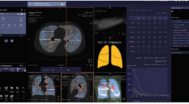
Nghiên cứu giá trị của chụp cắt lớp vi tính 128 định lượng trên bệnh nhân bệnh phổi tắc nghẽn mạn tính trước và sau ghép tế bào gốc tự thân
06/05/2021 15:35:43 | 0 binh luận
SUMMARY Background: Quantitative Computed Tomography (QCT) has been used for many years worldwide to evaluate and quantify lung parenchymal lesions in chronic obstructive pulmonary disease (COPD), including emphysema quantification (LAA-950), air-trapping assessment area (LAA- 856), bronchial wall area (WA), percentage of wall area (% WA), bronchial lumen area (LA), bronchial wall thickness (WT), studies show that QCT is highly accurate, strongly correlated with the respiratory function test (FEV1, FVC), grade classification according to GOLD. We applied this method to evaluate the indicators of emphysema (LAA-950), air-trapping (LAA-856), RVC¬856-950, bronchial wall area (WA), bronchial lumen (LA) and bronchial wall thickness (WT), percentage pulmonary vascular (%HAV) of COPD patients before and after autologous stem cell transplant from adipose tissue and bone marrow. Method: The study was conducted from 10.2019 - 10.2020 on 32 COPD patients diagnosed with COPD according to GOLD 2018 standards, patients with FEV1 <60% were selected for the autologous stem cell transplant study at The Respiratory Center - Bach Mai Hospital (4 GOLD II patients, 17 GOLD III patients, 11 GOLD IV patients). The patient was given quantitative CT scans 2 times, the first time before transplant and the second after 6 months after transplantation with a 128-detectors scanner of Siemens (Somatom Definition Egde) at Dien Quang Center - Bach Mai Hospital. Results: Percentage of emphysema (LAA-950) before grafting 31.49% ± 8.19, after grafting 32.8% ± 7.13), percentage of air-trapping in then exhalation (LAA-856) before grafting 63.65% ± 8.74, after transplant 61.41% ± 7.4 (statistically significant difference p = 0.026), RVC856- 950 before transplant 0.83 ± 1.82, post transplant 3.58 ± 1.76 (significant difference p = 0.000), these indicators are linearly correlated with FEV1, BODE and GOLD classification. The percentage of wall area (%WA) after transplantation was changed in the bronchial branch of segment 1 (70.74% before transplantation, 67.59% after transplantation, p = 0.02) and in branch of subsegment 1 (79.19% before transplantation, after transplantation 75.90%, p = 0.01), lumen area (LA), inner diameter (ID) of the posttransplant bronchial all increased in the segmental and subsegmental bronchial branches RB1, RB4, RB10, wall thickness (WT) decreased in the sub-branches RB1-1, RB4-1, RB10-1 (however the difference was not statistically significant with p <0.05). Conclusion: Emphysema (LAA-950), air-trapping (LAA-856, RVC856-950), percentage of bronchial wall (% WA), lumen area (LA), inner diameter (ID), thickness bronchial wall (WT) measured on QCT correlated with FEV1, FVC, GOLD, BODE before and after stem cell transplantation, can be used to assess the extent and stage of Chronic Obstructive Pulmonary Disease, pre- and post-assessment of autologous stem cell transplant therapy. Keywords: Quantitative CT COPD, Quantitative CT after autologous stem cell tranplantation
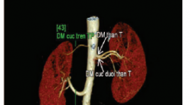
Vai trò của chụp cắt lớp vi tính 128 dãy trong đánh giá giải phẫu động - tĩnh mạch đoạn ngoài thận ở người cho sống
06/05/2021 16:27:48 | 0 binh luận
SUMMARY Objective: The purpose of this study was to determinate the accuracy of multidetector computed tomography (MDCT) angiography for imaging evaluation of renal arterial and venous structures of living donor kidney Materials and Methods: Two hundred twenty-eight potential living donor candidates were included to this study, who had CTA for the assessment of their renal vessels in our hospital between January 2018 and June 2020 and one hundred-eighty donors who underwent open surgical in this time. The number, course, and drainage patterns of the renal vessel were retrospectively observed from the scans. Anomalies of renal arteries, veins and inferior vena cava (IVC) were recorded. Multiplanar reformations (MPRs), maximum intensity projections, and volume rendering were used for analysis. The results obtained were correlated surgically. Results: A total of 228 potential live kidney donors underwent renal CTA (112 of the patients were male, and 116 were female. Mean age of donors was 38,32 ± 12,34 years. among them 180 patients had donor nephrectomy. There were 151 kidneys had single renal artery , here were 29 kidneys had at least one accessory or polar artery. There were 45 early branching renal arteries, two kidneys were retroaortic left renal veins. 16 kidneys had multiple renal veins. 55 kidneys wewe late venous confluence of renal veins. Sn, Sp, NPV, PPV compare open oparative nephrectomy 87,5%- 100%. Conclusion: Renal CTA is an accurate, safe and noninvasive diagnostic tool for determination of renal artery and vein abnormalities in living kidney donors preooparative. It helps with surgery planning, choosing operation side and exclusion of donors. Keyword: CTA, MSCT, living donor
Bạn Đọc Quan tâm
Sự kiện sắp diễn ra

THÔNG BÁO SINH HOẠT KHOA HỌC 19/01/2024: CẬP NHẬT CHẨN ĐOÁN VÀ ĐIỀU TRỊ UNG THƯ TRỰC TRÀNG
19-01-2024 09:47:01 0 bình luận

Thông tin đào tạo
- Những cạm bẫy trong CĐHA vú và vai trò của trí tuệ nhân tạo
- Hội thảo trực tuyến "Cắt lớp vi tính đếm Photon: từ lý thuyết tới thực tiễn lâm sàng”
- CHƯƠNG TRÌNH ĐÀO TẠO LIÊN TỤC VỀ HÌNH ẢNH HỌC THẦN KINH: BÀI 3: U não trong trục
- Danh sách học viên đạt chứng chỉ CME khóa học "Cập nhật RSNA 2021: Công nghệ mới trong Kỷ nguyên mới"
- Danh sách học viên đạt chứng chỉ CME khóa học "Đánh giá chức năng thất phải trên siêu âm đánh dấu mô cơ tim"


Đơn vị hợp tác

