


U thận trong bệnh cảnh phakomatosis, những điều cần lưu ý: nhân hai trường hợp lâm sàng
02/06/2020 10:35:14 | 0 binh luận
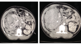
Renal tumors in the Phakomatosis disease, what to note: two case report SUMMARY Phakomatosis are a group of neurocutaneous disorders characterised by involvement of structures that arise from the embryonic ectoderm , consist of central nervous system, skin ,eyes and others: kidney, heart, lung… In this article we focus on the incidence of renal tumors in Tuberous sclerosis, one of the common diseases in Phakomatosis. Tuberous sclerosis is a rare neurocutaneous disorder (phakomatosis) characterized by the development of multiple benign tumours in various organs, including the kidneys. Tuberous sclerosis complex has several renal manifestations including angiomyolipomas (AML) and renal epithelial neoplasms. Angiomyolipomas is found in 40% of patients with tuberous sclerosis, and its most common complication is ruptures due to aneurysms. We present two cases was diagnosed with bilateral angiomyolipomas in tuberculosis and emphasizes the importance of the diagnosis and choice of appropriate treatment for each patient. Key words : Phakomatosis, renal tumor
Nhân hai trường hợp Schizencephaly
31/03/2020 22:28:11 | 0 binh luận
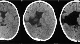
Schizencephaly – 2 cases report SUMMARY Introduction: Schizencephaly is a congentital brain malformation, which is characterized by clefts extending from pial surface of cerebral mantle to ventricle. This morphologically malformation divides into 2 types “closed-lip” and “open-lip”. Clinical symptoms ranged from simple to complicated depend on the location of clefts. This is a rare malformation, according to statistics, the incidence of the disease is approximately 1-2: 100.000 populations. Therefore, we are describing 2 cases encountered in Hue UniversityHospital within 2 years. Approach :Case 1: 2-year-old male patient, presenting the clinical symptom of half body weakness since 8 months old. Pregnant history of his mother were been unexplained fever in 8th month pregnancy, untreated. He underwent brain CT scanner and MRI. Case 2: 33-year-old male, hospitalization by presenting the epilepsy. He has been the half body weakness since neonatal phase. He underwent brain MRI. Result : Case 1: Presenting a large cleft lined by grey matter between right ventricle and pial surface of right hemisphere. Right parietal, temporal, and frontal lobeswere atrophic; the septum pellucidum is absent. Posterior part of corpus callosum was atrophic Case 2: Large size cleft lined by grey matter between right ventricle and pial surface of right hemisphere. Right frontal lobe is small. Right frontal gyri and lined-cleft gyri are small, hyperdensity. Medial line deviates toward left side. Corpus callosum is normal. Conclusion : Schizencephaly malformation occurs in any ages with different clinical symptoms. Typical character is the cleft, which is lined by grey matter from ventricle to arachinoid space. This accompanies other abnormalities such as the absence of septum pellucidum, polygyri, atrophy of corpus callosum. Diagnostic imaging, especially MRI, is crucially important to define this malformation and accompied abnormalities. Key words: Schizencephaly, MRI.
Bước đầu nghiên cứu vai trò của chụp cắt lớp vi tính 128 dãy trong kiểm tra sau phẩu thuật trong phình động mạch não vỡ
11/04/2020 23:13:09 | 0 binh luận
Phình động mạch não là một loại tổn thương thường gặp của hệ thống động mạch não, chiếm khoảng 1-8% dân số Đa số phát hiện khi có biến chứng vỡ gây chảy máu nội sọ Có hai cách điều trị hiệu quả là phẫu thuật và can thiệp nội mạch. Phẫu thuật kẹp cổ túi phình bằng clip cho đến nay vẫn được coi là phương pháp điều trị triệt để tuy nhiên không thể khẳng định tình trạng túi phình tồn dư, hay còn tồn tại túi phình trong quá trình mổ. Kiểm tra sau phẫu thuật là quan trọng nhằm đánh giá và theo dõi tình trạng túi phình và nhu mô não Chụp mạch số hóa xóa nền được xem là phương pháp tốt nhất về đánh giá mạch máu tuy nhiên đây là phương pháp xâm nhập đồng thời phương pháp này không đánh giá được tình trạng nhu mô não. Chụp CT scanner 128 dãy là phương pháp không xâm nhập, giá thành tương đối rẻ cho kết quả nhanh, chính xác, đánh giá được cả về mạch máu và tổn thương nhu mô não.
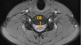
Ca lâm sàng: Bệnh lý Hirayama
22/12/2019 15:37:07 | 1 binh luận
Báo cáo mô tả một ca bệnh Hirayama của 1 nam thanh niên 16 tuổi bị yếu, teo cơ bàn tay kèm theo hình chụp cộng hưởng từ ở tư thế cổ thẳng có hình ảnh teo dẹt tủy cổ khu trú ngang mức đốt sống C5, C6.
Bạn Đọc Quan tâm
Sự kiện sắp diễn ra

THÔNG BÁO SINH HOẠT KHOA HỌC 19/01/2024: CẬP NHẬT CHẨN ĐOÁN VÀ ĐIỀU TRỊ UNG THƯ TRỰC TRÀNG
19-01-2024 09:47:01 0 bình luận

Thông tin đào tạo
- Những cạm bẫy trong CĐHA vú và vai trò của trí tuệ nhân tạo
- Hội thảo trực tuyến "Cắt lớp vi tính đếm Photon: từ lý thuyết tới thực tiễn lâm sàng”
- CHƯƠNG TRÌNH ĐÀO TẠO LIÊN TỤC VỀ HÌNH ẢNH HỌC THẦN KINH: BÀI 3: U não trong trục
- Danh sách học viên đạt chứng chỉ CME khóa học "Cập nhật RSNA 2021: Công nghệ mới trong Kỷ nguyên mới"
- Danh sách học viên đạt chứng chỉ CME khóa học "Đánh giá chức năng thất phải trên siêu âm đánh dấu mô cơ tim"


Đơn vị hợp tác

