


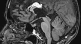
Lipoma of Corpus Callosum A case report in Quang Tri General Hospital
22/11/2020 19:13:47 | 0 binh luận
Đặc điểm hình ảnh cộng hưởng từ của u sợi và u vỏ - sợi buồng trứng
18/12/2019 09:32:38 | 0 binh luận
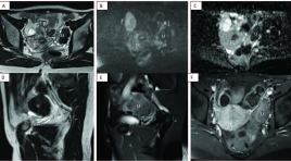
Value of magnetic resonance imaging in the diagnosis of ovarian thecomas/fibrothecomas
SUMMARY
Purpose: Our study aims to study the value of conventional magnetic resonance imaging (MRI) combined with DWI and Dynamic technique in the diagnosis of thecomas/fibrothecomas and differential diagnosis benign with malignant ovarian tumors.
Material and method: In total, 68 thecomas/fibrothecomas, 63 malignant ovarian tumors were included in our study. All patients underwent conventional MRI, DWI in 79 cases and Dynamic enhancement (DCE) in 14 cases. The clinical features and characteristics of conventional MRI, DWI and DCE of these two groups were analyzed. Apparent diffusion coefficient (ADC) values, Tmax, MRE were measured and compared between groups. Univariate analysis, multivariate logistic regression analysis were analyzed. Sensitivity (Se), Specificity (Sp), Positive Predictive Value (PPV), Negative Predictive Value (NPV) were included.
Results: All the fibromas/fibrothecomas showed hypo-isointensity on T1 weighted imaging (T1WI) and 77.9 % lesions showed hypo- to isointensity on T2 weighted imaging (T2WI). After administration of contrast medium, 82,3% tumors appeared as minor to mild enhancement, 71,4% benign tumor had type 1 curve, Tmax cutoff were 230s with Se and Sp 71,4%. MRE were not already measured because of few cases. On DWI, 68,4% fibromas/fibrothecomas manifested no signal intensity or low signal intensity. The ADC cutoff were 1.07 x 10⁻³ mm2/s to differentiate benign from malignant ovarian tumors. Multivariate logistic regression analysis showed that only T2WI and ADC were the important indicators in discriminating fibromas/fibrothecomas or benign tumors from malignant ovarian tumors.
Conclusion: The combination of DWI, DCE with conventional MRI is of great value in the diagnosis of fibromas/fibrothecomas and differentiation benign ovarian tumors from malignant ovarian tumors
Keywords: Fibromas/fibrothecomas, Conventional magnetic resonance imaging, Diffusion-weighted imaging, Apparent diffusion coefficient value, Dynamic contrast enhancement.
Bạn Đọc Quan tâm
Sự kiện sắp diễn ra

THÔNG BÁO SINH HOẠT KHOA HỌC 19/01/2024: CẬP NHẬT CHẨN ĐOÁN VÀ ĐIỀU TRỊ UNG THƯ TRỰC TRÀNG
19-01-2024 09:47:01 0 bình luận

Thông tin đào tạo
- Những cạm bẫy trong CĐHA vú và vai trò của trí tuệ nhân tạo
- Hội thảo trực tuyến "Cắt lớp vi tính đếm Photon: từ lý thuyết tới thực tiễn lâm sàng”
- CHƯƠNG TRÌNH ĐÀO TẠO LIÊN TỤC VỀ HÌNH ẢNH HỌC THẦN KINH: BÀI 3: U não trong trục
- Danh sách học viên đạt chứng chỉ CME khóa học "Cập nhật RSNA 2021: Công nghệ mới trong Kỷ nguyên mới"
- Danh sách học viên đạt chứng chỉ CME khóa học "Đánh giá chức năng thất phải trên siêu âm đánh dấu mô cơ tim"


Đơn vị hợp tác

