


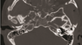
ĐẶC ĐIỂM HÌNH ẢNH CLVT ĐA DÃY VỠ XƯƠNG HÀM MẶT DO TAI NẠN GIAO THÔNG
13/10/2023 16:55:55 | 0 binh luận
SUMMARY Objectives : study the multi-slice CT (MSCT) imaging in the diagnosis of maxillofacial fractures by traffic accident. Subjects and methods: A cross-sectional descriptive study of 63 patients with maxillofacial trauma due to traffic accidents who underwent MSCT scan at Viet Duc Hospital in April 2022. Results: 46 male and 17 female. The mean age was 28.4±12.6 (from 15 to 75 years old). The most common traffic accidents are motorbikemotorcycle accidents with 21/63 (33.3%), self-motorbike-riding accidents are 18/63 (28.6%), car-motorcycle accidents account for 16 /63 (25.4%), pedestrian - car/motorcycle accidents are 5/63 (7.9%) and car-car accidents are 3/63 (4.8%). There were 28/63 (44.4%) cases associated with cranial fracture and 32/63 (50.8%) skull base fracture. In cases of maxillofacial fracture, orbital wall fracture was the most common with 41/63 (65.1%), maxillary fracture 39/63 (61.9%), zygomatic fracture 32/63 (50.8%), nose bone 19/63 (30.2%), frontal sinus wall 13/63 (20.6%), palatal and mandibular fracture were 11/6 (17.5%). The most common type of maxillary fractures is Lefort 1 with 11/63 (17, 5%) on both right and left side, Lefort 2 and 3 fractures are less common with 1.6% to 6.3%, respectively. Conclusion : MSCT is a reliable method in diagnosing maxillofacial fractures. Keywords: maxillofacial trauma, traffic accident, computer tomography.

ĐÁNH GIÁ SỐNG THÊM TRUNG HẠN ĐIỀU TRỊ NÚT MẠCH HÓA CHẤT BỆNH NHÂN UNG THƯ BIỂU MÔ TẾ BÀO GAN TẠI BỆNH VIỆN BẠCH MAI
13/10/2023 15:44:22 | 0 binh luận
SUMMARY Purpose: Evaluation the survival of the patients with hepatocellular carcinoma (HCC) who underwent transcatheter arterial chemoembolization (TACE). Subjects and methods: Retrospective study on transcatheter arterial chemoembolization in patients with hepatocellular carcinoma from July 2017 to July 2020 at Radiology Center - Bach Mai Hospital. Results:. 70 patients including 64 men (92.9%) and 6 women (7.1%) had a confirmed diagnosis of HCC with a mean age of 61.5 ± 11.34 years (from 32 to 83 years). The mean number of treatment sessions were 4.5±0.4 lần (1-16). Progression-free survival after initial TACE and overall survival median was 29.7±3.3 months, 43.3±2.5 months, respectively. Cumulative survival time at 1 year, 2 years, 3 years and 5 years is 91.4%, 77.1%, 63.7% and 29.4%, respectively. Patients treated with cTACE were 67.1% and Deb-TACE were 32.9%. mRECIST after the first TACE: complete response, partial response, stable disease and advanced disease were 30.0%, 55.7%, 4.3%, 10.0%, respectivel. Patients with objective response had a survival of 47.8±2.3 months, and patients with poor response had a survival of 12.2±2.7 months, respectively (p = 0.001). mRECIST response predicted survival among the patients on univariate analysis (HR=15.13, p=0.000). The independent predictors for survival were mRECIST response (p=0.001). Conclusion : Use of TACE in intermediate stage HCC patients gives a significant survival advantage when objective response is achieved as per mRECIST. Key words: TACE, Progression-free survival, overall survival..
ĐÁNH GIÁ KẾT QUẢ CỦA DẪN LƯU Ổ DỊCH QUA DA TRONG ĐIỀU TRỊ VIÊM TUỴ CẤP NẶNG
13/10/2023 15:35:24 | 0 binh luận
SUMMARY Objectives: Evaluating the result and complications of percutaneous drainage under the guidance of imaging in the treatment of severe acute pancreatitis. Subjects and methods: A total of 51 patients diagnosed with severe acute pancreatitis at Bach Mai hospital from 7/2021 to 7/2022 were treated by percutaneous drainage. Results: 51 patients included 41 men and 10 women, with the age from 27 to 72 years old, the success rate of drainage technique is 97,3%, the mortality rate of patients in the study is 7,8% and there were no cases of complications after drainage. Clinical and laboratory symptoms were significantly reduced compared with pre-drainage. Conclusions: Percutaneous drainage under the radiology guidance with ascite, inflammatory fluid in acute pancreatitis is a safe technique with a high success rate. Keywords : severe acute pancreatitis, percutaneous drainage
NGHIÊN CỨU KẾT QUẢ SỚM ĐIỀU TRỊ UNG THƯ BIỂU MÔ TẾ BÀO GAN Ở VỊ TRÍ NGUY CƠ BẰNG ĐỐT SÓNG CAO TẦN
13/10/2023 15:24:36 | 0 binh luận
SUMMARY Background: Radiofrequency ablation is a radical treatment with high efficiency in the very early stages (BCLC 0) and early stages (BCLC A). However, with tumors in risk locations, radiofrequency ablation has many difficulties in determining the tumor on ultrasound and the risk of damage to adjacent organs. Objectives: To evaluate early outcomes of RFA in treating HCC patients in risk locations. Subjects and methods: 50 HCC patients in risk locations underwent ablation at the radiology center of Bach Mai hospital. Follow up 3 months to evaluate the effectiveness after the treatment. Results: 50 patients male/ female=41/9, average age = 61.63 years old (35–86). 94% patients with HCC have a history of cirrhosis and 88% patients with HCC have a history of hepatitis. Among the tumors in risk locations, the tumors adjacent to the diaphragmatic accounted for the highest rate of 44%. We infused the artificial ascites in 20 cases(40%) and the pleural effusion in 14 cases(28%). Follow-up 3 months after treatment, there was no the major complication. 7 patients had the atelectasis. The rate of tumor complete response was 92%, the rate of tumor incomplete response was 4%. 2 patients appeared new lesions(4%). Conclusion: Radiofrequency ablation is a minimally invasive, safe and effective method for tumors in risk locations. Keywords: Radiofrequency ablation (RFA) for HCC in risk locations , the image-guided techniques, the outcome after the treatment.
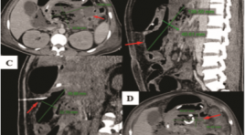
KẾT QUẢ ĐIỀU TRỊ CAN THIỆP NỘI MẠCH GIẢ PHÌNH ĐỘNG MẠCH TẠNG BỤNG KHÔNG DO CHẤN THƯƠNG
13/10/2023 15:18:32 | 0 binh luận
SUMMARY Objective: to apply and estimate the efficacy of embolization in non traumatic visceral arterial pseudoaneurysm. Subject and method: Twenty-seven patients underwent angiography and embolization at Bachmai hospital from 7/2020 to 7/2022. Result: In 27 patients (20 males & 07 females), 09 patients show extravasation in DSA. The splenic artery and the superior mesenteric artery are the arteries most commonly with visceral arterial pseudoaneurysms. Of the 27 patients studied, 24 had technical success in the first intervention, one required secondary intervention because of rebleeding and it was successfully treated by reintervention, one required ileectomy after embolisation due to ongoing hemorrhage, one patient died from uncontrolled blood transfusion shock. Conclusion: In hemodynamically stable and controlled patients, selective and super selective embolization is a safe and effective method for managing non traumatic visceral arterial pseudoaneurysm. Keywords: Visceral arterial pseudoaneurysm (VAPA), embolization, extravasation.
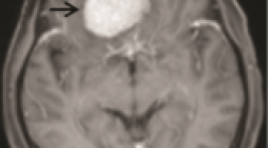
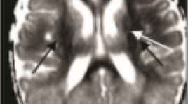
GIÁ TRỊ CỦA CỘNG HƯỞNG TỪ KHUẾCH TÁN TRONG PHÂN BIỆT U MÀNG NÃO ĐIỂN HÌNH VÀ KHÔNG ĐIỂN HÌNH
13/10/2023 12:54:47 | 0 binh luận
SUMMARY Objective: To determine the value of Diffusion-weighted imaging in distinguishing typical and atypical meningiomas. Subjects and methods: The study was performed at Choray Hospital from January 2021 to August 2022, with 66 typical and atypical meningiomas patients. The descriptive retrospective cross-sectional study was performed. Compare the tumor's signal intensity and mean ADC value with the histopathological results to distinguish typical and atypical meningiomas. Results: In 66 patients, there were 13(19.7%) male patients and 53(80.3%) female patients. The mean age of patients was 60.53 years old. The number of typical and atypical meningiomas cases was 43 (65.2%) and 23 (34.8%). There was no significant signal intensity difference between typical and atypical meningiomas (p=0.56). The mean ADC value for typical meningiomas is 0.843x10⁻³mm2/s and atypical is 0.737x103mm2/s, the difference is statistically significant with p=0.003. On the ROC curve, with an ADC cutoff of 0.780x10⁻³mm2/s, DWI can distinguish typical and atypical meningioma with sensitivity, specificity, PPV, NPV, and accuracy is 83.8%, 51.7%, 67.4%, 71.4%, 66.6% respectively. Conclusion: The mean ADC value of typical meningiomas is higher than that of atypical meningiomas. DWI is valuable in the differential diagnosis of typical and atypical meningiomas. Keywords: meningiomas, typical, atypical, diffusion-weighted imaging, ADC.
ĐÁNH GIÁ PHÂN BỐ DƯỢC CHẤT PHÓNG XẠ 18F-FLUORINE-FLUOROTHYMYDINE TRÊN CHUỘT THÍ NGHIỆM GÂY KHỐI U
13/10/2023 12:49:57 | 0 binh luận
SUMMARY PET (positron emission tomography) techniques and radiopharmaceutical tracers are becoming increasingly common. Since 1991, 3’-deoxy-3’-18Fluorine-Fluorothymidine (18F-FLT) has been studied as a PET tracer for tumor proliferation assessment. The purpose of this study was to assess tracer uptake (%ID/g) in tumor-bearing mice. Materials and methods: Two cell lines (4T1 and LLC) were inoculated into BALB/c mice. The radiotracer was then injected, and tumor resection and organ samples were performed at 20, 40, 60, 90 and 120 minutes later. A gamma-ray spectrometer was used to calculate uptake values. Results: The data show that the tumor’s %ID/g were 3.68 ± 0.29 and 5.30 ± 0.44 at 60-minute post-injection for the 4T1 and LLC tumors, respectively. In other organs, the high uptake was observed in the kidneys, spleen, and bone marrow, but a low level of tracer was seen in the brain. Keywords: 3’-deoxy-3’-18Fluorine-Fluorothymidine, positron emission tomography, tumor-bearing mice.
NGHIÊN CỨU ĐẶC ĐIỂM HÌNH ẢNH CỘNG HƯỞNG TỪ SỌ NÃO Ở TRẺ SƠ SINH ĐỦ THÁNG CÓ BỆNH LÝ NÃO DO THIẾU MÁU CỤC BỘ/THIẾU OXY
13/10/2023 12:40:53 | 0 binh luận
SUMMARY Objectives: to describe the intracranial injuries on brain MRI of term newborns with hypoxic ischemic encephalopathy (HIE) Methods: A prospective and retrospective study based on medical records and brain MRIimages of term newborns with HIE. Brain MRI was performed within 2 weeks after birthon T1, T2, and DWI, SWI sequences Results: There was 94 eligible cases included in the study (male/ female:61/33). The mean gestational age was 39 weeks; mean age of brain MRI was 8 days, mean birth weight was 3100gr. The infants were classified by Sarnat criteria as mild/moderate/severeHIE by ratio: 22.3%/64.9%/12.8%. The infants were mainly given birth by vaginaldelivery 51%, which was followed by cesarean section 45.7%. The sentinel events weredefined mostly as prolonged labor causing fetal distress and placental abruption,umbilical cord accidents. Therapeutic hypothermia was performed in 80,9% cases.Intracranial injury was present in 82 (87.2%) infants. The brain lesions on MRIdiversified. The most common brain injuries were in deep gray nuclei and posterior limbof the internal capsule: thalamus (62.8%), the lenticular nucleus (60.6%), posterior limbof the internal capsule (47.9%), caudal nucleus (26.6%). The following was cortical andsubcortical white matter abnormalities 44.7%. The periventricular and punctate whitematter injuries are less common: 23.4% and 15.9%.The other brain injuries include the corpus callosum (35.1%), the optic radiation(30.8%), hippocampus (12.8%). The infratentorial structures injuries are less common,predominantly in severe HIE cases: brainstem injury 23.1% and cerebellar injury 9.6%.Cortical abnormalities were seen in 42 of 94 (44.7%) predominantly in central sulcus(26.6%), interhemispheric sulcus (21.3%). The cortex injuries in other regions includedinsular region (8.5%), frontal region (27.7%), parietal region (23.4%), occipital region (13.8%), temporal region (8.5%) Intracranial hemorrhage was present in 36/94 (38.3%) infants, including 32 (34%) with subdural hemorrhage, followed by intraventricular hemorrhage (5.3%), intraparenchymal hemorrhage (5.3%) Deep gray nucleus injuries, optic radiation, corpus callosal, hippocampal,brainstem, periventricular and subcortical white matter injuries were significantlyassociated with severity of HIE. The cortical injuries in interhemispheric, insular, frontaland temporal regions were more common in the infants with severe HIE Conclusion: Intracranial injury in the term newborn with HIE diversify, predominantly insupratentorial injuries. The most common brain injuries were in the deep gray nuclei andposterior limb of the internal capsule, followed by cortical and subcortical white matter.The corpus callosum, optic radiation, and hippocampus abnormalities were alsofrequently seen. The brainstem injury is significantly less common and cerebellar injuryis rarely seen. Posterior subdural hemorrhage was the most common intracranialhemorrhage, which was not severe clinically and frequently resolve without treatment Key words: newborn, hypoxic ischemic encephalopathy (HIE), brain MRI Abbreviations: HIE: hypoxic ischemic encephalopathy. MRI: Magnetic Resonance Imaging
Bạn Đọc Quan tâm
Sự kiện sắp diễn ra

THÔNG BÁO SINH HOẠT KHOA HỌC 19/01/2024: CẬP NHẬT CHẨN ĐOÁN VÀ ĐIỀU TRỊ UNG THƯ TRỰC TRÀNG
19-01-2024 09:47:01 0 bình luận

Thông tin đào tạo
- Những cạm bẫy trong CĐHA vú và vai trò của trí tuệ nhân tạo
- Hội thảo trực tuyến "Cắt lớp vi tính đếm Photon: từ lý thuyết tới thực tiễn lâm sàng”
- CHƯƠNG TRÌNH ĐÀO TẠO LIÊN TỤC VỀ HÌNH ẢNH HỌC THẦN KINH: BÀI 3: U não trong trục
- Danh sách học viên đạt chứng chỉ CME khóa học "Cập nhật RSNA 2021: Công nghệ mới trong Kỷ nguyên mới"
- Danh sách học viên đạt chứng chỉ CME khóa học "Đánh giá chức năng thất phải trên siêu âm đánh dấu mô cơ tim"


Đơn vị hợp tác

