


NGHIÊN CỨU ĐẶC ĐIỂM HÌNH ẢNH SIÊU ÂM NỘI SOI Ở BỆNH NHÂN UNG THƯ THỰC QUẢN
13/10/2023 12:07:09 | 0 binh luận
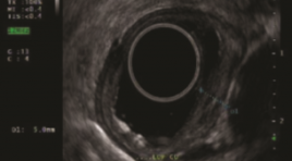
SUMMARY Purpose: The aim of study was decribed the imaging characteristics of endoscopic ultrasound in esophageal cancer. Methods and materials: A cross-sectional descriptive study on 40 patients with esophageal cancer were assigned to endoscopic ultrasound. Results: All study subjects were male with mean age 58.33 ± 7.24. 97.5% of SCC; 2.5% were high-grade dysplasia, none of the patients had adenocarcinoma. Moderately differentiated dominate 55%, 25% highly differentiated and 20% poorly differentiated. .Location of lesions on endoscopy is common in the middle and lower thirds of the esophagus. 85% of esophageal cancer lesions are hypoechoic, 7.5% hyperechoic, and 7.5% mixed. Of which, 70% are heterogeneous, the remaining are homogenous. The lesion size is less than half the circumference accounting for 62.5% and larger than half the circumference accounting for 32.5%. TNM classification on endoscopic ultrasound according to AJCC 8th stage T: Tis (7.5%), T1a (45%), T1b (30), T2 (10%), T3(7.5%). Stage N: N0 (70%), N1 (27.5%), N2 (2.5%). TNM stage: Stage 0 (5%), IA (15%), IB (47.5%), IIB (25%), IIIA (5%), IIIB (2.5%). Conclusion: Endoscopic ultrasound plays an important role in diagnosis and staging in patients with esophageal cancer Key words: endoscopic ultrasound, esophageal cancer, squamous cell carcinoma (SCC), adenocarcinoma.
NHẬN XÉT ĐẶC ĐIỂM LÂM SÀNG VÀ HÌNH ẢNH CẮT LỚP VI TÍNH PHỔI Ở BỆNH NHÂN SAU NHIỄM SARS-COV-2 TẠI CÁC CƠ SỞ KHÁM CHỮA BỆNH CỦA MEDLATEC
13/10/2023 12:01:05 | 0 binh luận
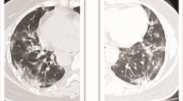
SUMMARY A cross-sectional descriptive study was conducted on 1436 patients after SARS-CoV-2 infection who visited MEDLATEC medical facilities from March 1, 2022 to March 31, 2022. The results showed that 39% were male and 61% female, with a mean age of 39 and the majority of patients in the working age range from 18-60 years old. 74% of patients infected with SARS-CoV-2 have clinical symptoms, of which cough is the most common symptom, followed by chest pain and shortness of breath. The group of patients with a healthy history accounted for 76% of cases; the remaining group of underlying diseases with hypertension accounted for the most, followed by the group with chronic lung disease. Up to 49% of patients have suspected COVID-related lung lesion; with the most important lesions being interstitial thickening and most of them being mild according to the CT-score scale. Keywords: clinical characteristics, CT scanner, SARS-CoV-2
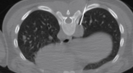
GIÁ TRỊ DỰ BÁO TỬ VONG CỦA ĐỘ NẶNG TỔN THƯƠNG PHỔI TRÊN X-QUANG TẠI THỜI ĐIỂM NHẬP VIỆN Ở BỆNH NHÂN COVID-19
13/10/2023 11:44:33 | 0 binh luận
SUMMARY Background: We aimed to investigate the performance of a chest X-ray (CXR) scoring scale of lung injury in prediction of death among patients with COVID-19 admitted at Vinmec Central Park hospital (HCM City, VN) during the peak epidemic in 2021. Method: Retrospective design; X-ray images and clinical data were collected from all hospitalized patients with SARS-CoV-2 PCR positive from July to September 2021. Three radiologists independently assessed the CXR score at admission which consists of measuring both severity and extent of lung injuries on four lung quadrants (scale o to 24). Association between CXR and mortality risk was estimated using a survival regression with log-log distribution. Result: The study included 219 patients (mortality rate = 28). There was a high consensus for CXR scoring among 3 radiologists (κ = 0.90; CI95%: 0.89-0.92). PCA analysis revealed that CXR has a similar role as CRP score for predicting risk of mortality. CXR score was the strongest predictor of mortality (tdAUC 0.85 CI95% 0.69–1) within the first 3 weeks after admission, compared with other conventional clinical features. After adjusting for the patient’s age, there was a significant effect of increased CXR score on mortality risk (HR = 1.15, CI95%:1.04-1.27, p=0.009). At a critical threshold of 16 points, the CXR score allows for predicting inhospital mortality with good sensitivity (0.82; CI95%: 0.78 to 0.87) and specificity (0.89; CI95%: 0.88 to 0.90). Conclusion: The day-one CXR score is an independent and effective predictor of the risk of death in COVID-19 and could be used to identify high-risk patients in countries like Vietnam where CXR is more readily available than CT scans. Keywords: COVID-19, Chest X-ray, Lung injury score, Mortality prediction
ĐÁNH GIÁ HÌNH THÁI TIỂU NHĨ TRÁI TRÊN CẮT LỚP VI TÍNH ĐA DÃY Ở BỆNH NHÂN TRIỆT ĐỐT RUNG NHĨ QUA ỐNG THÔNG
13/10/2023 11:29:50 | 0 binh luận
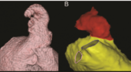
SUMMARY Purpose: to assess left atrial appendage morphology by multidetector computed tomography in atrial fibrillation (AF) patients for catheter ablation Materials and Methods: This retrospective and cross-sectional study included 45 patients diagnosed with AF and treated by catheter ablation and 45 patients without AF undergoing multi-detector computed tomography in E hospital from 1/2020 to 7/2022 Results: The mean age 56.16 ± 11.83, median of left atrium volume in the AF group was 118.13 (96 - 145.56) ml > the control group 72.88 (60.53 - 95.74) ml, (p < 0.001). The most common left atrium morphology in both groups is "cactus" with 46.67% in the AF group and 33.33% in the control group, the middle left atrial appendage orifice was the most common in the AF group (53,33%), while in the control group, the lower left atrial appendage orifice was the most common (44.44%), the length of the left atrial appendage in the AF group was 43.15 ± 8.11 mm, the control group was 44.29 ± 9.76 mm. There is a positive correlation with the mean level between left atrial appendage length and left atrial volume (r = 0.573, p < 0.001). Conclusion: The most common left atrium morphology in both groups is "cactus". There is a positive correlation with the mean level between left atrial appendage length and left atrial volume. Keywords: atrial fibrillation, catheter ablation, left atrial, left atrial appendage, multi-detector computed tomography
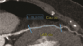
ĐÁNH GIÁ ĐẶC ĐIỂM HÌNH ẢNH CỦA CẦU CƠ ĐỘNG MẠCH VÀNH BẰNG CHỤP CLVT 256 DÃY
13/10/2023 11:00:20 | 0 binh luận
SUMMARY Purpose: to evaluate and analyze the ratio of position, depth and length of the coronary myocardial bridge, the status of pre-myocardial atherosclerosis and the degree of vascular stenosis during systole by multislice-computed tomography (MSCT) images series. Material and method : Computed tomography scan for 175 patients using a 256-row MSCT machine at Hanoi Friendship Hospital between July 2021 and August 2022. Select the best quality images in systole and diastole. Measure the length and thickness of the bridge. For each myocardial bridge segment, assess for the presence of anterior atherosclerotic plaque no more than 2 cm in length of the artery. Assess the degree of vascular stenosis during systole caused by the myocardial bridge. Result: Most of the myocardial bridges were located in the anterior interventricular artery (LAD) (98.3%). The average values of the length and thickness of the bridge are 23.1 ± 10.5mm and 1.0 ± 1.0mm. Atherosclerosis was detected in 77 cases (accounting for 43%). The average degree of tunneling artery stenosis was 23.0% systolic. Conclusion: MSCT 256 series has a high value in detecting bridges, determining the location, there by measuring the length, the thickness of the pons, the status of anterior atherosclerotic plaques as well as the degree of narrowing of the vessel lumen systole of the musculature. Keywords: myocardial bridge, coronary artery, multislice computed tomography.
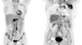
ĐÁNH GIÁ BƯỚC ĐẦU GIÁ TRỊ CỦA INTERIM PET/CT SAU 2 CHU KỲ ĐIỀU TRỊ HOÁ CHẤT ABVD CỦA BỆNH NHÂN U LYMPHO HODGKIN
13/10/2023 10:44:46 | 0 binh luận
SUMMARY Purpose: The study aimed to initial evaluate the value of interim PET/ CT after 2 cycles of ABVD chemotherapy in Hodgkin lymphoma patients. Material and Methods: The study was carried out retrospective and prospective descriptive study on 56 patients at Tan Trieu K Hospital from March 2020 to June 2022. Patients were examined clinically, paraclinically, had a CT scan or PET/CT film before treatment, then received ABVD chemotherapy, after 2 cycles of being taken and evaluated Interim PET/CT, and continued treatment based on the results. Result of iPET/CT leads to continue or change of regimen continued treatment or change of regimen and follow-up. Results: In a total of 56 patients, male/female ratio: 1/1.6, mean age 30±12.6 (youngest age 7 years old, oldest age 67 years old). 35/56 patients (62.5%) had CT scan and 21/56 patients (37.5%) had background PET/CT scan before treatment. 30.4% had 2 lymph node sites and 28.6 patients had 3 lymph node sites on the body, lymph nodes above the diaphragm 67.9% and lymph nodes both above and below the diaphragm 12,5%. 49 patients had lymph node lesions, short axis size of lymph nodes maximum 27.10±8.3 mm. Lesion to the mediastinum in 15/58 patients (25.8%), spleen in 5 patients. Patients with early stage I-II accounted for 82.1%, advanced stage III-IV accounted for 17.9%. After 2 cycles of ABVD chemotherapy, the response rate on PET/CT was iPET/CT2 (-) 43/56 patients (76.8%, of which Deuville score 1 point 93%) and iPET/CT2 (+) in 13/56 patients (23.3%). The prognostic index of IPS low-risk group (0-2) complete metabolic response was 86.4%. In the high-risk group IPS (4-7) partial metabolic response and complete metabolic response was 54.5% according to Lugano classification. Conclusion: Interim PET/CT has an important role in providing prognostic information in the treatment of ABVD chemotherapy in Hodgkin Lymphoma patients, reducing the toxicity of chemotherapy and determining the next treatment modalities. Keywords: lymphoma Hodgkin, interim PET/CT.
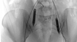
BÁO CÁO CA LÂM SÀNG: PHỐI HỢP CHẸN BÓNG ĐỘNG MẠCH CHẬU TRONG VÀ NÚT ĐỘNG MẠCH TỬ CUNG DỰ PHÒNG XUẤT HUYẾT Ở SẢN PHỤ MẮC RAU CÀI RĂNG LƯỢC KẾT HỢP RAU TIỀN ĐẠO
12/10/2023 15:53:35 | 0 binh luận
SUMMARY Placenta acrreta spectrum is a condition in which the placenta partially or completely invades and cannot be separated from the uterus muscle. Placenta previa is a condition in which the placenta partially or completely covers the cervix. Both phenomena increase the risk of postpartum haemorrhage, hemostasis disorders, and threaten the life of the mother and the fetus. Combine of the two phenomena increase blood loss in cesarean delivery as well as postpartum period, and is a great challenge required multidisciplinary for successful management. Prophylactic endovascular intervention is a minimally invasive treatment method, which plays an important role in prevent haemorrhage in the management of placenta accreta spectrum. This report describes a case of placenta percreta combine with placenta previa, which received prophylactic endovascular intervention using balloon occlusion of internal illiacs and uterine arteries embolization and underwent a cesarean delivery at 36 weeks. The patient had a successful delivery and preserved the uterus after surgery. Key word : Placenta acrreta spectrum, prophylatic balloon occlusion, uterine artery embolization
SARCOMA CƠ VÂN DI CĂN PHỔI TỔNG QUAN TÀI LIỆU VÀ BÁO CÁO CA BỆNH
12/10/2023 15:49:15 | 0 binh luận
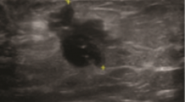
SUMMARY Sarcoma is a general term for a type of cancer found in the connective tissue cells (the cells of the mesenchyme). There are many types of sarcoma because connective tissue cells are present everywhere in the body (bone, cartilage, blood vessels ...) but this type of cancer is divided into 2 main groups, bone sarcoma, and soft tissue sarcoma. Rhabdomyosarcoma (RMS) belongs to the group of soft tissue sarcoma of skeletal muscle, is a common malignancy, and is one of the leading causes of cancer death in children. Alveolar rhabdomyosarcoma (ARMS) is a subtype of RMS that is extremely rare in adults. We present a pediatric case of ARMS, primary in the butt area with pulmonary metastases, confirmed by histopathology and immunohistochemistry. Reported data and literature review will help physicians have a better diagnostic approach when encountering similar cases. Keyword: Rhabdomyosarcoma; alveolar rhabdomyosarcoma, soft tissue tumor lung metastasis.
Bạn Đọc Quan tâm
Sự kiện sắp diễn ra

THÔNG BÁO SINH HOẠT KHOA HỌC 19/01/2024: CẬP NHẬT CHẨN ĐOÁN VÀ ĐIỀU TRỊ UNG THƯ TRỰC TRÀNG
19-01-2024 09:47:01 0 bình luận

Thông tin đào tạo
- Những cạm bẫy trong CĐHA vú và vai trò của trí tuệ nhân tạo
- Hội thảo trực tuyến "Cắt lớp vi tính đếm Photon: từ lý thuyết tới thực tiễn lâm sàng”
- CHƯƠNG TRÌNH ĐÀO TẠO LIÊN TỤC VỀ HÌNH ẢNH HỌC THẦN KINH: BÀI 3: U não trong trục
- Danh sách học viên đạt chứng chỉ CME khóa học "Cập nhật RSNA 2021: Công nghệ mới trong Kỷ nguyên mới"
- Danh sách học viên đạt chứng chỉ CME khóa học "Đánh giá chức năng thất phải trên siêu âm đánh dấu mô cơ tim"


Đơn vị hợp tác

