


Đánh giá kết quả bước đầu về cộng hưởng từ đàn hồi gan tại trung tâm y khoa Medic
11/04/2020 14:58:33 | 0 binh luận
Evaluate the initial result of liver mr elastography at the medic medical center SUMMARY Purpose: To evaluate the initial result of liver magnetic resonance elastography was performed in the patients and to compared with fibroscan. Methods and Materials: We retrospectively 210 patients was performed MR elastography, with 80 patients was performed MRE and Fibriscan at Medic medical center from July 2016 to June 2017, age range, 31-71 years. All the patients executed MRE on the GE signa explorer 1,5T MRI, and on Fibroscan of Echosens. Results: I n 210 patients was executed MRE, No. of patients no Fibrosis (F0): 29; Light fibrosis (F1): 39; Significant fibrosis (F2): 36; progressive fibrosis (F3): 41; Cirrhossis (F4): 65. Had detected 12 tumors (5 HCC, 1 FNH, 6 Hemangioma) and 14 cysts in liver. In 80 patients was executed MRE and Fibroscan: 9 patients F0 with MRE, the same 3F0 and 6F1 with Fibroscan; 12 patients F1 with MRE, same 5F1 and 7F2 with Fibroscan; 20 patients F2 with MRE, same 16F2 and 4F3 with Fibroscan; 15 patients F3 with MRE, same 13F3 and 2F4 with fibroscan; 24 patients F4 with MRE, same 24F4 with Fibroscan. Conclusion: MRE is a reliable non-invasive technique, safer, less expensive for evaluating hepatic fibrosis. Can be carried out at the same time as MRI examination with a search for hepatic tumor, and performed in most patients with liver disease including those with ascites or obesity. Can replace the method of liver biopsy unnecessary, risk of complications.
Kỹ thuật kết hợp hình ảnh siêu âm với hình ảnh cắt lớp vi tính hoặc cộng hưởng từ
30/03/2020 22:56:08 | 0 binh luận
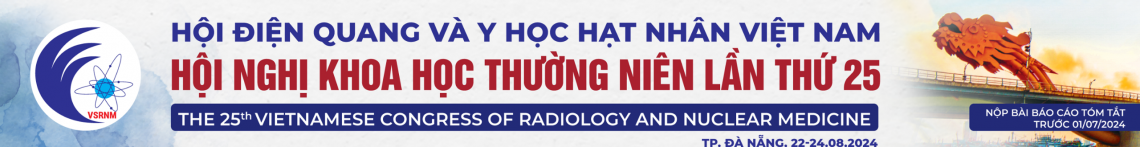
Fusion imaging of ultrasonography with CT or MRI ABSTRACT: Fusion imaging of ultrasonography with CT or MRI is the technique that creates hybrid images of two imaging modalities and combines advantages of ultrasound such as dynamic real-time imaging with advantages of CT of MRI such as high spatial and contrast resolution. The fusion imaging of ultrasound with other imaging modalities is a recently developed technique of this decade and expected to establish new and useful application in clinical practice. Key word: Fusion imaging of ultrasonography with CT or MRI
Mô tả đặc điểm hình ảnh cộng hưởng từ và hình ảnh18F-FDG PET/CT não ở các bệnh nhân mắc bệnh Alzheimer tại Bệnh viện Lão khoa Trung ương
01/04/2020 16:23:25 | 0 binh luận
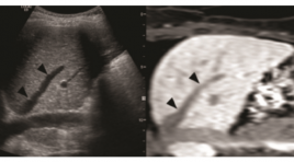
Description of MR and brain 18F-FDG PET/CT imaging characteristics in Alzheimer’s Disease patients in National Hospital of Geriatrics SUMMARY Purpose : Study realised is aimed to describe MRI features as well as brain 18F-FDG PET/CT characteristics of Alzheimer’s Disease patients in the National Hospital of Geriatrics. Methods: From 2014 to 2015, it is the first time in Vietnam, brain 18F-FDG PET/CT scan applied in studying Alzheimer disease with 32 cases including 16 Aalzheimer patients in pathologic group and 16 non-demential elderly individuals (“Normals” or NLs) in control group, brain 18F-FDG PET/CT scans performed at the Center of Nuclear Medicine and Oncology - Bach Mai Hospital. Brain MRI investigation was also realized for all Alzheimer patients. 18F-FDG PET/CT data in Alzheimer group was confronted to Nls group. Results: Mean age in AD patients is 65.1± 8.2 years old. Most of AD patients examined at moderate to severe stage (90%). Brain MRI shows 93% AD patients with brain atrophy (mild to severe level), 75% with pathological medial temporal lobe atrophy (abnormal MTA scale) and 81.3% having parietal cortical atrophy. Evan index is higher than normal in 65% of cases. No abnormal regional cortical glucose hypometabolism on brain 18F-FDG PET/ CT imaging seen in all persons in Nls group. On the other hand, in Alzheimer group, 93,8% of cases having a hypometabolism occurred in temporo-parietal association region and 81.3% of cases on right side, meanwhile all Alzheimer patients suffering from a glucose hypometabolism affecting bilateral posterior cingulate gyrus as well as left hippocampus. Occipital glucose metabolism in Alzheimer group is principally well reserved and hypometabolism extending to frontal regions in about a half of all cases. Conclusion : Most of Alzheimer patients having cerebral degenerative abnormalities on brain MRI presented by multi-grade cerebral parenchymal atrophy. Characteristic atrophic regions in Alzheimer patients include medial temporal and parietal lobes. Glucose hypometabolisme imaging in 18F-FDG PET/CT scans in Alzheimer group is quite specific with anatomically regional hypometabolism patterns seen in temporo-parietal association regions as well as in posterior cingulate gyri, with dominance on left side. Brain MRI and brain 18F-FDG PET/CT scan are wellknown as imaging technics with a high sensibility, high medical safety which are objective valued imaging technics and applied more and more in clinical and recherch Alzheimer disease in particular as well as in dementia in general, helping and taking part in to improve definitive and discriminative diagnostic ability of Alzheimer disease and other dementia. Key words: Alzheimer’s Disease (AD), MRI, Brain 18F-FDG PET/CT, dementia.
Gía trị cộng hưởng từ khuếch tán trong chẩn đoán ung thư tuyến tiền liệt
01/04/2020 16:10:33 | 0 binh luận
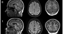
The value of diffusion-weighted imaging in the detection of prostate cancer SUMMARY Purpose: The aim of study was to determine the value of DWI in diagnosis of prostate carcinoma especially for differentiating benign from malignant lesion of the prostate. Materials and methods : During a period of 4/2014-3/2015, 41 consecutive patients with elevated PSA level were evaluated with DWI of the prostate. The results were confirmed by TRUSguided biopsy. We compare two groups (prostate carcinoma/PCa and prostate non-carcinoma/PNCa) by variants: tissue diffusivity, mean of ADC… Analyzing ROC curve to find the value of DWI in differentiating benign from malignant tissue of the prostate. Results: Patients ranged in age from 50 to 94 years (mean 73±10year). 18 patients were confirmed to have PNCa (44%), whereas 23 patients had Pca (56%). The mean of ADC values for PNCa and PCa were 829.2±119.2 and 544.6±102.7x10⁻³ mm2/s respectively. The mean ADC value of PCa was significantly lower than PNCa (p<0.05). On ROC curve, using the discrimination threshold of ADC is 633x10-3 mm2/s, the DWI provided a sensitivity of 82.6%, specificity of 94.4% and accuracy of 96%. Conclusions: Diffusion-weighted imaging of the prostate can be used to differentiate benign from malignant tissue of the prostate with high accuracy. Key words: Prostatic carcinoma, non-carcinoma, diffusion magnetic resonance imaging, ADC, TRUS.
Đặc điểm hình ảnh và vai trò cộng hưởng từ trong chẩn đoán rò hậu môn
01/04/2020 15:59:05 | 0 binh luận
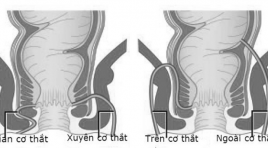
Imaging characteristic and role of MRI in diagnostic perianal fistula SUMMARY The aims of this study: To determine imaging chacteristic and the concordance in evaluation of perianal fistula between MRI image and operative result. Materials and methods: Descriptive study was performed from January 2014 to September 2015 at Ha Noi Medical University Hospital. 95 patients with clinically suspected fistula-inano underwent preoperative MRI and operated at HMU hospital. MRI scanning was performed on GE 1.5Tesla system with pelvis phased-array coil. Results: There were 95 patients including 81 males and 14 female. Value of MRI sequences in detection of perianal fituas is high. The ability of T2W, STIR and T1W fat sat +Gado in detection of fistulas are 94.7%, 95.7% and 98.9% respectively. In the classification of primary tracts, the concordance rate between MRI image and operative result was 84.2%, most of which were transphinteric fistulas. The concordance rate in identification of the internal opening and the extensive lesions were 96.6% and 94% respectively. Conlusions : MRI is a good imaging modality in preoperative evaluation of perianal fistulas with high accuracy (classification of primary tracts, detection of internal opening and assessment extensions). Keywords : Anal fistula, MRI, classification of fistulas.
Đặc điểm hình ảnh và vai trò của chụp cắt lớp vi tính trong chẩn đoán và theo dõi điều trị hóa chất ung thư phổi không tế bào nhỏ
01/04/2020 15:55:07 | 0 binh luận
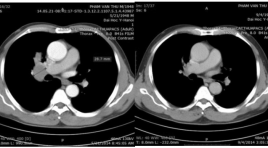
The image characteristic and the value of CT-Scanner in diagnosis of non-small cell lung cancer and follow-up post chemotherapy SUMMARY Lung cancer is non-small cell type common , accounting for 80% of all lung cancers and is the kind of good response to chemotherapy. The problem of early diagnosis and follow-up after treatment chemicals tumors is essential. Objective : Describe some features computerized tomography images of lung cancer non-small cell and rating the value of computerized tomography in monitoring chemotherapy lung cancer non -small cell. Subjects and Methods: The study conducted on 47 patients diagnosed with lung cancer non -small cell had computed tomography movie multiple slices at Medical University Hospital from 5/2012 to Hanoi in May / 201. Study design: descriptive cross-sectional, prospective combined with retrospective. Results : In 47 patients with 28 patients with tumors in the (R) 59.6%, 19 patients with tumors in the (L) 40,46%. Smallest tumor size 2,4cm, the largest 8.2 cm, medium 5.3 cm. Before average injection of contrast 100% of tumors with low levels < 50 Hu, after injecting drug absorbed 100% u > 20 HU. Bo irregular tumor (lobe segments) or fringed or hemp (100%). In 38 patients with metastases that 100% cost- compartment shaft size > = 15 mm and soaked after dye injection drugs. Which necrotic nodes, adhesive 9 patients (23.7%), Lymphadenopathy mere 29 patients (76.3%). Results after 6 round treatment chemicals according to RECIST Version 1.1: Complete response of 25 patients (53.3%), can meet 17 patients (36.1%), Stable 5 patients (10.6%), Progress: 0 patients (0%) Conclusion : CLVT valuable in the diagnosis and monitoring of cancer chemotherapy -small cell lung. Keywords: Lung cancer, lung cancer non-small cell, computed tomography.
Nghiên cứu đặc điểm hình ảnh cắt lớp vi tính trong chẩn đoán chảy máu não thất
01/04/2020 15:50:40 | 0 binh luận
The role of CT-Scanner in diagnosis of intraventricle hemorrhage SUMMARY Objective : To describe the feature of intraventricular hemorrhage (IVH) image in CT scanner and beginning describe some reasons related to IVH in adults. Methods: In a cross - sectional using quantitative methods, we chose 52 patients who definitive diagnosed IVH by CT scan in Bach Mai hospital from August/2014 to September/2015. Results: The most common location hematoma in primary parenchyma is hippocampal central gray (55.55%), the most common location of IVH is occipital horn ventricular, the more greater hematoma size, the more high risk of poor prognosis, the more higher level of IVH, the high risk of poor prognosis. In people over 50 years old, the most common causes of IVH is hypertension and hypertension coordinate with other causes (51%),while people under 50 years old, they usually caused by vascular abnormalities (30%), the main causes of thalamus lobe bleeding and hippocampal central gray bleeding is hypertension (25%), lobe of brain bleeding caused by aneurysm (42.86%). Conclusion: When patients are diagnosed stroke, they must be take CT scanner image immediately, when the results showed IVH image, they must be take cerebrovascular CT scanner with multiple array receiver, with intravenous injection of contrast dye to initially find the causes. Key words: CT scanner, intraventricular hemorrhage, cause.
Đánh giá các chỉ số tưới máu trong ung thư phổi trên chụp cắt lớp vi tính tưới máu
01/04/2020 14:06:24 | 0 binh luận
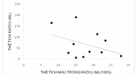
Assessement changing in haemodynamics and contrast enhancement pattern of lung cancer lesions on perfusion CT SUMMARY Introduction : Lung tumour perfusion CT may help to evaluate characteristics and haemodynamics of lung cancer. This research was carried out to assess changes in haemodynamics and contrast enhancement pattern of lung cancer lesions. Methods: 12 patients were enrolled to the study underwent perfusion CT of lung tumour. Perfusion paramerters were obtained from perfusion CT maps. Results: perfusion paramerters increased in malignant tumour with measured blood flow of 33.15 ±15.7 ml/100g/min, blood volume of 17ml/100g and tumour volume ranged from 7 – 577ml. blood volume in small tumours was higher than that in larger lesions. The higher homogeneity in blood flow was found in small tumours. Conclusion : Lung tumour perfusion CT could be used to assess changes in tumour haemodynamics that may help in pretreatment of unresectable lung cancers. Keyword: Lung cancer, perfusion.
Bạn Đọc Quan tâm
Sự kiện sắp diễn ra

THÔNG BÁO SINH HOẠT KHOA HỌC 19/01/2024: CẬP NHẬT CHẨN ĐOÁN VÀ ĐIỀU TRỊ UNG THƯ TRỰC TRÀNG
19-01-2024 09:47:01 0 bình luận

Thông tin đào tạo
- Những cạm bẫy trong CĐHA vú và vai trò của trí tuệ nhân tạo
- Hội thảo trực tuyến "Cắt lớp vi tính đếm Photon: từ lý thuyết tới thực tiễn lâm sàng”
- CHƯƠNG TRÌNH ĐÀO TẠO LIÊN TỤC VỀ HÌNH ẢNH HỌC THẦN KINH: BÀI 3: U não trong trục
- Danh sách học viên đạt chứng chỉ CME khóa học "Cập nhật RSNA 2021: Công nghệ mới trong Kỷ nguyên mới"
- Danh sách học viên đạt chứng chỉ CME khóa học "Đánh giá chức năng thất phải trên siêu âm đánh dấu mô cơ tim"


Đơn vị hợp tác

